Time-of-flight optical measurement and decoding of fast-optical signals

a technology of optical measurement and optical signal, applied in the field of noninvasive measurement methods and systems in the human body, can solve the problems of difficult prediction without detailed microscopic knowledge of the scattering characteristics of the brain volume of interest, difficult to obtain fast-optical signals by conventional optical detectors, and difficult to achieve the effects of sufficient temporal resolution and sensitivity

Active Publication Date: 2020-02-27
HI LLC
View PDF0 Cites 25 Cited by
  • Summary
  • Abstract
  • Description
  • Claims
  • Application Information

AI Technical Summary

Benefits of technology

[0011]There, thus, remains a need to provide a relatively simple optical measurement system for measuring the intensity of biologically intrinsic signals, such as fast-optical signals, in the brain at a sufficient temporal resolution and sensitivity.

Problems solved by technology

However, because optical measurement techniques rely on light, which scatters many times inside brain, skull, dura, pia, and skin tissues, the light paths occurring in these techniques comprise random or “diffusive” walks, and therefore, only limited sensitivity of fast-optical signals can be obtained by a conventional optical detector.
The reason for this limited detection sensitivity of fast-optical signals is that the paths of photons striking the detector in such schemes are highly variable and difficult, and even impossible, to predict without detailed microscopic knowledge of the scattering characteristics of the brain volume of interest, which is typically unavailable in practice (i.e., in the setting of non-invasive measurements through skull for brain imaging and brain interfacing).
In summary, light scattering has presented challenges for optical measurement techniques in achieving high sensitivity with regard to detecting fast-optical signals.
Moreover, the diffusive nature of light propagation also creates challenges for measurements of fast changes in optical scattering inside tissue, since essentially all photons traveling between source and detector are highly diffused to begin with.
Nearly all diffusive optical measurement techniques to date offer relatively poor temporal resolution (100 ms-1 sec per sample), as they are primarily designed to detect hemodynamics that vary on a similarly slow time scale.
Furthermore, some diffusive optical measurement techniques require relatively complicated circuitry for detecting a minute signal within the background light exiting the head.
Known diffusive optical measurement techniques, although potentially capable of detecting the occurrence of a fast-optical signal, are not capable of effectively, reliably, and accurately measuring the intensity of a fast-optical signal (see Syre F, et al., “Are VEP Corelated Fast Optical Signals Detectable in the Human Adult by Non-Invasive Nearinfrared Spectroscopy (NIRS)?”, Advances in Experimental Medicine and Biology. Vol. 530, pp.
However, because FD-NIRS modulates the light source at a frequency that is slower than the timescale of certain temporal features in fast-optical signals, i.e., slower than the timescales of time of flight variations in light propagation through the skin, skull and brain are that have been induced by fast-optical signals, FD-NIRS cannot efficiently or reliably detect these features, thereby limiting its detection sensitivity to fast-optical signals.
There are several orders of magnitude more of these shallow photons than signal photons, giving FD-NIRS an enormous background that adds shot noise, increases necessary dynamic range on the photon detector, etc.
However, this solution results in a significant loss of detected photons and significantly decreases the spatial resolution of the measurement.
In particular, these holographic methods mix the detected light, which takes the form of a spatially disordered speckle interference pattern, against a reference beam, thereby requiring a relatively complicated and expensive arrangement of components.
Further, while the iNIRS or SS-OCT approaches are very sophisticated, they require the detection and measurement of speckles, presenting challenges in a highly attenuating medium, such as the human body, due to the very low number of photons that reach each detector.
Thus, a camera with very large number of detectors (or pixels) are required to sufficiently detect many dim speckles, thereby further increasing the complexity and expense of the system.
Furthermore, even the fastest cameras, required to detect the speckles, are much slower than a single detector, and thus, the detection speed of the speckle interference pattern is limited in the iNIRS approach.
The iNIRS approach has also not yet been demonstrated to detect fast-optical signals.
However, this approach requires a complicated electronic circuit and is limited on the number of photons that can be counted as a unit of time.
Furthermore, this DOT approach has not yet been demonstrated to detect fast-optical signals.

Method used


Image

  • Time-of-flight optical measurement and decoding of fast-optical signals
  • Time-of-flight optical measurement and decoding of fast-optical signals
  • Time-of-flight optical measurement and decoding of fast-optical signals

Examples


Embodiment Construction

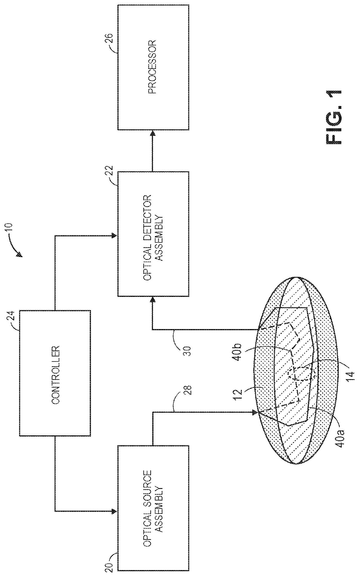
[0079]Referring to FIG. 1, one embodiment of an optical measurement 10 (and variations thereof) constructed in accordance with the present inventions will be described. The optical measurement system 10 is designed to non-invasively acquire physiological-encoded signal light (i.e., signal light representative of a physiologically-dependent optical parameter) in an anatomical structure 12 via one or more optical paths 14, processing the physiological-encoded signal light, and distinguishing between states of a physiological activity within the anatomical structure 12.

[0080]It should be appreciated that the term “state,” in the absolute sense, is arbitrary, and is only meaningful in the relative sense when compared to other states. For example, the physiological activity may have either an active state or an inactive state in a binary sense (either the physiological activity is present or the physiological activity is absent), or the physiological activity may have many (even an infin...


PUM


Abstract

An optical measurement system comprising an optical source configured for delivering sample light in an anatomical structure, such that the sample light is scattered by the anatomical structure, resulting in physiological-encoded signal light that exits the anatomical structure, an optical detector configured for detecting the physiological-encoded signal light, and a processor configured for acquiring a TOF profile derived from the physiological-encoded signal light, the initial TOF profile having an initial contrast-to-noise ratio (CNR) between a plurality of states of a physiological activity in the anatomical structure. The processor is further configured for applying one or more weighting functions to the initial TOF profile to generate a weighted TOF profile having a subsequent CNR greater than the initial CNR between the plurality of states of the physiological activity. The processor is further configured for processing the weighted TOF profile, and identifying one of the plurality of states of the physiological activity.

Description

RELATED APPLICATION DATA[0001]This application claims the benefit of U.S. Provisional Patent Application 62 / 722,152, filed Aug. 23, 2018, which is expressly incorporated herein by reference.FIELD OF THE INVENTION[0002]The present inventions relate to methods and systems for non-invasive measurements in the human body, and in particular, methods and systems related to detecting physiological activity in the human body, animal body, and / or biological tissue.BACKGROUND OF THE INVENTION[0003]Measuring neural activity in the brain is useful for medical diagnostics, neuromodulation therapies, neuroengineering, or brain-computer interfacing. Conventional methods for measuring neural activity in the brain include diffusive optical imaging techniques, which employ moderate amounts of near-infrared or visible light radiation, thus being comparatively safe and gentle for a biological subject in comparison to X-Ray Computed Tomography (CT) scans, positron emission tomography (PET), or other met...

Claims


Application Information

Patent Timeline
Patent Type & Authority Applications(United States)
IPC IPC(8): A61B5/00
CPCA61B5/0004A61B5/0261A61B5/0084A61B5/0066
Inventor ALFORD, JAMUMARBLESTONE, ADAMVELLEKOOP, IVOSOBEK, DANIELHENNINGER, MICHAELROBINSON, BRIANSHEN, YUECHENGHORSTMEYER, ROARKE
Owner HI LLC
Why Eureka
Try Eureka