

Methods of Using Multilayer Magnetic Micelle Compositions
a technology of magnetic micelle composition and composition, applied in the direction of capsule delivery, microcapsules, gene material ingredients, etc., can solve the problems of hampered clinical applications, low chitosan transfection efficiency, and difficult gene delivery
- Summary
- Abstract
- Description
- Claims
- Application Information
AI Technical Summary
Benefits of technology
Problems solved by technology
Method used
Image

Examples
example 1
Preparation of Multilayered Mag-Micelles (4MNPs)
[0065]The 4MNPs were prepared and conjugated with DNA as described by Wang et al. [C. Wang et al., Journal of Controlled Release 2012, 163, 82], MPEG-PLA-OH diblock copolymers were synthesized from DL-dilactide and methoxypolyethylene glycol (mPEG) of various molecular weights using stannous 2-ethyl-hexanoate as a catalyst by catalyzed ring-opening polymerization [A. Lucke et al. Biomaterials 2000, 21, 2361]. First, DL-dilactide was vacuum-dried at room temperature for 4 hours and mPEG (MW 20 K Da) was vacuum-dried at 80° C. for 3 hours. Then 0.5 g of dried mPEG, 3 g of dry DL-dilactide, the stannous 2-ethyl-hexanoate (3% w / w) and 20 mL of toluene were added into a two-neck flask and mixed. The reaction solution was refluxed for 5 hours at 140° C. under argon gas protection and precipitated with cold diethyl ether. The purified product was kept under vacuum at room temperature for 24 hours. SPIONs were prepared according to the procedu...
example 2
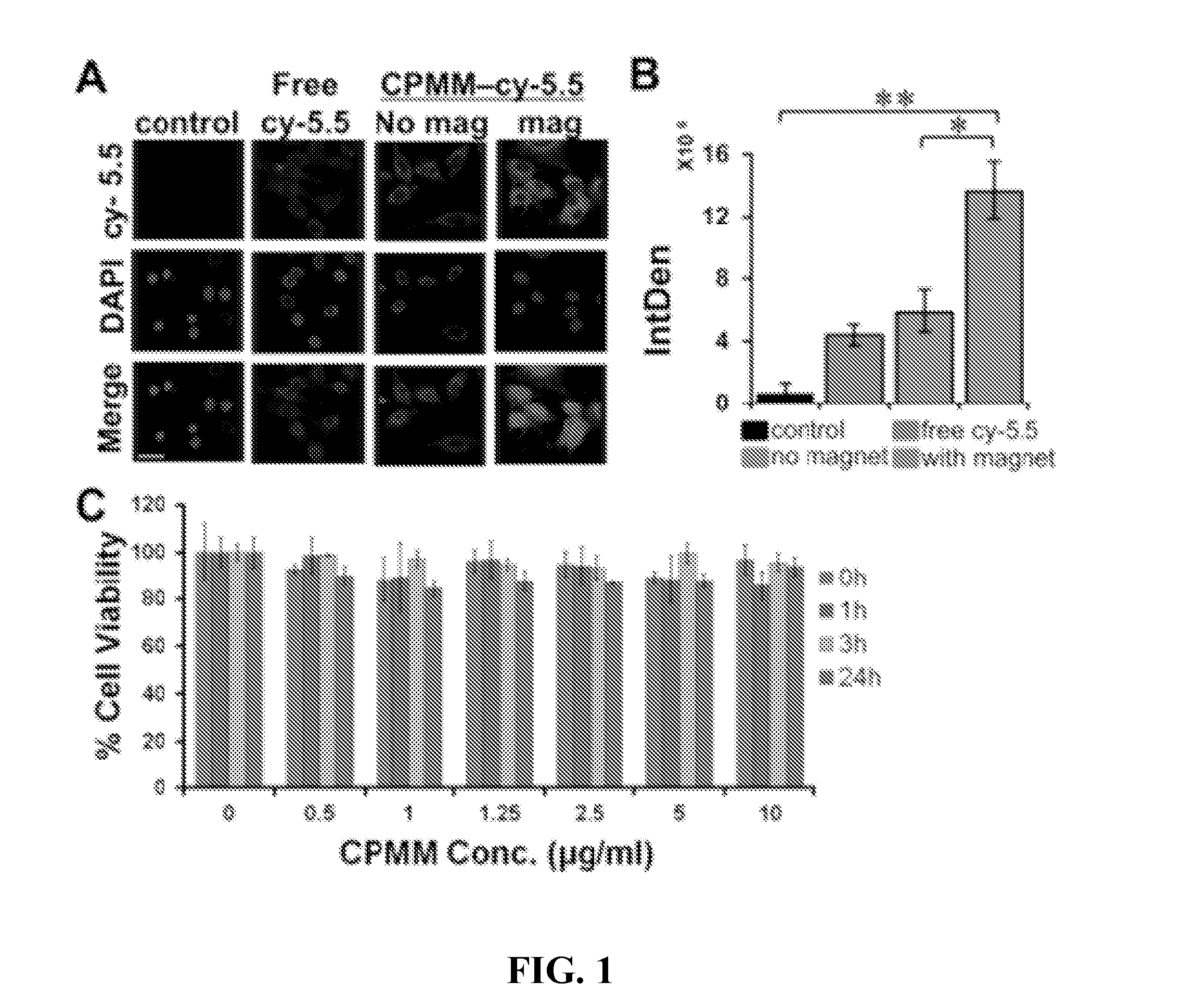
Cellular Uptake of 4MNP and its Effect on Cell Viability
[0066]HT22 cells were treated with free cy-5.5 dye or cy-5.5 conjugated 4MNP with or without magnet for 1 hour. HT22 cells (donated by Dr. Bruce Citron, Bay Pine VA Hospital, Fla.) were cultured in DMEM with 10% FBS and 1% penicillin / streptomycin in an atmosphere of 5% CO2. Cells were plated at a density of 20,000 per well in 8 well-chamber slides 24 hours prior to the experiment. cy-5.5 was conjugated to the 4MNP nanoparticles at a ratio of 4MNP:cy-5.5 of 1:10 and dialyzed for 24 hours to remove excess cy-5.5. 4MNP-cy-5.5 conjugate equivalent to 2.5 μg / ml 4MNP was added to the cells and incubated at 37° C. for 1 hour with or without a bar magnet underneath the wells. An equal amount of free cy-5.5 was used as a control. After 1 hour, cells were washed 3 times with sterile PBS and fixed with 4% paraformaldehyde for 10 minutes, washed with sterile PBS and cover slipped using DAPI containing mounting medium. Cells were observed u...
example 3
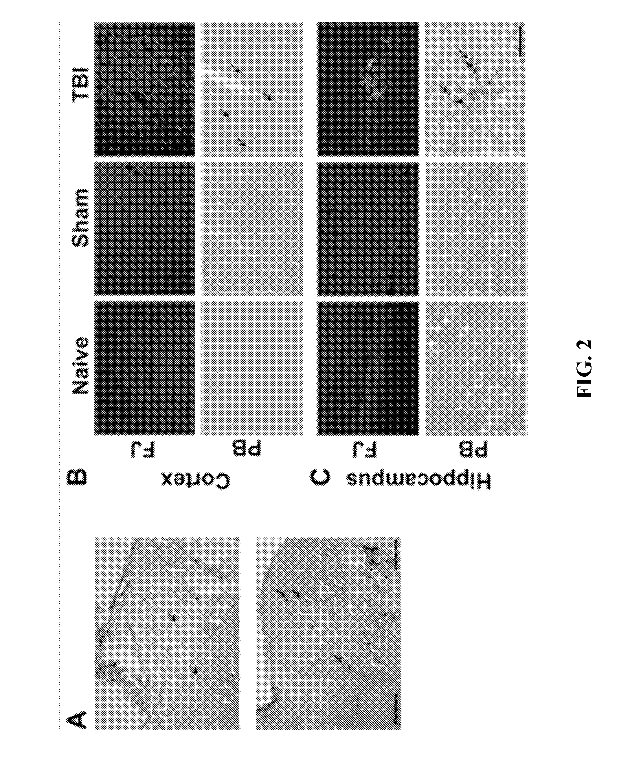
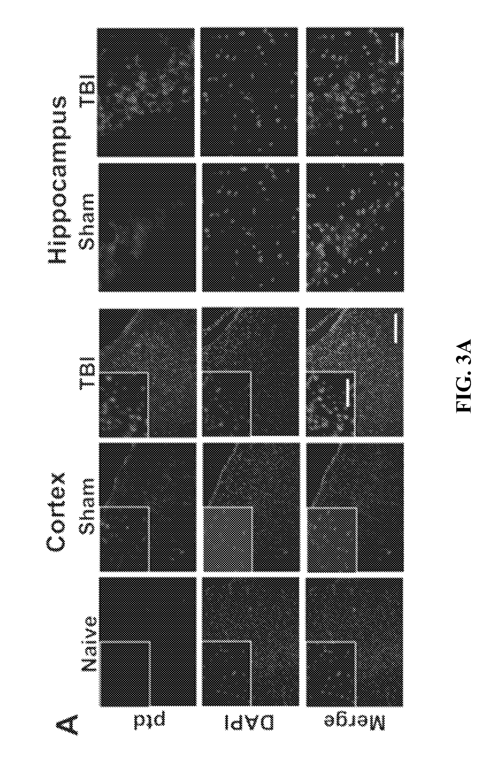
Effect of Magnetic Field on the Concentration of 4MNP in the Brain after Mild TBI
[0071]To observe whether magnetofection causes any differences in concentrating 4MNP nanoparticles in the brain or not, rats were subjected to mild TBI (mTBI) to the cerebral cortex or were sham operated and, immediately thereafter, td Tomato plasmid-complexed 4MNPs were administered intranasally (i.n.). One group of 6 rats, including 3 sham and 3 mTBI animals, were subjected to a magnetic field for 1 hour. Another group of 6 rats, also including 3 sham and 3 mTBI animals, were not. The degree of Prussian Blue (PB) staining was compared between the two groups.
[0072]All animal procedures were conducted in accordance with the NIH Guide for the Care and Use of Laboratory Animals following a protocol approved by the Institutional Animal Care and Use Committee at the University of South Florida. Male Sprague-Dawley rats (Harlan, Indianapolis, Ind.) weighing 250 to 300 g were housed in a climate-controlled ro...
PUM
| Property | Measurement | Unit |
|---|---|---|
| Time | aaaaa | aaaaa |
| Weight ratio | aaaaa | aaaaa |
| Hydrophobicity | aaaaa | aaaaa |
Abstract
Description
Claims
Application Information