Self-assembling nanoparticle drug delivery system
a nanoparticle and nanoparticle technology, applied in the direction of granular delivery, virus, dna/rna fragmentation, etc., to achieve the effect of improving the binding affinity of bioactive agents
- Summary
- Abstract
- Description
- Claims
- Application Information
AI Technical Summary
Benefits of technology
Problems solved by technology
Method used
Image
Examples
example 1
77C His-Tagged Core Protein
[0142]The 77C His-tagged Core Protein was cloned into the NdeI / XhoI restriction sites of vector pET21b (Novagen). This plasmid was transformed into E. coli BL21 (DE3) PlysS cells (Stratagene) for protein expression via normal methods. The nucleic acid and amino acid sequences are below.
[0143]77C His-tagged Core Protein has the following nucleic acid sequence:
(SEQID NO: 28)ATG GAT ATC GAT CCG TAT AAA GAA TTT GGC GCC ACC GTG GAA CTG CTG AGC TTTCTG CCG AGC GAT TTC TTT CCG AGC GTG CGT GAT CTG CTG GAT ACC GCG AGC GCGCTG TAT CGC GAA GCG CTG GAA AGC CCG GAA CAT TGT AGC CCG CAC CAT ACC GCCCTG CGT CAG GCG ATT CTG TGC TGG GGT GAA CTG ATG ACC CTG GCG ACC TGG GTTGGC AAC AAC CTG TGT GAT CCG GCG AGC CGC GAT CTG GTT GTG AAC TAT GTG AATACC AAC ATG GGC CTG AAA ATT CGT CAG CTG CTG TGG TTT CAT ATC AGC TGC CTGACC TTT GGC CGC GAA ACC GTG CTG GAA TAT CTG GTG AGC TTT GGC GTT TGG ATCCGT ACC CCG CCG GCG TAT CGT CCG CCG AAT GCG CCG ATT CTG AGC ACC CTG CCGGAA ACC ACC GTT GTG CGT CGC...
example 2
[0150]The various wild type and modified core proteins described herein can be expressed and purified according to Protocol 1 or Protocol 2 as follows:
Protocol 1:
[0151]A pET-11a vector containing the full-length HBV C-protein gene, is transformed into E. coli DE3 cells and grown at 37° C. in LB media, fortified with 2-4% glucose, trace elements and 200 ug / mL carbenicillin. Protein expression is induced by the addition of 2 mM IPTG (isopropyl-beta-D-thiogalactopyranoside). Cells are harvested by pelleting after three hours of induction. SDS-PAGE is used to assess expression of C-protein.
[0152]Core protein is purified from E. coli by resuspending in a solution of 50 mM Tris-HCl, pH 7.4, 1 mM EDTA, 5 mM DTT, 1 mM AEBSF, 0.1 mg / mL DNasel and 0.1 mg / mL RNase. Cells are then lysed by passage through a French pressure cell. The suspension is centrifuged at 26000×G for one hour. The pellet is discarded and solid sucrose added to the supernatant to a final concentration of 0.15 M and centrif...
example 3
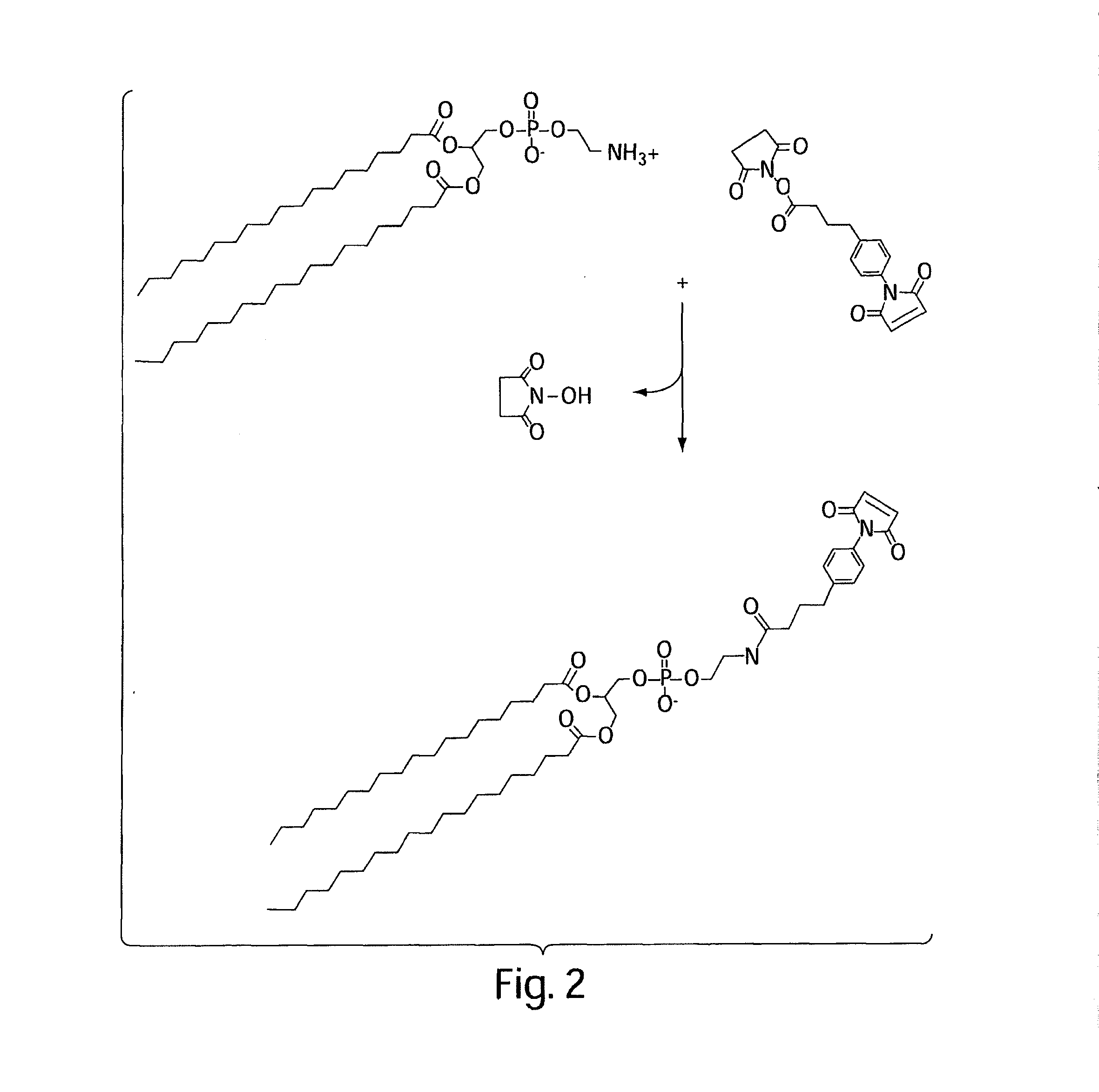
[0159]The following protocol describes conjugation of phospholipids via a SMPB (succinimidyl-4-(p-maleimidophenyl)butyrate) intermediate. This is shown schematically in FIG. 2.
[0160]1. Dissolve 100 micromoles of phosphatidyl ethanolamine (PE) in 5 mL of argon-purged, anhydrous methanol containing 100 micromoles of triethylamine (TEA). Maintain the solution under an argon or nitrogen atmosphere. The reaction can also be done in dry chloroform.
[0161]2. Add 50 mg of SMPB (Pierce) to the PE solution. Mix well to dissolve.
[0162]3. React for 2 hours at room temperature, while maintaining the solution under an argon or nitrogen atmosphere.
[0163]4. Remove the methanol from the reaction solution by rotary evaporation and redissolve the solids in chloroform (5 mL).
[0164]5. Extract the water-soluble reaction by-products from the chloroform with an equal volume of 1% NaCl. Extract twice.
[0165]6. Purify the MPB-PE derivative by chromatography on a column of silicic acid (Martin F J et al, Immuno...
PUM
| Property | Measurement | Unit |
|---|---|---|
| ionic strength | aaaaa | aaaaa |
| size | aaaaa | aaaaa |
| ionic strength | aaaaa | aaaaa |
Abstract
Description
Claims
Application Information
 - R&D
- Intellectual Property
- Life Sciences
- Materials
- Tech Scout
- Unparalleled Data Quality
- Higher Quality Content
- 60% Fewer Hallucinations
Browse by: Latest US Patents, China's latest patents, Technical Efficacy Thesaurus, Application Domain, Technology Topic, Popular Technical Reports.
© 2025 PatSnap. All rights reserved.Legal|Privacy policy|Modern Slavery Act Transparency Statement|Sitemap|About US| Contact US: help@patsnap.com


