
Methods and Apparatus for Optoacoustic Guidance and Confirmation of Placement of Indwelling Medical Apparatus

a technology of optoacoustic guidance and confirmation, which is applied in the field of apparatus and methods for guiding and confirming the placement of medical devices, can solve the problems of affecting the placement or positioning of endotracheal tubes, and affecting the safety of patients

Active Publication Date: 2014-02-27
BOARD OF RGT THE UNIV OF TEXAS SYST
View PDF18 Cites 34 Cited by
  • Summary
  • Abstract
  • Description
  • Claims
  • Application Information

AI Technical Summary

Benefits of technology

The patent describes a system and method for tracking the placement of medical devices placed through a body channel or cavity. The system includes an indwelling device with an optical component that emits light into the tissue near the placement site. The light is absorbed by the tissue and converted into an acoustic signal by an acoustic component. The acoustic signal is then received by external detectors and analyzed by an analyzer to produce information about the placement of the device. The system can provide real-time tracking and can be used with different types of medical devices, such as endotracheal tubes. The technical effects of the patent include improved placement accuracy and reduced risk of complications during medical procedures.

Problems solved by technology

For example, improper placement or positioning of an endotracheal tube may be lethal.
However, endotracheal tubes are often misplaced, particularly when placed in emergency circumstances, and endotracheal tube misplacement contributes to morbidity and mortality.
Placement in the esophagus rather than in the trachea results in failure to effectively provide oxygen and remove carbon dioxide.
Even a single breath administered while a tube is improperly positioned in the esophagus risks gastric inflation and promotes regurgitation and aspiration of gastric contents.
Positioning of an endotracheal tube insufficiently far into the trachea risks laryngeal damage from cuff pressure on the structures in the larynx and, of greater immediate concern, risks accidental withdrawal into the pharynx.
Positioning of an endotracheal tube too deeply may result in intubation of a main-stem bronchus, usually the right, causing hypoxemia because of failure to ventilate the opposite, usually the left, lung.
Patients requiring emergency intubation usually have severe physiologic compromise, such as respiratory failure and cardiac arrest, and often must be intubated under poorly controlled circumstances by personnel with highly variable experience and expertise.
These patients are particularly vulnerable to episodic hypoxemia.
In out-of-hospital situations, placement is usually performed by emergency medicine technicians or paramedics, whose experience and training often are limited.
The inevitable disparities in experience and expertise between ED physicians, respiratory therapists, anesthesiologists and out-of-hospital emergency responders are compounded in emergency circumstances by less than optimal conditions and limited monitoring equipment.
These important factors further reduce the chances of proper initial placement and subsequent maintenance of proper positioning of endotracheal tubes.
Currently available technology is unsuitable for monitoring of endotracheal position, especially by personnel of limited experience.
Although each is relatively reliable, each also is associated with errors, the consequences of which can be grave.
In some patients, visualization of the larynx is inadequate to confirm endotracheal tube placement.
Direct visualization of an endotracheal tube passing the cords requires expertise in laryngoscopy, is sometimes difficult or impossible to achieve, and cannot be performed repeatedly.
Fiberoptic bronchoscopy requires technical expertise, interferes with ventilation, and cannot be performed continuously.
Chest radiography is intermittent, requires movement of a patient to perform radiography, and does not provide rapid feedback.
Auscultation is prone to both false-positive and false-negative findings.
The dramatic decrease in respiratory complications of anesthesia during the past thirty years is certainly attributable in part to expeditious recognition and correction of esophageal intubation, although anesthesia personnel continue to be challenged by difficulty in confirming endotracheal tube placement in the mid-trachea, especially in circumstances in which post-intubation movement of a patient can result in movement of the tube within the trachea.
The challenges of recognizing esophageal intubation and endotracheal tube movement are much greater in emergency circumstances outside the operating room.
However, in emergency circumstances, especially during cardiac arrest, capnography is less reliable because carbon dioxide exhalation is highly variable and requires ventilation.
In patients during cardiac arrest, minimal carbon dioxide may be exhaled through the lungs and use of this method provides a substantial incidence of false-positive and false-negative results in emergency intubations.
However, the SCOTI device requires disconnection from the ventilator, only differentiates esophageal from tracheal intubation, has an appreciable false-positive and false-negative rate and does not indicate proper position within the trachea.
Ultrasound-based techniques require expertise in ultrasonography and are not suitable for continuous monitoring.
Although all approaches offer advantages and provide feedback that can be helpful, no single device is sufficiently reliable to be considered the standard of care and some, such as fiberoptic bronchoscopy, require substantial skill and training.

Method used


Image

  • Methods and Apparatus for Optoacoustic Guidance and Confirmation of Placement of Indwelling Medical Apparatus
  • Methods and Apparatus for Optoacoustic Guidance and Confirmation of Placement of Indwelling Medical Apparatus
  • Methods and Apparatus for Optoacoustic Guidance and Confirmation of Placement of Indwelling Medical Apparatus

Examples


example 1

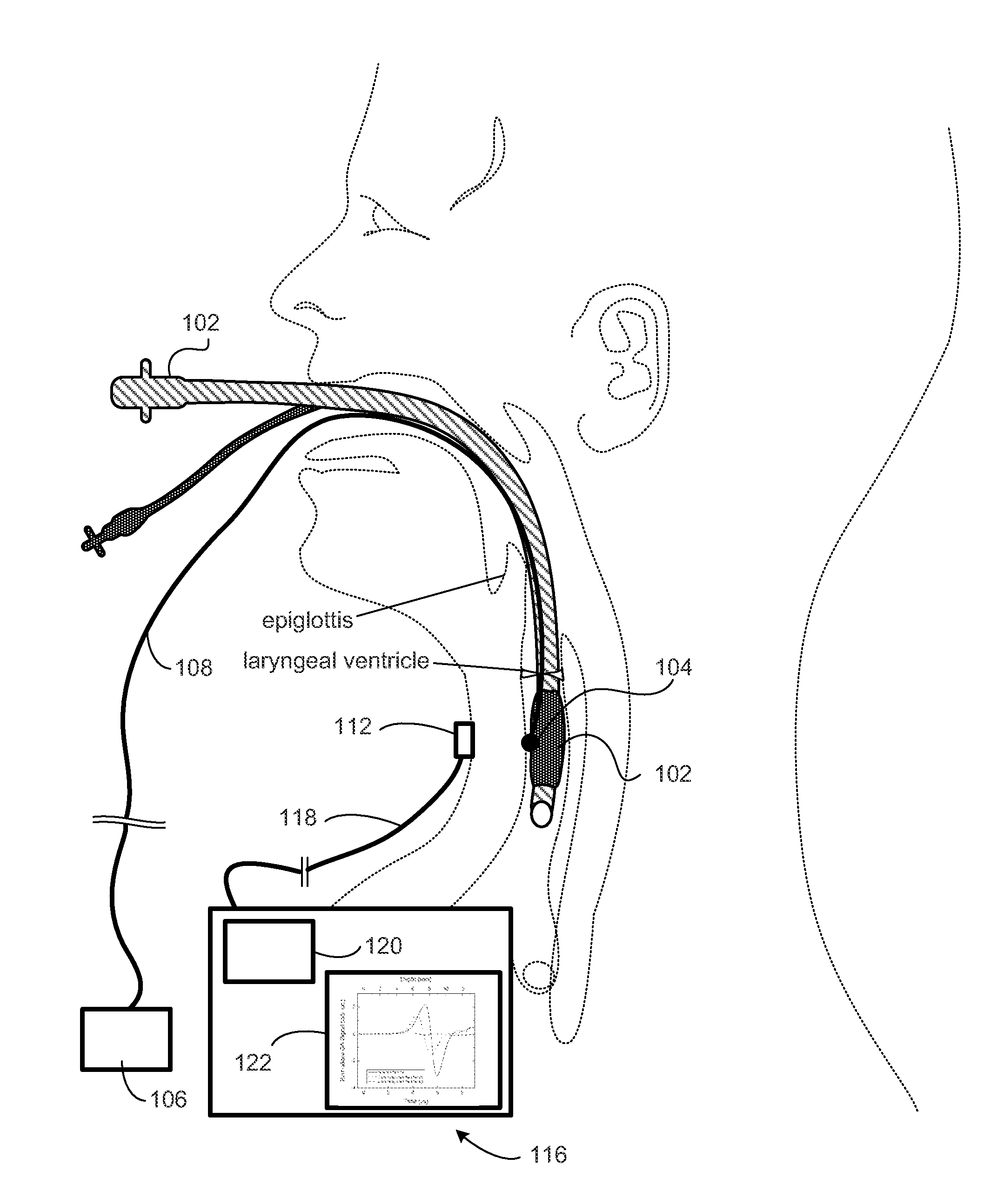
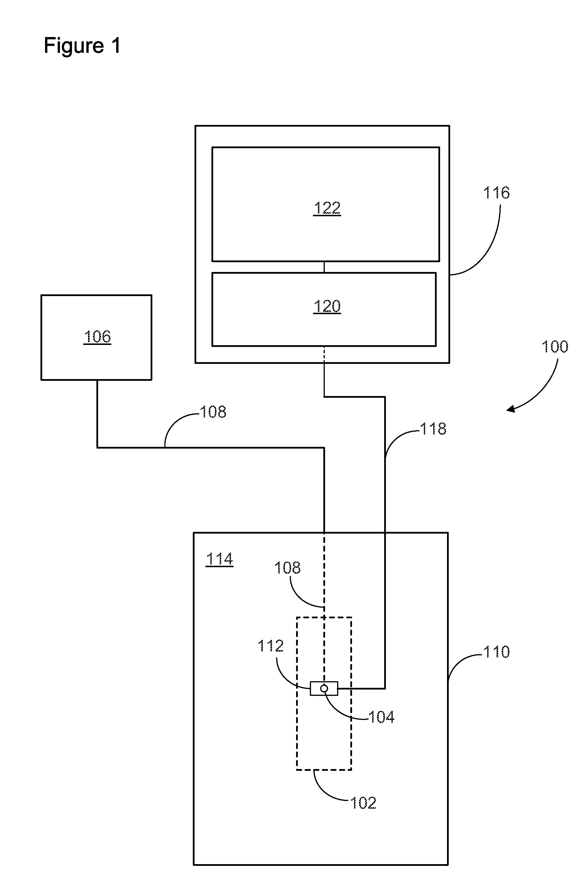
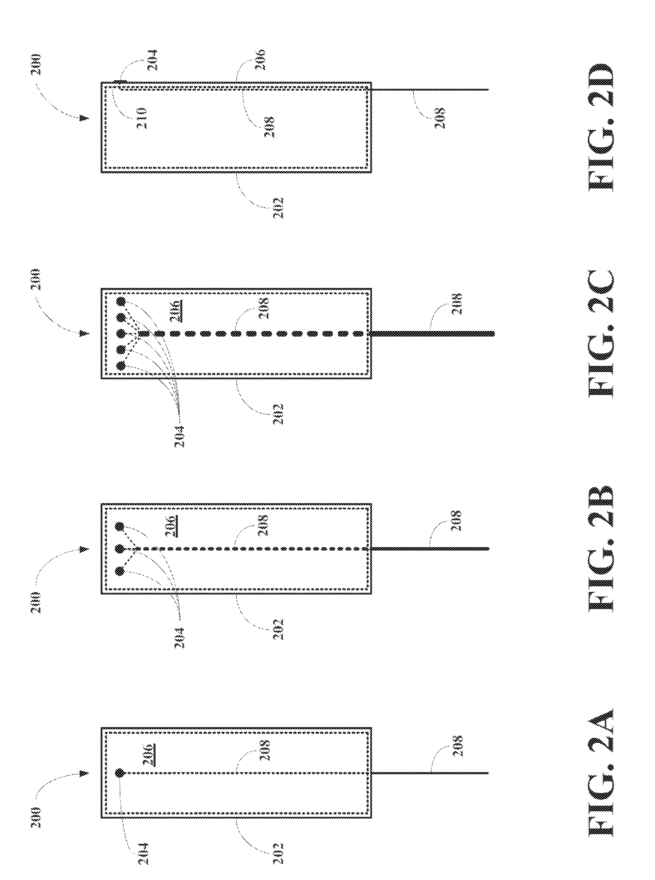
[0056]An embodiment of an endotracheal tube apparatus as disclosed herein including a pulsed laser light source, a light conduit, a light exit port and an acoustic detector was tested to demonstrate that a signal obtained by positioning a pulsed laser light exit port within an endotracheal tube in a sheep with an acoustic detector positioned on an anterior neck to permit non-invasive accurate confirmation and maintenance of the tube location in the sheep.

[0057]Laser optoacoustic imaging combines the merits of optical tomography (high optical contrast) and ultrasound imaging (minimal scattering of acoustic waves) to yield high contrast, sensitivity, and resolution. Certain of the present inventors have developed laser optoacoustics as a technique for tissue characterization and diagnostic imaging. See e.g. Esenaliev R O, et al. “Laser opto-acoustic tomography for medical diagnostics: Experiments with biological issues”SPIE Proc. 1996; 2676: 84-90. Optoacoustic techniques utilize sens...


PUM


Abstract

Medical apparatus are disclosed for optoacoustic monitoring of an indwelling unit of the apparatus, where the indwelling unit includes one or more optical components capable of directing pulsed light into an overlying tissue. The apparatus also include one or more acoustic components in contact with an exterior surface of the tissue to detect induced pressure waves producing an acoustic output analyzed with an optoacoustic unit to monitor and confirm proper placement of the indwelling unit. Methods for using the apparatus are also disclosed.

Description

CROSS REFERENCE TO RELATED APPLICATIONS[0001]This application claims priority based on U.S. Provisional Application Ser. No. 61 / 480,482 filed Apr. 29, 2011, which is incorporated herein by reference.FIELD OF THE INVENTION[0002]This invention relates generally to apparatus and methods for guiding and confirming placement of medical devices. More particularly, embodiments of this invention relate to medical apparatus and methods that utilize optoacoustic guidance and confirmation of placement of endotracheal tubes and other indwelling medical devices.BACKGROUND OF THE INVENTION[0003]Without limiting the scope of the invention, its background is described in connection with existing methods and apparatus for guiding and confirming proper placement of medical apparatus that are inserted through a body channel or cavity.[0004]Medical diagnosis and treatment frequently involve insertion of medical devices that are passed through naturally occurring channels and cavities in the patient's b...

Claims


Application Information

Patent Timeline
Patent Type & Authority Applications(United States)
IPC IPC(8): A61B5/06A61B1/06A61M16/04A61B5/00A61B1/07
CPCA61B5/061A61B5/0095A61M16/0434A61M16/0488A61B1/0676A61B1/07A61B5/06A61B2505/05A61M2205/3375A61M2205/581A61M2205/583A61M2205/502A61M16/0411
Inventor PROUGH, DONALD S.ESENALIEV, RINAT O.PETROV, YURIYPETROV, IRENE
Owner BOARD OF RGT THE UNIV OF TEXAS SYST
Features
  • Generate Ideas
  • Intellectual Property
  • Life Sciences
  • Materials
  • Tech Scout
  • Unparalleled Data Quality
  • Higher Quality Content
  • 60% Fewer Hallucinations
Learn More