Compositions and methods for culturing cells from normal human tubo-ovarian epithelium and human tubo-ovarian tumors
a technology of ovarian epithelium and ovarian tumors, which is applied in the field of compositions and methods for culturing cells from normal human tubo-ovarian epithelium and human tubo-ovarian tumors, can solve the problems of inability to distinguish between subtypes of ovarian cells, and many cell types are difficult to culture using available media and existing techniques
- Summary
- Abstract
- Description
- Claims
- Application Information
AI Technical Summary
Benefits of technology
Problems solved by technology
Method used
Image

Examples
example 1
Establishment of Normal Ovarian and Fallopian Tube Cultures
[0156]a. WIT-fo Medium:
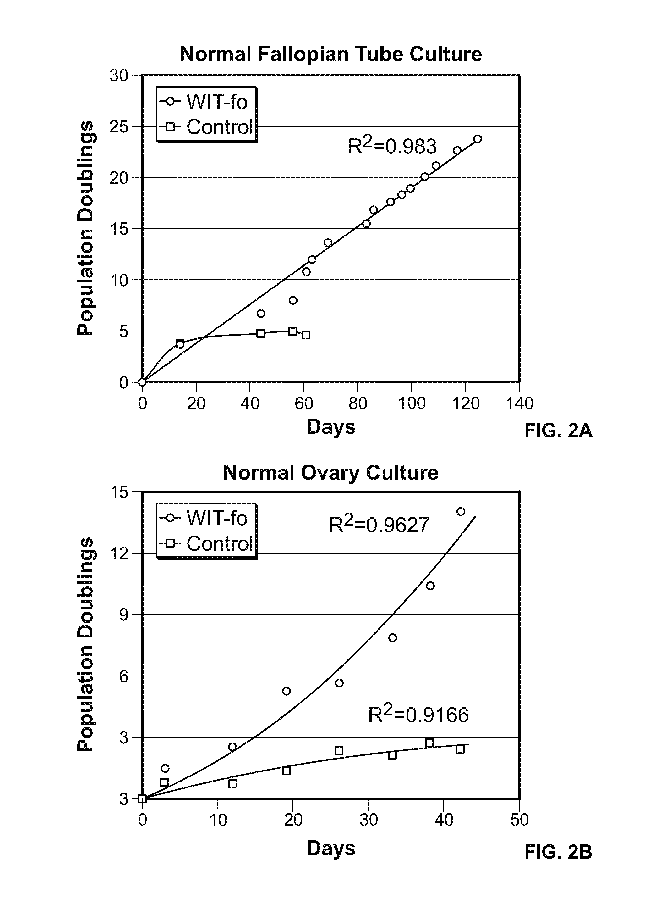
[0157]The normal ovarian epithelial and fallopian tube epithelial cells were cultured in WIT-fo nutrient medium at least 15 population doublings (FIG. 2 A-B, blue line), while replica plates of the same cells under standard media conditions stopped growing after a few passages (FIG. 2 A-B, red line).
[0158]b. Standard Ovarian Epithelial Culture Medium:
[0159]For ovarian epithelial cells, a control medium composed of a 1:1 mixture of MCDB 105 / Medium 199 supplemented with a range of 5-10% fetal bovine serum, 2 mm 1-glutamine and 10 ng / ml epidermal growth factor was used. Dulbecco's modified Eagle's medium (DMEM) / Ham's F-12 (1:1 mixture) with 10-15% fetal bovine serum as a control medium produced similar results. These two media have been used by many other investigators over the past two decades for short term culture of ovarian cells. Ovarian cells were not propagated beyond a few population doublings in ...
example 2
Characterization of Normal Ovarian and Fallopian Tube Cells Cultured in WIT-Fo
[0168]In order to identify the lineage and origin of the cultured cells, immunohistochemical characterization of normal human ovary and fallopian tube tissues was performed and a marker panel that distinguishes different subsets of epithelial cells in normal fallopian tube and ovarian tissues in vivo was developed.
[0169]The immunohistochemical examination of formalin-fixed paraffin embedded (FFPE) sections of normal human ovarian and fallopian tube tissues with antibodies that recognize different cells defined normal cell subsets. After screening a number of antibodies, a panel of three antibodies—PAX8, FOXJ1 and Keratin 7 (CK7)—was determined to allow distinguishing between surface vs. inclusion cyst epithelium in the ovary, and ciliated vs. non-ciliated cells in the fallopian tube. All the ovary epithelium and a subset of fallopian tube epithelium were determined to be Keratin 7 positive (+). While the o...
example 3
Establishment of hTERT Immortalized Normal Ovarian and Fallopian Tube Cultures
[0172]a. Immortalization of normal ovarian and fallopian tube cultures. Normal human epithelial cells do not grow well in cell culture and have a finite life span in conventional culture media. In contrast, it is more straightforward to culture all rodent cells and non-epithelial human cell types.
[0173]In order to establish continuous normal human epithelial cell cultures, oncogenic transformation is used to create cell lines that can be cultured continuously. In this approach, the oncogenic transformation of normal epithelial cells is achieved by exposing normal human cells to chemical mutagens, radiation or other carcinogenic agents. These agents cause widespread mutations, DNA breaks and chromosomal rearrangements, and cells may no longer be considered as fully normal cells. An alternative method for transforming normal cells uses viral oncogenes to transform normal cells. Among these Human Papilloma Vi...
PUM
| Property | Measurement | Unit |
|---|---|---|
| v/v | aaaaa | aaaaa |
| concentration | aaaaa | aaaaa |
| concentration | aaaaa | aaaaa |
Abstract
Description
Claims
Application Information
 - R&D
- Intellectual Property
- Life Sciences
- Materials
- Tech Scout
- Unparalleled Data Quality
- Higher Quality Content
- 60% Fewer Hallucinations
Browse by: Latest US Patents, China's latest patents, Technical Efficacy Thesaurus, Application Domain, Technology Topic, Popular Technical Reports.
© 2025 PatSnap. All rights reserved.Legal|Privacy policy|Modern Slavery Act Transparency Statement|Sitemap|About US| Contact US: help@patsnap.com