Methods and Devices for Detection and Acquisition of Biomarkers

a biomarker and detection method technology, applied in the field of biomarker detection and acquisition methods and devices, can solve the problems of not directly sampling clinically relevant biomarkers from the site, unable to achieve rapid and highly sensitive detection of changes in a biomarker, and unable to directly sample clinically relevant biomarkers, etc., to achieve the effect of avoiding misdiagnosis risks, avoiding misdiagnosis risks, and avoiding misdiagnosis risks

Active Publication Date: 2014-09-25
THE SCRIPPS RES INST +1
View PDF6 Cites 10 Cited by
  • Summary
  • Abstract
  • Description
  • Claims
  • Application Information

AI Technical Summary

Benefits of technology

[0005]In one aspect, the invention provides devices for detecting or extracting one or more biomarkers from a tissue or biological sample of a subject in situ. The devices can comprise an array of microneedles and a plurality of probes specific for the biomarkers, wherein the probes are covalently attached to the microneedles. In some of

Problems solved by technology

Rapid and highly sensitive detection of changes in a biomarker is often technically impossible, or may require a cumbersome procedure involving multiple processing steps, necessitating large sample volumes and a prolonged diagnosis/prognosis timeline.
The sample from a patient is often of a limited volume and not amenable to processing or to procedures requiring multiple steps that ex

Method used


Image

  • Methods and Devices for Detection and Acquisition of Biomarkers
  • Methods and Devices for Detection and Acquisition of Biomarkers
  • Methods and Devices for Detection and Acquisition of Biomarkers

Examples


example 1

Detecting ssDNA in Solution with Polycarbonate Immobilized Probes

[0162]This Example describes polycarbonate functionalized by a DNA probe and its utilizations for specific capture of ssDNA from solution.

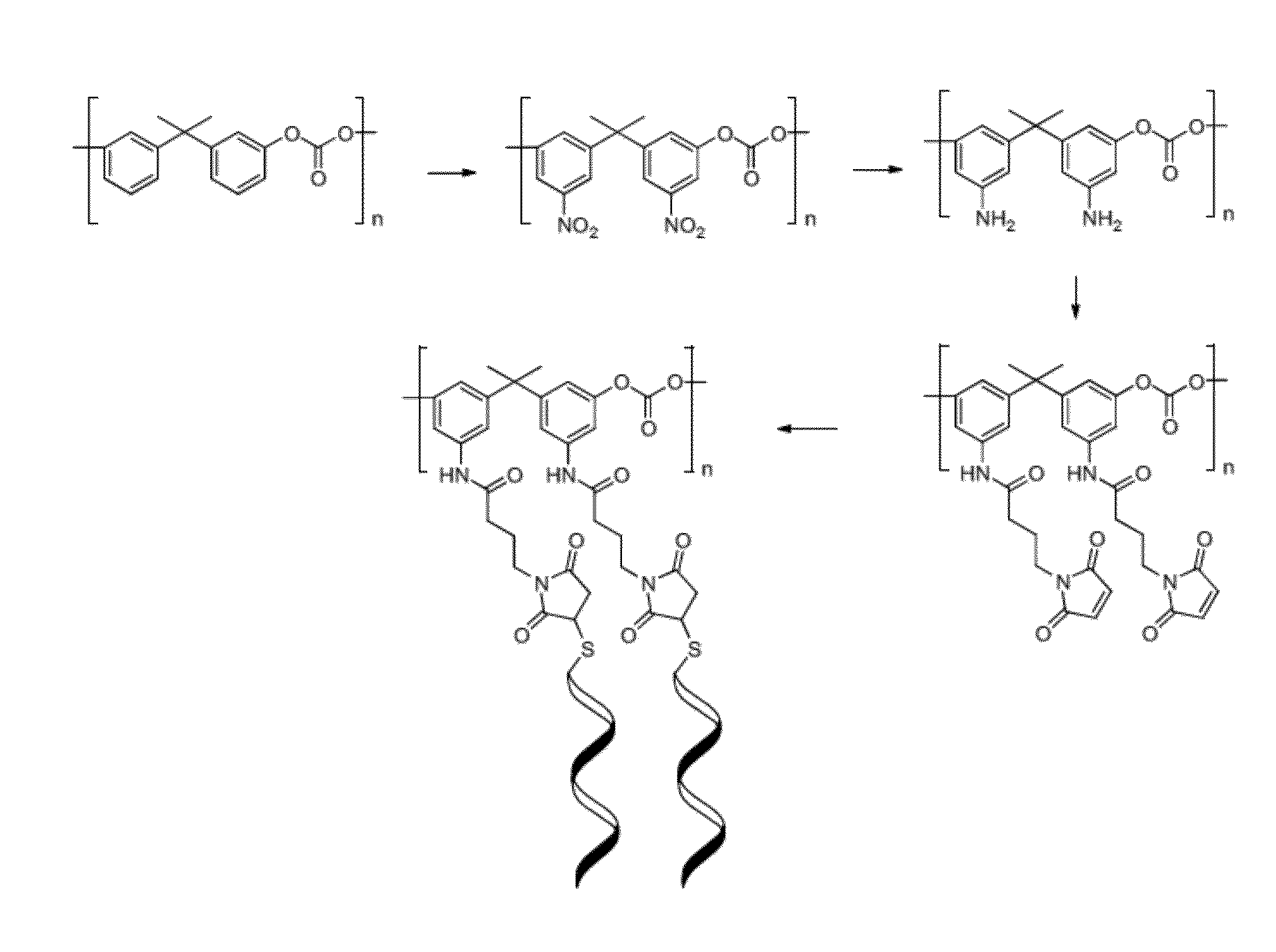
[0163]Attachment of DNA probes to polycarbonate: The surface of polycarbonate was first nitrated and reduced to introduce amino groups which were used further to attach commercially available thiol / amino bifunctional linker (FIG. 1). DNA with 3′ thiol modification was subsequently coupled to the linker tethered to the polycarbonate.

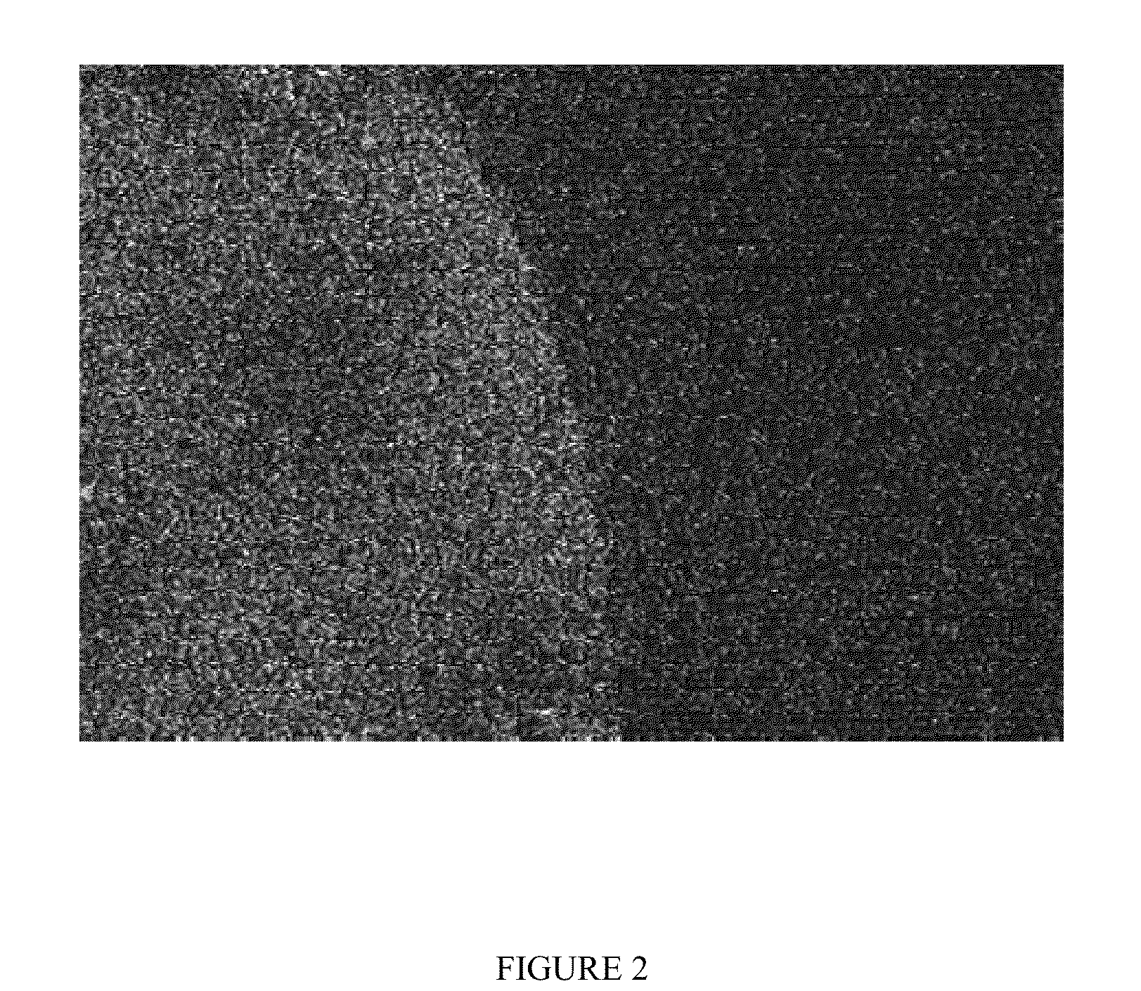
[0164]Visualization of 5′-Cy5 modified DNA attached to polycarbonate: To visualize the DNA on the surface of the polycarbonate, DNA modified with one Cy5 at the 5′ and thiol at the 3′ was attached using chemistry described above. Single layer 5′Cy5-DNA was imaged with confocal microscopy (FIG. 2) showing successful functionalization of the surface. In vitro capture of ssDNA on polycarbonate modified with specific DNA probe: In order to test the ability of ...

example 2

Conjugation of DNA Probes to Stainless Steel Surfaces

[0171]This Example describes attachment of DNA oligomer probes containing a 3′ thiol modification to stainless steel surfaces coated in gold. Attachment of DNA probe to gold-coated stainless steel: Gold surfaces was readily modified by attaching thiol-derivatized single-stranded DNA. The sulfur atom of the thiolated DNA forms a covalent bond with gold. Visualization of DNA to gold-coated stainless steel: To visualize the DNA on the gold-coated surface of the stainless steel sample, a fill-in PCR reaction was performed to synthesize the complementary strand of the single-stranded DNA using fluorescently labeled dUTP. The resulting double-stranded oligomers contained multiple copies of Chromatide® Alexa Fluor® 488-5-dUTP, which fluoresces in the green channel, similar to fluorescein. Fluorescence microscopy demonstrates successful functionalization of the gold-coated stainless steel surface (FIG. 3).

[0172]Materials and methods used ...

example 3

Enhanced Binding of Oligonucleotides to the Surface of Metal Microneedle Arrays

[0178]To further facilitate the attachment of DNA oligomer probes containing a 3′ thiol modification to stainless steel surfaces coated in gold, a NaCl titration was introduced. The protocol in EXAMPLE 2 described above was performed with the addition of increasing concentrations of NaCl. The amount of ssDNA coupled to the surface increased proportionally to the amount of NaCl added, and an approximately 10-fold increase was observed at the 1M NaCl concentration (FIG. 7). The quantitation of DNA bound to gold surface was determined using a Quanti-iT Green ssDNA Reagent Kit (Invitrogen).


PUM


Abstract

The present invention provides devices and methods for detecting and capturing molecular biomarkers from a subject in situ. Specifically, the devices contain an array of microneedles to which are attached probes specific for one or more biomarkers of interest. The devices can be used directly on a subject (e.g., via skin piercing) in detecting the biomarkers in the body of the subject (e.g., tissues, blood stream).

Description

CROSS-REFERENCE[0001]This application claims the benefit of U.S. Provisional Patent Application Ser. No. 61 / 737,237 filed on Dec. 14, 2012, which is incorporated herein by reference in its entirety.BACKGROUND[0002]Analysis of biomarkers is fast becoming the preferred method for early detection of disease, patient stratification and monitoring efficacy of treatment. Rapid and highly sensitive detection of changes in a biomarker is often technically impossible, or may require a cumbersome procedure involving multiple processing steps, necessitating large sample volumes and a prolonged diagnosis / prognosis timeline. The sample from a patient is often of a limited volume and not amenable to processing or to procedures requiring multiple steps that extend the processing time.[0003]Current methods for detecting molecular biomarkers or biological analytes of interest for diagnostic applications rely primarily on extraction of body fluid (e.g., blood, interstitial tissue fluid) from a patien...

Claims


Application Information

Patent Timeline
IPC IPC(8): C12Q1/68
CPCC12Q1/6837C12Q1/6841C12Q1/6876A61B5/150015A61B5/150022A61B5/150984A61B5/685A61B5/14514A61B5/1468A61B2562/046A61K9/0021C12Q1/6806A61B10/0045A61B10/0233A61B2010/008C12Q2531/113C12Q2565/501C12Q2565/607C12Q2565/629C12Q2543/101G01N33/54366G01N33/553G01N2800/20G01N2800/16C12Q2543/10A61B5/14503C12Q2600/16G01N1/08
Inventor MAHMOOD, TAHIR A.DICKERSON, TOBIN J.MAHMOOD, NADIR A.CAPEK, PETR
Owner THE SCRIPPS RES INST
Why Eureka
Try Eureka