Device and system for mechanical measurement of biomaterial

a mechanical measurement and biomaterial technology, applied in the field of biomaterial mechanical measurement devices and systems, can solve the problems of difficult measurement of forces and mechanical characteristics on a cellular scale, and achieve the effects of accurate localized measurement of mechanical properties, accurate quantification of mechanical characteristics and interactions of cells, and high accuracy

Inactive Publication Date: 2014-10-02
BROWN UNIVERSITY
View PDF7 Cites 13 Cited by
  • Summary
  • Abstract
  • Description
  • Claims
  • Application Information

AI Technical Summary

Benefits of technology

[0007]The present invention provides an apparatus and methods for accurate localized measurement of the mechanical properties and/or processes or interactions of a biomaterial such as a cell, a cultured tissue or a supporting or surrounding matrix medium forming the growth matrix of a cell or tissue culture, and for measuring growth, activity or responses of cells in a deformable or mechanically loaded environment, such as an environment having an applied load, strain distribution or deformation field. In one aspect the apparatus includes an actuator attachable to or integrated with a digital volume imaging device, such that the device images the specimen and derives measurements local strain or mechanical properties of imaged cells, cellular processes or interactions with the surrounding medium while the actuator creates a defined strain field in the specimen. Measurements of specimen properties may also be taken under controlled or known hydration, growth or nutrient media or other applied conditions or parameters to determine their effect on the cell and its responses to the strain field. Mechanical properties of the medium and mechanical interactions of a cell are measured with high accuracy on a micrometer- or sub-micrometer scale.
[0008]An embodiment of the invention includes a press assembly that provides a piston force, to create a uniform deformation or strain field in a medium containing or supporting a soft biospecimen. The device may be calibrated by operating on control specimens, such as acrylamide gels of different degrees of cross-linking and having known modulus, having nanoparticle markers distributed in the medium. The markers are tracked in each volume element or voxel by the volume imaging device to map the strain field induced by a given load, and may be re-imaged at one or more times to observe and quantify a response (such as movement of a cell) in the strain field, or to measure mechanical effects induced by or associated with movement of a cell in the medium, such as the magnitude, direction or distribution, and range of tensile or other forces exerted by the cell. The device is adapted to also make or permit dynamic observations in the field of view, of the effect of a defined load or impulse applied by the piston upon cellular processes of the biospecimen in the imaged volume.
[0009]According to this aspect of the invention, the press or actuator assembly includes, mounts upon or is otherwise adapted for integration with the specimen

Problems solved by technology

However the measurement of forces and of mechanical characteristics on a cellular scale remains difficult and

Method used


Image

  • Device and system for mechanical measurement of biomaterial
  • Device and system for mechanical measurement of biomaterial
  • Device and system for mechanical measurement of biomaterial

Examples


Embodiment Construction

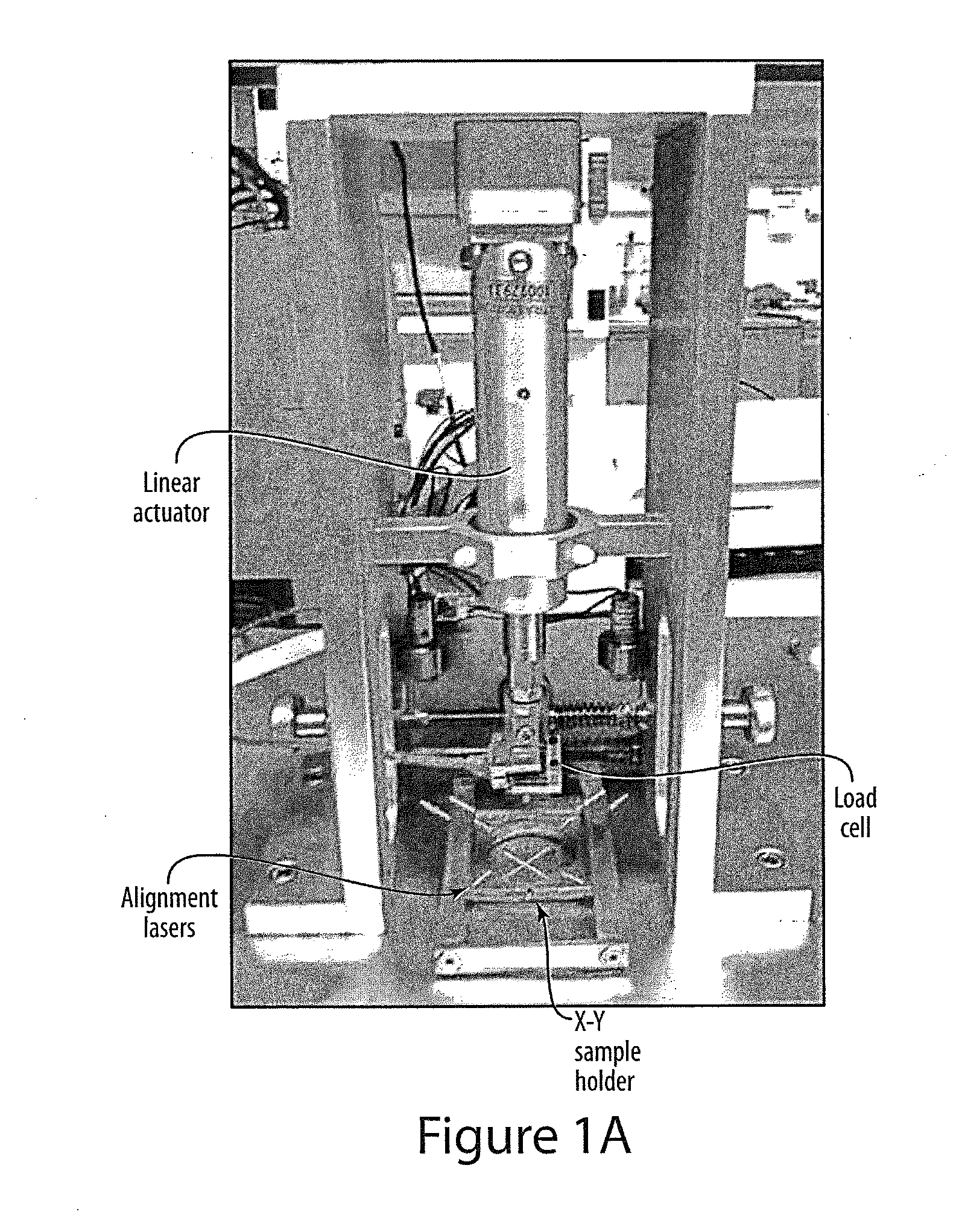
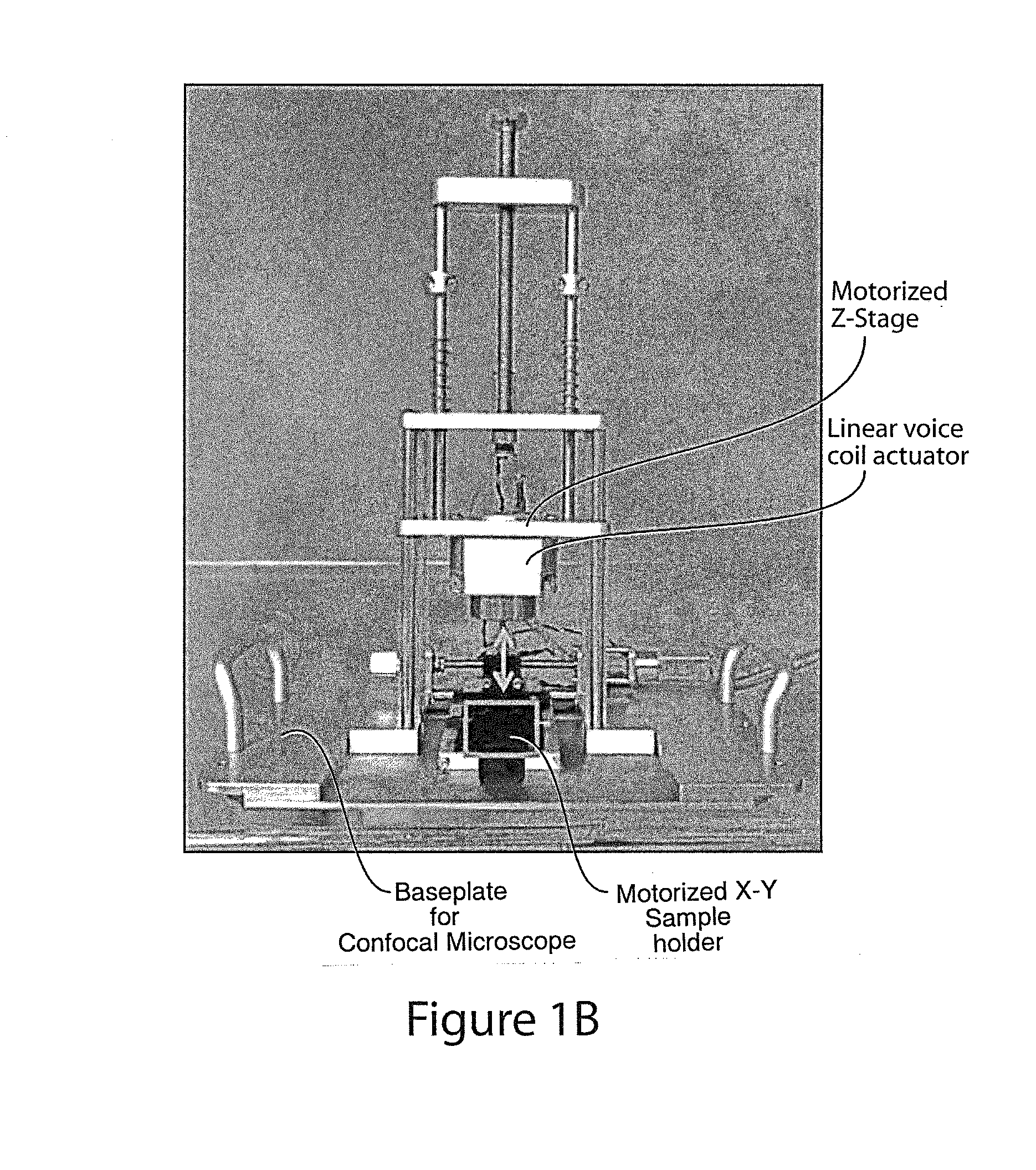
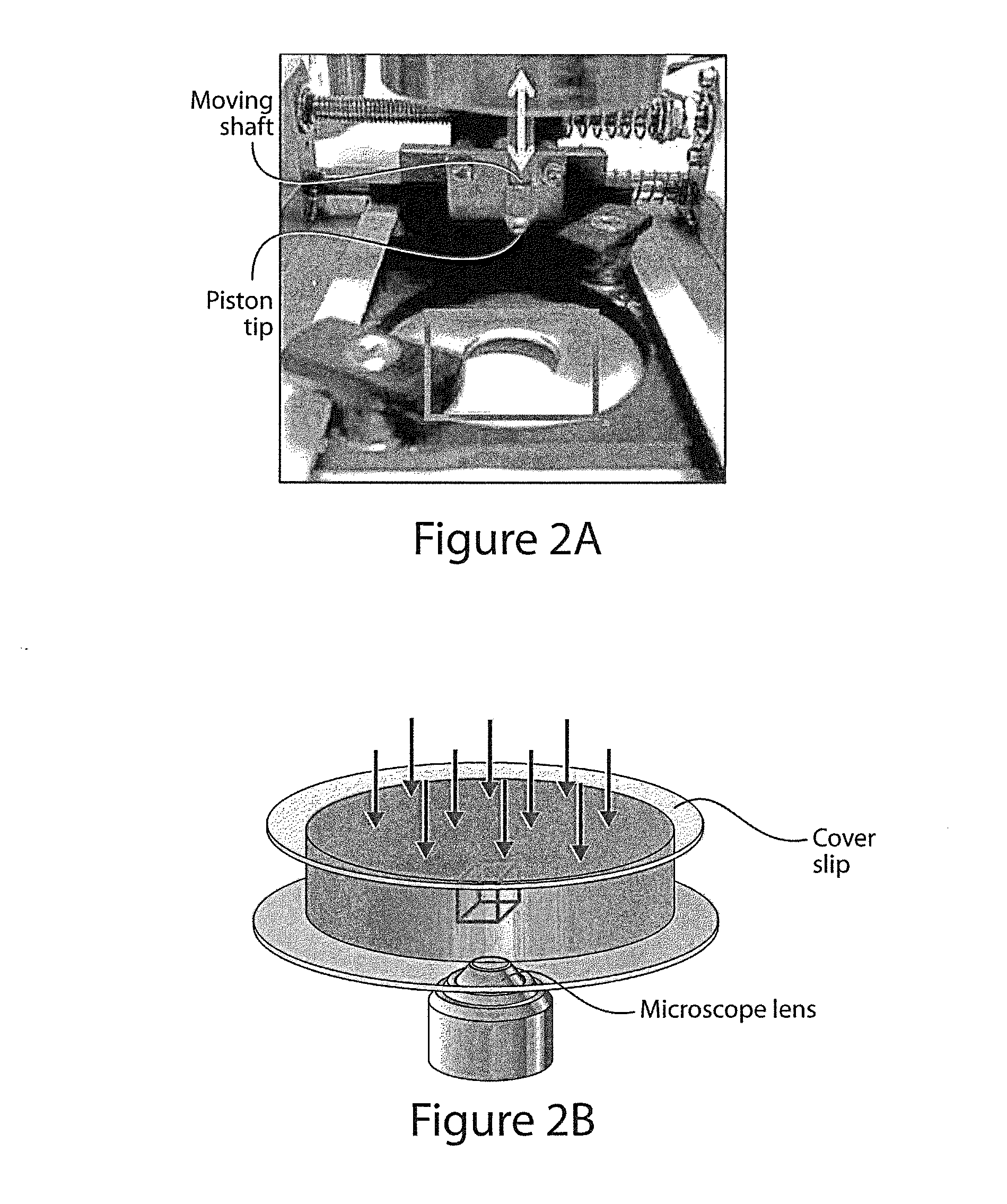
[0021]In accordance with one aspect of the invention, actuator device applies axial compression to a transparent medium or culture containing nanoparticles to derive a measurement of a mechanical parameter of interest. A system operates with a scanning laser confocal microscope (SLCM) that performs microscopic volume imaging and correlation of the nanoparticles in digital volume images (voxels) to map a uniform deformation of the medium. As applied to a biomaterial specimen, the system provides an accurate measure of the mechanical parameter of the matrix or cellular material appearing in the volume image and collectively gives an accurate tomographic map of the actual strain at depth. The defamation may be a result of an impulse applied by the actuator, a static compressive load or other deformation-causing actuation applied at the surface of the medium to introduce a uniform axial strain field over a region of the specimen. Furthermore, cell-matrix interactions, such as traction f...


PUM


Abstract

A test device applies a defined mechanical load to a soft biomaterial such as a cell culture and a microscope forms a volume image data set showing the strain field or displacement field occurring in the medium. The data set is processed to determine fundamental mechanical properties of the cell, its interaction with the surrounding medium, or its responses loading or deformation of its surrounding medium. The device may also be used to calibrate or determine fundamental mechanical properties of the medium. The device includes a linear actuator that bears against the specimen, and adapted for a volume imaging device such as a scanning laser confocal microscope that forms a volume image data set of the specimen. The specimen may be supported in a Petri dish and is preferably imaged from below by an inverted microscope. Preferably the actuator device attaches to or forms the specimen stage of microscope. Digital correlation of volumes in the data set allow computation of modulus, stress distribution and other mechanical characteristics of cell-matrix interactions, as well as mechanical properties of the cell and the matrix in response to changing loads, evolving chemical or ionic environment and growth phases. The test device may be operated to measure local mechanical parameters, to evaluate or design tissue-engineered implants, and to explore the mechanical properties of tissues, cells and cellular processes a micrometer scale with high accuracy.

Description

RELATED APPLICATION[0001]This international application claims the benefit of U.S. provisional application Ser. No. 61 / 576,008 filed Dec. 15, 2011 in the U.S. Patent and Trademark Office, entitled “Test device for mechanical properties of soft biomaterial”, which is incorporated by reference herein in its entirety.FIELD OF INVENTION[0002]Equipment for measurement, investigation, screening, or observation of biomaterial.BACKGROUND[0003]The present invention relates to equipment for investigation, screening or observation of biomaterial such as tissue, cells and biomatrix, and to methods of using the equipment.[0004]Cell mechanics and adhesion are widely recognized to play a central role in cell-tissue interactions and cellular functioning but their exact nature and mechanism are far from understood. Cells have been shown to mechanically probe and feel their surrounding microenvironment by applying displacements and exerting physical forces on their extracellular matrix. These phenome...

Claims


Application Information

Patent Timeline
IPC IPC(8): G01N33/50
CPCG01N2203/0286G01N3/08G01N3/32G01N2203/0019G01N33/5005G01N2203/0089G02B21/0052G02B21/0076G01N2203/005G01N2203/0405
Inventor FRANCK, CHRISTIANBAR-KOCHBA, EYAL
Owner BROWN UNIVERSITY
Why Eureka
Try Eureka