
Topographic genotyping for determining the diagnosis, malignant potential, and biologic behavior of pancreatic cysts and related conditions

a pancreatic cyst and genotyping technology, applied in the field of topographic genotyping for determining the diagnosis, malignant potential, and biologic behavior of pancreatic cysts and related conditions, can solve the problems of inability to distinguish malignant cysts from pre-malignant cysts on diagnostic needle aspiration or biopsy specimens, and inability to reliably define. , to achieve the effect of improving the analysis of pancreatic cyst fluid specimens and enhancing the definitive diagnosis

Inactive Publication Date: 2014-10-02
REDPATH INTEGRATED PATHOLOGY INC
View PDF0 Cites 2 Cited by
  • Summary
  • Abstract
  • Description
  • Claims
  • Application Information

AI Technical Summary

Benefits of technology

[0008]The invention described here introduces novel methods, reagents and scientific approaches not previously reported to significantly improve the analysis of pancreatic cyst fluid specimens. The invention also provides for enhancing a definitive diagnosis and prediction of early pancreatic cancer, and clearly separating this from the diagnosis of similar appearing but distinct pancreatic abnormalities and pre-cancerous conditions.

Problems solved by technology

Pancreas cancer is invariably fatal with less than 5% survival beyond five years for patients currently diagnosed with the disease.
Pathology evaluation takes the form of microscopic examination of such specimens with an indeterminate diagnosis rate of over 50% and an inability to reliably define microscopic criteria for early pancreatic cancer that can distinguish between similar appearing neoplastic and non-neoplastic lesions.
However, not every pancreatic cyst is necessarily early pancreatic cancer.
However, tools to differentiate malignant cysts from pre-malignant cysts on diagnostic needle aspiration or biopsy specimens currently do not exist.
In the absence of an associated mass representing advanced and frequently incurable disease, there are no reliable radiological criteria to separate these categories.
However, recent studies of this technique of differentiating malignant from pre-malignant pancreatic cysts have been disappointing.
However, the results provide no information as to the presence of cancer (Brugge, et al., 2004).

Method used


Image

  • Topographic genotyping for determining the diagnosis, malignant potential, and biologic behavior of pancreatic cysts and related conditions
  • Topographic genotyping for determining the diagnosis, malignant potential, and biologic behavior of pancreatic cysts and related conditions
  • Topographic genotyping for determining the diagnosis, malignant potential, and biologic behavior of pancreatic cysts and related conditions

Examples


example 1

[0178]A study was undertaken spanning 19 months from November of 2002 to May of 2004. All patients presenting to Presbyterian University Hospital with a pancreatic cyst were eligible for inclusion. EUS was performed with a Pentax CLA echo-endoscope. Pancreatic cyst aspiration was performed according to clinical need (Wilson Cook N1 (22 gauge or 25 gauge needles)) and fluid sent for cytological evaluation and CEA (carcinoembryonic antigen) tumor marker analysis. Intravenous (IV) antibiotics (Levofloxacin, 500 mg) were administered at the time of cyst aspiration.

[0179]Cyst aspirate cytology exams were performed in a routine fashion, familiar to the skilled artisan in the medical profession. The primary tissue and fluid targets were from the head of the pancreas. Cytopathologic criteria for malignancy included nuclear enlargement, pleomorphism (minimum of 3 to 4 fold variation in nuclear size), elevated N / C ratio, nuclear membrane irregularity, and coarse chromatin (Solcia, et al., “Tu...

example 2

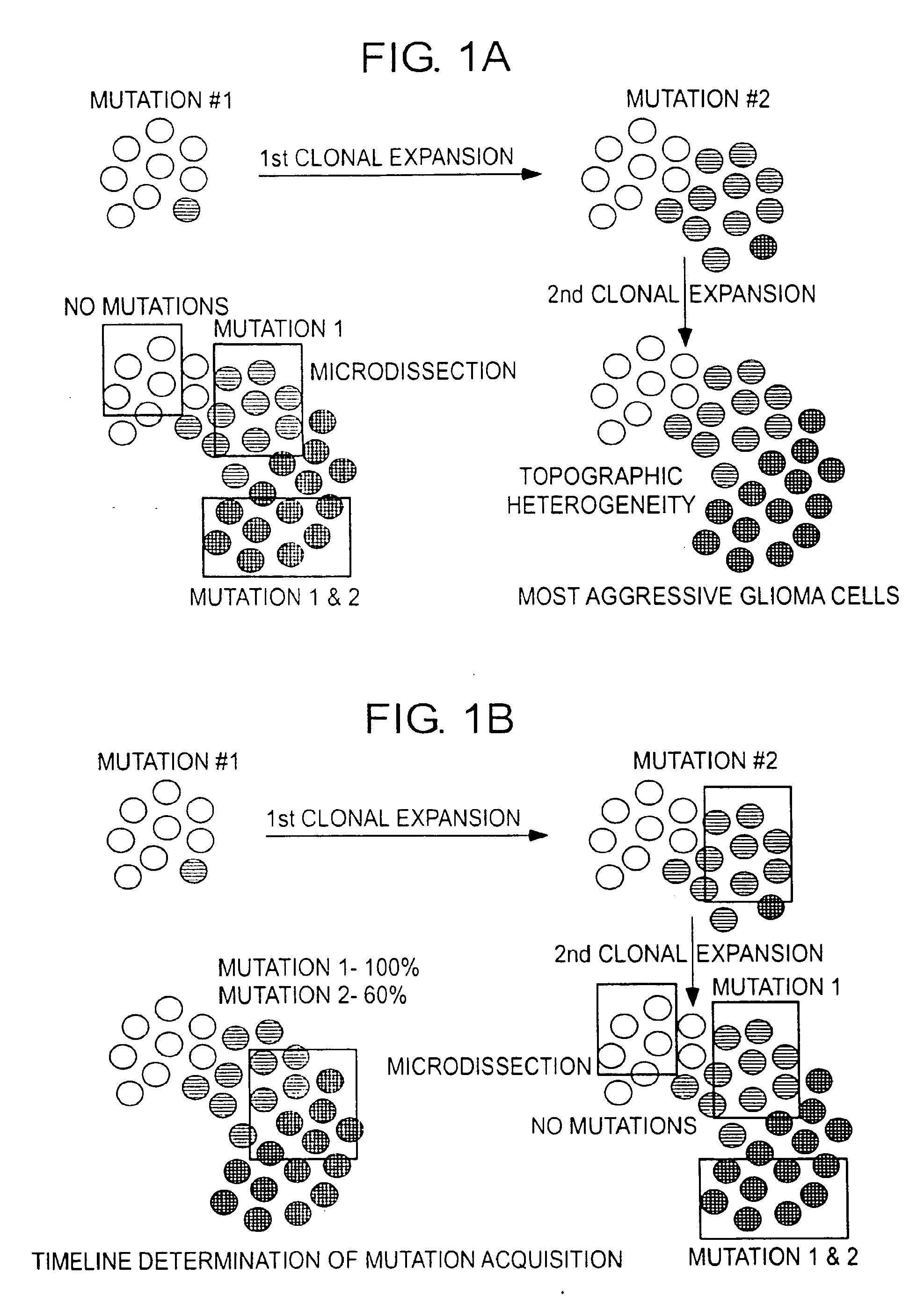
[0197]An interesting observation, consistently displayed by most metastatic neoplasms, is the preservation of the time course of mutation acquisition in metastatic deposits of tumor (FIG. 6, Table 8). In this representative example, a pancreatic carcinoma was associated with an isolated recurrence of cancer in the lip after a two year latency interval. The possibility of new primary occurrence was considered in view of the unusual location for metastatic spread (i.e., to the lip). No other tumor deposits were noted in the patient. The pancreatic and lip neoplasms were each microdissected at two sites within each specimen. Tissue microdissection was designed to capture purified representative cellular samples of each tumor deposit in pancreas and in lip. Tissue microdissection was performed manually using a scalpel under stereomicroscopic observation (Olympus SZ-PT). The neoplasms were found to be highly concordant with respect to the specific markers and alleles affected by mutation...

example 3

[0202]Additional experiments were performed to validate the approach of calculating the mutation acquisition pattern from the ratio of allele peak heights on electropherograms. These involved 17 cases of bile duct cancer and pancreatic cancer with cytologic and surgical specimens previously studied for microdissection based genotyping (Table 9). The methodology is similar, it involves obtaining and analyzing multiple cells clusters from different parts of the tumor (cytology and surgical pathology) for k-ras point mutation and microsatellite loss analysis. The LOH analysis utilized fluorescent capillary electrophoresis. Allelic imbalance analysis is based on the use of fluorescent labeled primers (HEX, TET, FAM, and TAMRA) during PCR amplification. The primer labeled molecules are then separated by capillary electrophoresis according to manufacturer's recommendations (Applied Biosystems, ABI 3100; Mountainview, Calif.). The temporal sequence for each patient was calculated as discus...


PUM

PropertyMeasurementUnit
optical densityaaaaaaaaaa
concentrationaaaaaaaaaa
ODaaaaaaaaaa

Abstract

The application relates to a method of a predicting the presence of invasive pancreatic cancer or high grade dysplasia, pre-cancerous pancreatic states and non-neoplastic conditions comprising detailed molecular analysis incorporating DNA quality and quantity, K-ras mutational analysis and a broad spectrum of tumor suppressor gene linked microsatellite LOH. Methods of diagnosing, determining prognosis of and determining a course of treatment for pancreatic cancer or high grade dysplasia, pre-cancerous pancreatic states and non-neoplastic conditions are also provided.

Description

REFERENCE TO RELATED APPLICATIONS[0001]This application claims benefit of U.S. Provisional Application Nos. 60 / 620,926 filed Oct. 22, 2004, 60 / 631,240 filed Nov. 29, 2004, 60 / 644,568 filed Jan. 19, 2005, 60 / 679,968 filed May 12, 2005, and 60 / 679,969 filed May 12, 2005 which are incorporated herein in their entirety for all purposes.FIELD OF THE INVENTION[0002]The application relates to a method of a predicting the presence of invasive pancreatic cancer, high grade dysplasia, pre-cancerous pancreatic states and alternative neoplastic and non-neoplastic conditions comprising detailed molecular analysis incorporating DNA quality and quantity, K-ras mutational analysis, and a broad spectrum of tumor suppressor gene linked microsatellite loss of heterozygosity. The application provides a means to clearly separate cancer from non-cancer states such as those produced by inflammation, infection and trauma involving the pancreas. Methods of diagnosing, determining prognosis of, and determini...

Claims


Application Information

Patent Timeline
Patent Type & Authority Applications(United States)
IPC IPC(8): C12Q1/68G01N33/574
CPCC12Q1/6806C12Q1/6827C12Q1/6886C12Q2600/106C12Q2600/112C12Q2600/118G01N33/57438C12Q2600/156C12Q2600/154C12Q2545/114C12Q2527/125Y02A90/10G16H50/30
Inventor FINKELSTEIN, SYDNEY DAVIDSWALSKY, PATRICIA
Owner REDPATH INTEGRATED PATHOLOGY INC
Learn More