Enrichment & isolation of microbial cells & microbial nucleic acids from a biological sample

a technology of microbial cells and nucleic acids, applied in the field of methods, can solve the problems of reducing the efficiency of nucleic acid testing, limiting the use of amplification assays, and reducing the sensitivity of diagnostic assays

Active Publication Date: 2014-11-13
GENEOHM SCI CANADA
View PDF1 Cites 11 Cited by
  • Summary
  • Abstract
  • Description
  • Claims
  • Application Information

AI Technical Summary

Benefits of technology

[0015]The present invention relates to a method which advantageously allows the processing of large volumes of biological samples such as whole blood. Unlike previous methods, in which the sample volume that could be analyzed was often limited by the presence of inhibitors (especially in the case of blood), significantly larger volumes may be processed and nucleic acids from microorganisms reproducibly amplified by the method of the present invention. The ability to amplify from large sample volumes using fewer processing steps allows the practitioner to detect more rapidly rare target sequences which may be missed when a small aliquot of a sample or a diluted sample must be amplified to avoid interference from inhibitors.
[0017]The method may advantageously employ a single centrifugation step (e.g., between about 3200 g and about 10000 g) to concentrate microorganisms, as opposed to multiple centrifugations / washings. In addition, the method of the present invention may comprise the addition of a small volume of differential lysis solution to the initial volume of biological sample thereby avoiding multiple fold dilution of the sample and the need for additional concentrating steps or the use of smaller sample volumes. Advantageously, no specialized equipment is required to practice the present invention. The reagents are inexpensive and readily available, and none require special handling.

Problems solved by technology

Many biological samples and materials of interest contain substances that reduce the efficiency of nucleic acid testings.
For example, it is well known that many substances that inhibit enzyme activity are present in many types of cells and can limit the use of amplification assays such as PCR.
Furthermore, host cells (i.e., cells naturally present in the sample being tested for the presence of contaminating microbial cells e.g., a patient's red blood cells and white blood cells) in biological samples tested for the presence of microbial cells contribute to the “dilution” of microbial cells nucleic acids and leads to a decrease in sensitivity of the diagnostic assay.
Isolation of nucleic acids is cumbersome and requires that a high concentration of nucleic acid be present to be effective.
Dilution or use of small sample volumes significantly compromises the sensitivity of the nucleic acid analysis.
In addition, Saponin is a chemically complex substance made of various chemical compounds and Applicants have observed that it is prone to lot-to-lot variability which reduces reproducibility.
Moreover, at concentrations employed, Saponins do not lyse white blood cells such as macrophages, thereby contributing to the “dilution” of microbial cells which often results in a decrease in sensitivity.
A disadvantage of this approach is the presence of a larger proportion of blood cells nucleic acids than of microbial nucleic acids which reduces sensitivity of microbial detection in the sample.
In addition, systems like SeptiFast™ prep kit require numerous handling steps and take about two hours of treatment prior to extraction of human and microbial DNA.
Furthermore it was shown that a majority of blood samples collected from septicemic patients may contain as low as 10 colony Forming Unit (CFU) of microbial cells / ml of blood (Johnson et al., 1993, APMIS, 101:595-601), which may be insufficient to allow detection using these processing methods.

Method used


Image

  • Enrichment & isolation of microbial cells & microbial nucleic acids from a biological sample
  • Enrichment & isolation of microbial cells & microbial nucleic acids from a biological sample
  • Enrichment & isolation of microbial cells & microbial nucleic acids from a biological sample

Examples


example 1

Material and Methods

[0092]Strains, storage and growth conditions. The bacterial species used as a model in the following experiments is Staphylococcus aureus (ATCC 36232). The strains were kept cryopreserved in 50% glycerol at −80 ° C. Bacterial cells were cultured at 35° C. on blood agar plates (Quelab, Montreal). After overnight incubation, three colonies were transferred into 10 mL of Tryptic Soy Broth (TSB) and were incubated overnight and used as stationary phase culture. The number of viable bacteria was determined by the standard spread plate method.

[0093]Blood donations. Blood donations were drawn at the Minnesota Memorial Blood Center and kept at 4° C. After usual testing for presence of HBV, HIV, STS and HCV the blood bag was cleared and shipped to INO facility (GSI-Qc) in isolated box cooled with ice packs. Upon reception, the blood was kept at 4° C. until use. Blood donation was 2-4 week-old when tested. The blood bag was dispensed in 50 mL-aliquot into 50-mL Falcon™ tub...

example 2

New Differential Lysis Protocol for Concentrating Microbial Cells from Large Blood Sample Volumes

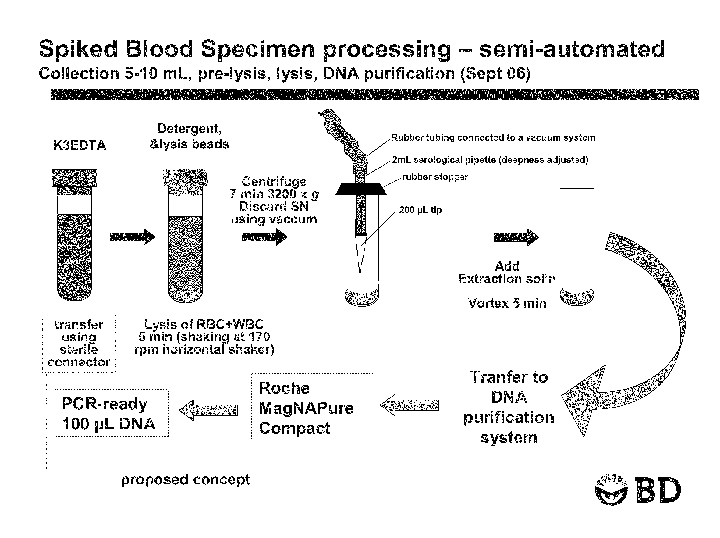
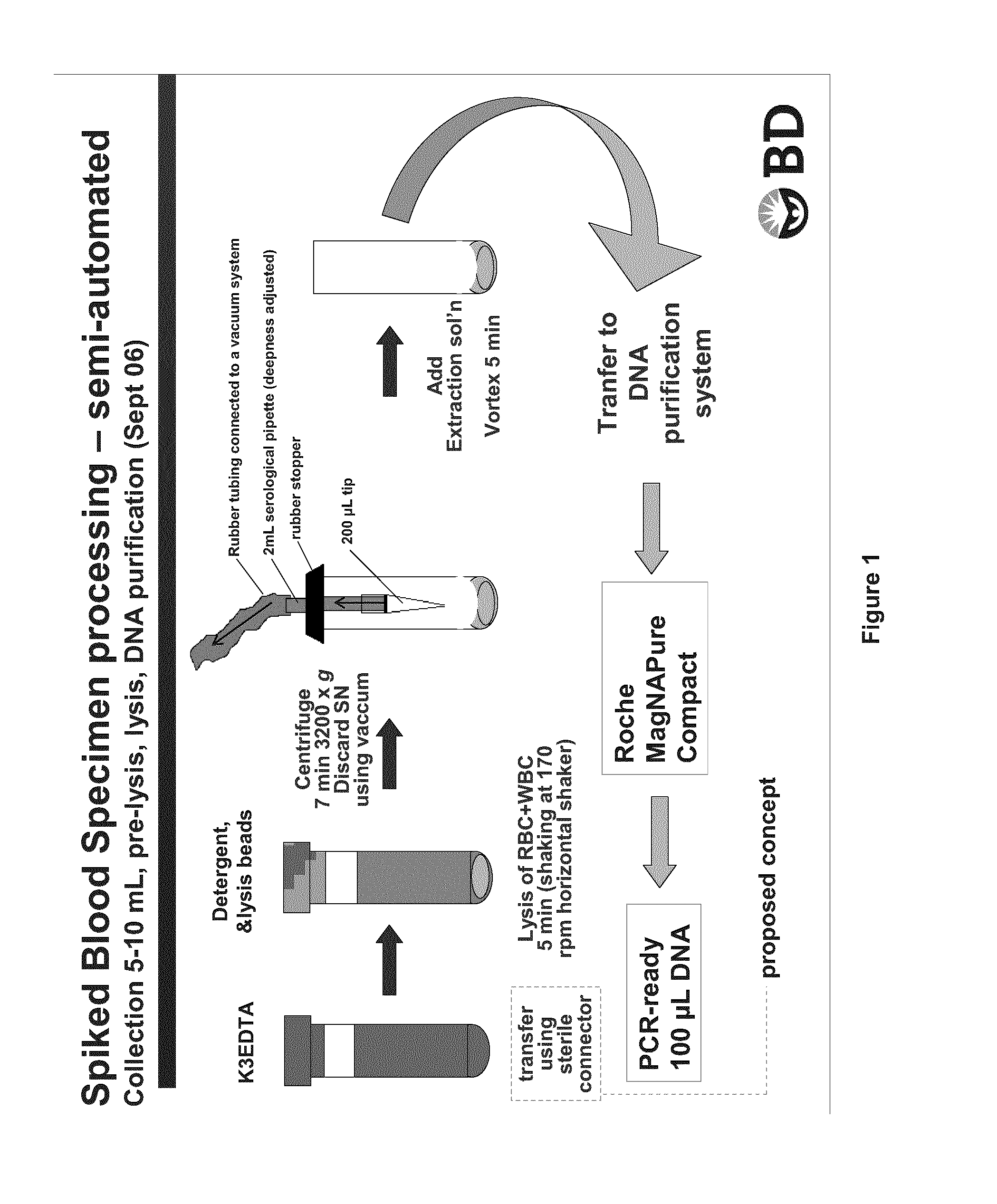
[0094]A K3-EDTA fresh blood sample (K3-EDTA used was a liquid EDTA solution) of 5-10 mL was transferred into Sarstedt™ 10 ml tubes with round bottom containing equivalent of three times the lysis matrix contained in IDI-Lysis tube (BD GeneOhm lysis tube cat # 441243), 0.1% SDS as blood cell lysing agent and 0.005% of silicone as antifoaming agent. Each tube was then agitated at 170 rpm for 5 min at 25° C. onto a horizontal shaker. Following this blood cell lysis step, blood samples were centrifuged at 3200×g for 7 min. at room temperature.

[0095]Supernatant was discarded by aspiration using the device shown in FIG. 1. A small volume of liquid was left over the pellet of microbial cells, cell debris and extraction matrix. A volume of 100 μL of PBS was added before performing lysis of bacteria by vortexing 5 min at high speed on a conventional Vortex Genie™ with multi-tube adaptor. After ly...

example 3

Comparison of Specimen Processing Performance Between 10 ml and 5 ml Blood Sample Volumes

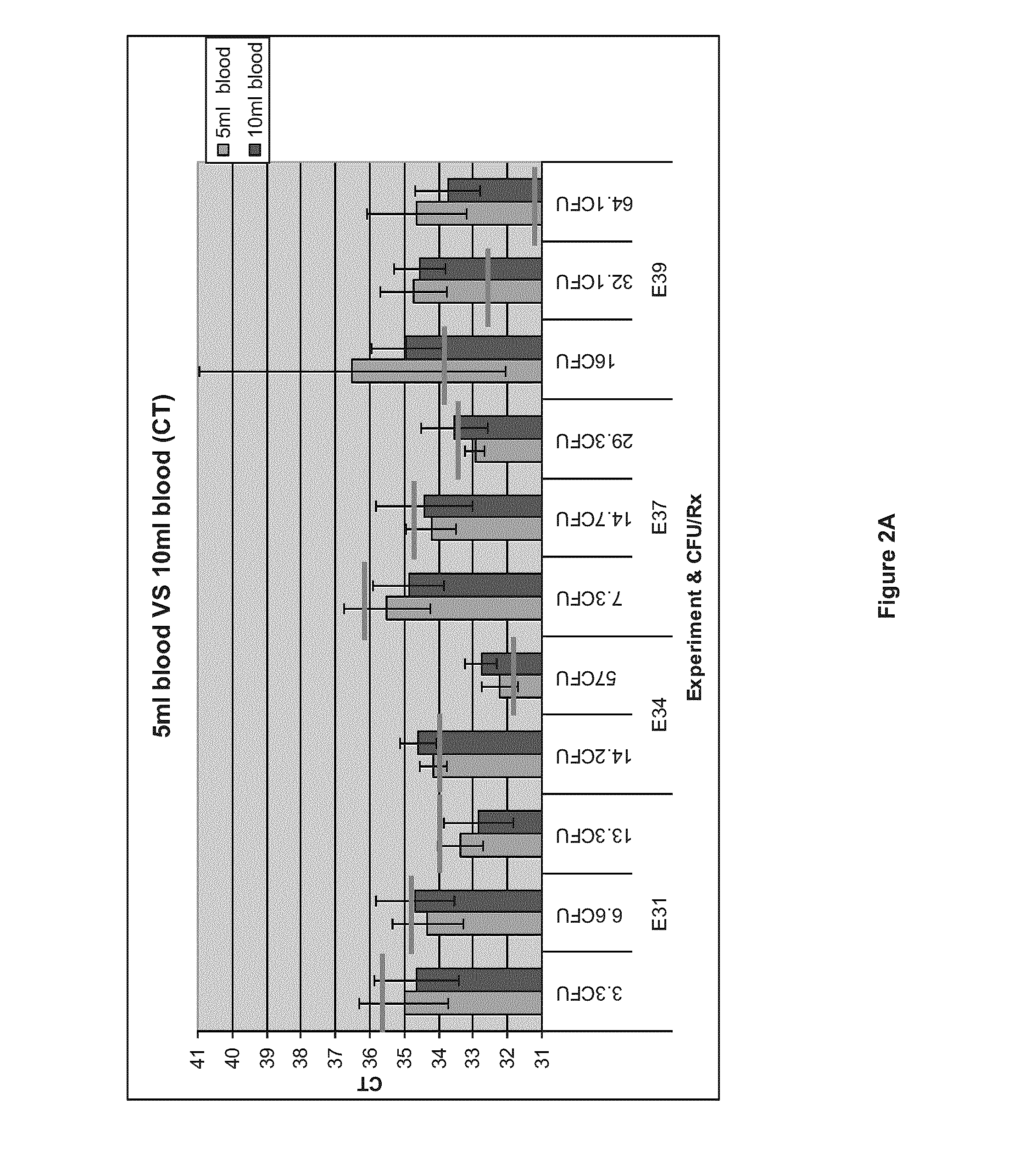
[0097]The objective of this experiment was to determine if processing 10 ml of blood samples could yield similar or better results than using 5 ml blood samples. Four experiments were conducted with four different blood donations (E31: 1472658, E34: 1475067, E37: 1477263 and E39: 1482868) according to the general procedure described in Example 1. Blood samples of 5 or 10 mL were spiked at approximately 100 CFU / volume to allow the comparison.

[0098]MPC purified DNA was then analyzed by Real-Time PCR, in 96-well plates, on a MyIQ™ (BioRad) instrument. PCR Mastermix™ (MM) was prepared as a 4×solution. S. aureus was amplified using genus specific primer set (TstaG—422 (GGCCGTGTTGAACGTGGTCAAATCA (SEQ ID NO:1)) and TstaG—765 (TIACCATTTCAGTACCTTCTGGTAA) (SEQ ID NO:2)) without internal control. Final reaction volume was 25 μL. Sample was added at two volumes (4.7 μL and 18.8 μL) to 6.25 μL of 4×MM with a...


PUM

PropertyMeasurementUnit
diameteraaaaaaaaaa
diameteraaaaaaaaaa
volumeaaaaaaaaaa

Abstract

A method for enrichment and isolation of microbial cells and microbial nucleic acids from a biological sample is described. The method comprises (i) adding to an initial volume of biological sample a differential cell lysis solution to obtain a final concentration of 0.1 to 1% of SDS in the sample; (ii) mixing the solution obtained in step (i) for a period of time sufficient to lyse the host cells present in the biological sample, while preserving the integrity of cells; and (iii) separating the microbial cells from the lysed host cells components. Differential cell lysis solutions and kits for practicing the method of the present invention are also provided.

Description

CROSS REFERENCE TO RELATED APPLICATIONS[0001]This application claims benefit, under 35 U.S.C. §119(e), of U.S. provisional application Ser. No. 61 / 578,352, filed on Dec. 21, 2011 which is incorporated herein by reference in its entirety.STATEMENT REGARDING FEDERALLY SPONSORED RESEARCH OR DEVELOPMENT[0002]N.A.FIELD OF THE INVENTION[0003]The present invention relates to methods for preparing biological samples for analysis. More particularly, the present invention relates to processing of biological samples for enrichment and isolation of microbial cells and their nucleic acids for subsequent analysis, including for example, nucleic acid amplification. The present invention also relates to the detection of microbial cells in biological samples.[0004]REFERENCE TO SEQUENCE LISTING[0005]This application contains a Sequence Listing in computer readable form entitled 787-Sequence listing_ST25, created Dec. 17, 2012 having a size of 1 Kbytes, which is incorporated herein by reference in its...

Claims


Application Information

Patent Timeline
Patent Type & Authority Applications(United States)
IPC IPC(8): C12Q1/68C12N15/10C12Q1/24
CPCC12Q1/6806C12Q1/24C12N15/1003C12N1/06
Inventor MENARD, CHRISTIANROY, ANNIEBOUCHER, PATRICKLETOURNEAU, STEVE
Owner GENEOHM SCI CANADA
Why Eureka
Try Eureka