
Detection and treatment of breast cancer

a breast cancer and detection technology, applied in the field of detection and treatment of breast cancer, can solve the problems of limited prognostic markers for breast cancer, insufficient function, and often different shapes of cancer cells, and achieve the effect of increasing endogenous pappa levels and focusing and effectiv

Inactive Publication Date: 2014-11-27
EURO LAB FUER MOLEKULARBIOLOGIE EMBL +2
View PDF0 Cites 0 Cited by
  • Summary
  • Abstract
  • Description
  • Claims
  • Application Information

AI Technical Summary

Benefits of technology

This patent is about finding a protein called Pregnancy-Associated Plasma Protein A (PAPPA) and how it is important for cells to grow and divide properly. The text also explains how PAPPA is commonly lost or impaired in cancer, making it a promising target for detecting and treating breast cancer. In simple terms, this patent provides a better understanding of breast cancer biology and suggests a more effective way to detect and treat the disease.

Problems solved by technology

Cancer cells are often shaped differently from healthy cells, do not function properly, and can spread into many areas of the body.
Unfortunately however, prognostic markers for breast cancer are limited.
A complexity for treatment is that not all pre-invasive cancers will progress to become invasive (or metastatic), for example as in DCIS, and so not all patients need to be treated in the same way.
A difficulty is that at present it is difficult to differentiate between patients who have a cancer (or abnormal cells) that will remain pre-invasive, and those who will progress to invasive cancer.

Method used


Image

  • Detection and treatment of breast cancer
  • Detection and treatment of breast cancer
  • Detection and treatment of breast cancer

Examples


example 1

Methods

Tissue Specimens

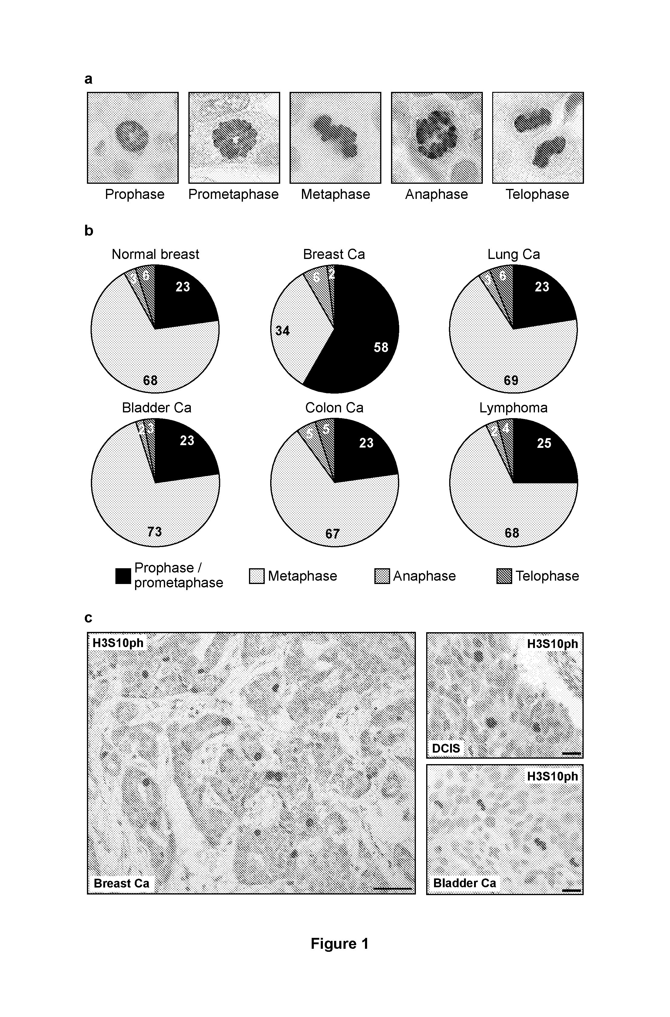
[0134]Formalin-fixed, paraffin-embedded tissue was retrieved from the archives of the Department of Pathology at UCL (UCL Hospitals, London, UK) and included: invasive breast cancer (n=182, 156 of which were included for analysis of mitotic phase distribution); ductal carcinoma in situ (DCIS; n=81, 69 evaluable); normal breast tissue from reduction mammoplasty specimens (n=33, evaluability not relevant); normal breast tissue from pregnant patients (n=5, all evaluable); colon adenocarcinoma (n=41, all evaluable); transitional cell carcinoma of the bladder (n=27, all evaluable); penile squamous cell carcinoma (n=33, all evaluable); gastric adenocarcinoma (n=21, all evaluable); malignant melanoma (n=21, all evaluable); small cell lung cancer (n=30, all evaluable); and non-Hodgkin lymphoma (n=29, all evaluable). Cases were selected on the basis of available histological material and clinico-pathological information. Histological specimens had been reviewed by a qu...

example 2

[0160]Exogenously added IGF-1 restores normal progression through mitosis in breast cancer cells displaying prophase / prometaphase delay phenotype.

[0161]A known function of PAPPA is to release the hormone IGF-1 from its sequestering inhibitory binding protein IGFBP-4. By increasing the local bioavailability of IGF-1 at the cell surface, PAPPA activity increases IGF-dependent signalling and promotes cell growth and proliferation. Thus it can be postulated that PAPPA is likely to affect mitotic progression in breast cancer cells by modulating signalling through the IGF pathway. It follows from this that treatment of T47D breast cancer cells, which closely resemble tumour cells in vivo (i.e. display PAPPA promoter methylation and the mitotic delay phenotype; FIG. 14), with recombinant IGF-1 should restore normal progression through mitosis in this cell line. For these experiments, T47D cells were serum starved for 48 hours. The viability of the cells was established using Trypan Blue vi...


PUM

PropertyMeasurementUnit
population doubling timeaaaaaaaaaa
pHaaaaaaaaaa
concentrationaaaaaaaaaa

Abstract

The present invention describes methods for determining the risk that a breast precursor lesion will progress to invasive breast cancer and / or the risk of recurrent non-invasive disease in a patient, comprising detecting the presence and / or level of PAPPA and / or PAPPA functional activity in a breast tissue sample obtained from the patient, wherein if PAPPA is not present, or is present at a reduced amount compared to a control, there is the risk of progression to invasive cancer and / or the risk or recurrent disease.The present invention also enables the chemosensitisation of mitotically delayed breast cancer cells to anti-proliferative agents, preferably anti-mitotic agents, by restoring normal progression through mitosis. In this embodiment a first drug is applied to release breast cancer cells from the mitotic block and, sequentially, a second drug affecting proliferating cells is administered for cancer cell killing.

Description

FIELD OF THE INVENTION[0001]This invention relates to the use of specific biological markers for the prognostic assessment of proliferative lesions in breast tissue, and for identifying the risk of proliferative lesions progressing to invasive breast cancer and / or the risk of developing recurrent disease, and subsequent treatment.BACKGROUND OF THE INVENTION[0002]Neoplasms and cancer are abnormal growths of cells. Cancer cells rapidly reproduce despite restriction of space, nutrients shared by other cells, or signals sent from the body to stop reproduction. Cancer cells are often shaped differently from healthy cells, do not function properly, and can spread into many areas of the body. Abnormal growths of tissue, called tumours, are clusters of cells that are capable of growing and dividing uncontrollably. Tumours can be benign (noncancerous) or malignant (cancerous). Benign tumours tend to grow slowly and do not spread. Malignant tumours can grow rapidly, invade and destroy nearby ...

Claims


Application Information

Patent Timeline
Patent Type & Authority Applications(United States)
IPC IPC(8): C12Q1/68A61K38/30A61K45/06G01N33/574
CPCC12Q1/6886G01N33/57415C12Q2600/154A61K45/06C12Q2600/118A61K38/30A61K38/1709A61K38/1754A61P15/00A61P35/00C07K14/4715C12Q2600/156C12Q2600/158A61K2300/00A61K38/17C07K14/4702G01N33/574
Inventor ELLENBERG, JANNEUMANN, BEATELODDO, MARCOWILLIAMS, GARETHSTOEBER, KAI
Owner EURO LAB FUER MOLEKULARBIOLOGIE EMBL
Learn More