

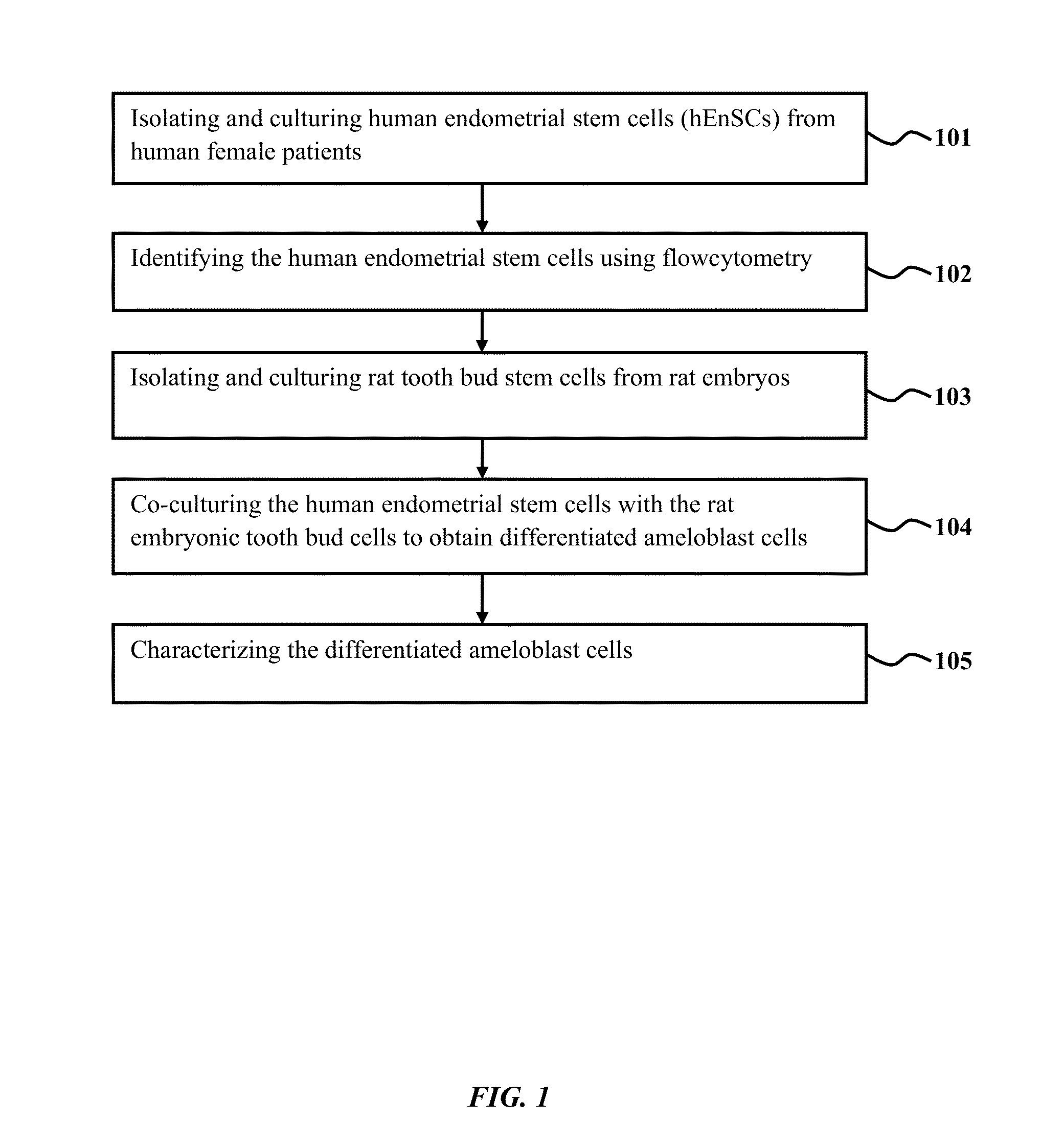
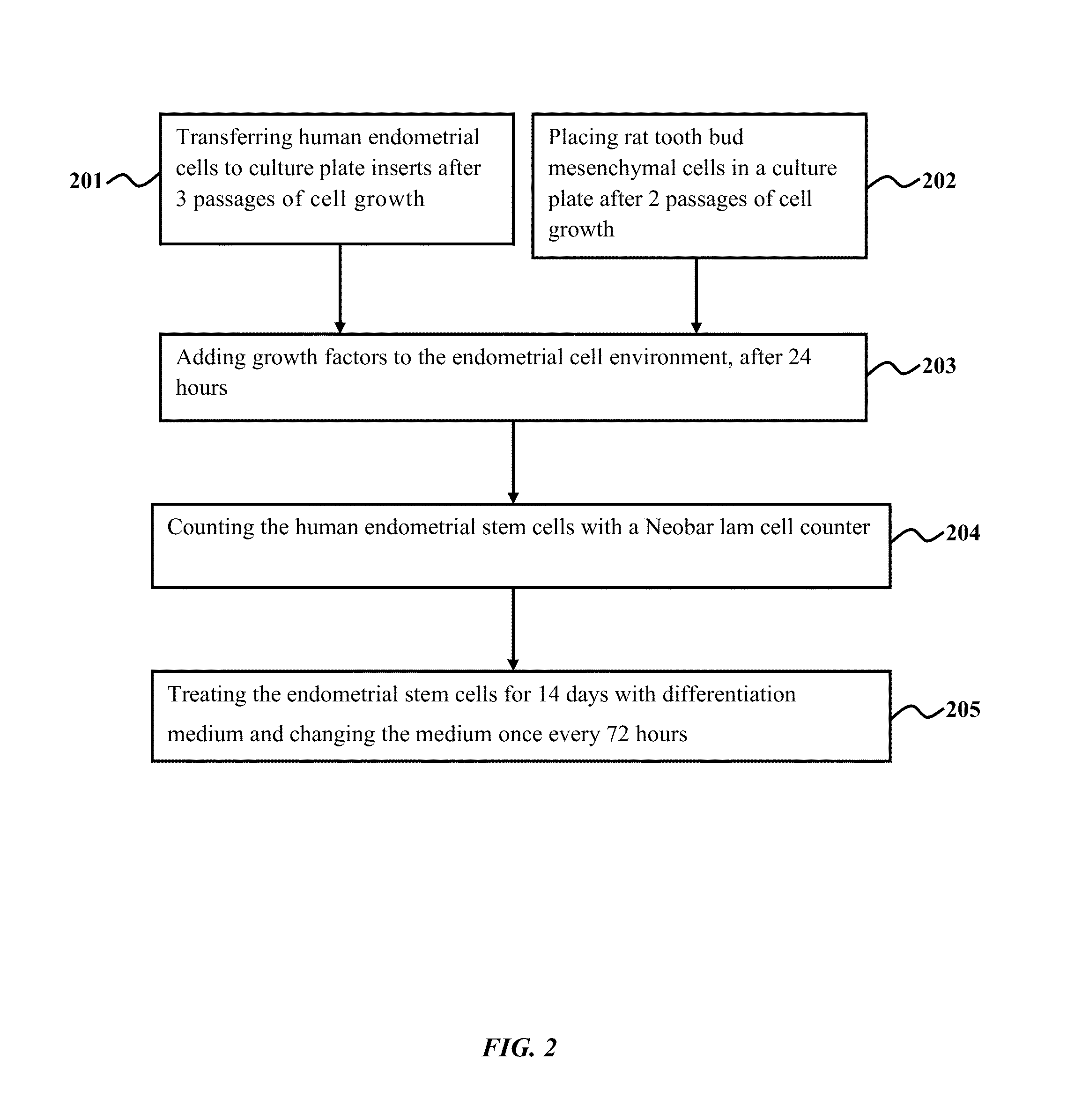
Method of co-culturing human endometrial stem cells and rat embryonic tooth bud cells to obtain ameloblast cells
- Summary
- Abstract
- Description
- Claims
- Application Information
AI Technical Summary
Benefits of technology
Problems solved by technology
Method used
Image

Examples
example 1.1
Isolation of hEnSCs
[0135]Ten endometrial biopsy samples were obtained from female patients between the age groups of 18-40. The biopsy samples were transferred into Hanks fluid and washed in PBS containing antibiotics. The biopsy samples were sliced into small fragments and immersed in collagenase Type IA and 2 mg / mL of DMEM (containing antibiotics) for 2 hours at 37° C. Epithelial stem cells and stromal cells present in the endometrial biopsy were separated using 45 μm and 70 μm filters. These separated cells were then centrifuged at 1000 rpm for 15 minutes and purified by Ficoll purification. The purified cells were placed in a flask and incubated at 37° C. in 5% CO2 and 95% moisture. The purified stem cells were then analyzed using flow cytometry.
example 1.2
Flow Cytometry Analysis of hEnSCs
[0136]Human endometrial stem cells were prepared for flow cytometry analysis by removing cell culture media from a cell plate. The cell plate was then rinsed once with 1×PBS. 5 mL of 0.2% EDTA (in PBS) was added to the cell plate. Cell surface staining was compromised when Trypsin / EDTA solution was used in the place of 0.2% EDTA. The hEnSCs were incubated in an incubator at 37° C. for 2 minutes. The hEnSCs were suspended in 5 mL of DMEM media that was added to neutralize EDTA. The suspended hEnSCs were collected in 15 mL tubes and centrifuged for 5 minutes at 1000 RPM. Prior to staining, the hEnSCs were transferred to the cell culture plate. 20 μL Human Fc Receptor Binding Inhibitor Purified was added (per 100 μL cell) to the culture plate. The culture plate was then incubated for 10-20 minutes at 2-8° C. or at room temperature. 50 μL of human endometrial cell suspension (from 2×10⁵ to 108 cells) was aliquoted to each well in a culture plate. A recom...
example 1.3
Isolation of Rat Embryonic Tooth Bud Cells
[0137]Eleven Wistar rat embryos aged 14 days were obtained from pregnant female rats that were anesthetized. The Rat mesenchymal tooth bud cells were isolated from the lower jaw of the rat embryo using a scalpel. The embryonic tooth bud tissues were placed in 1% trypsin at 4° C. for an enzymatic reaction called trypsinization. The trypsinization of rat embryonic tooth bud cells resulted in dental epithelial cells and mesenchymal cells. The dental epithelial cells and mesenchymal were completely separated under a stereo microscope. The dental epithelial and mesenchymal cells were rinsed in PBS and exposed to collagenase IA for 3 hours at 37° C. These dental epithelial and mesenchymal cells were then passed through a 70 mm Nylon filter, centrifuged at 2000 g for 15 min and rinsed again with PBS. The dental epithelial and mesenchymal cells were then transferred to a 6 well plate containing 0.5 mL DMEM culture medium (100× antibiotic, 10% FBS an...
PUM
| Property | Measurement | Unit |
|---|---|---|
| Temperature | aaaaa | aaaaa |
| Temperature | aaaaa | aaaaa |
| Length | aaaaa | aaaaa |
Abstract
Description
Claims
Application Information