ENCAPSULATION AND CARDIAC DIFFERENTIATION OF hiPSCs IN 3D PEG-FIBRINOGEN HYDROGELS

a technology of peg-fibrinogen hydrogel and encapsulation, which is applied in the field of encapsulation and cardiac differentiation of hipscs in 3d peg-fibrinogen hydrogel, can solve the problems of unclear boundaries between them, 20-30% drug withdrawal from the market, and not directly repair damaged myocardium

Active Publication Date: 2015-05-14
AUBURN UNIV
View PDF0 Cites 9 Cited by
  • Summary
  • Abstract
  • Description
  • Claims
  • Application Information

AI Technical Summary

Benefits of technology

[0016]The present invention relates to compositions and methods for culturing and differentiating pluripotent stem cells into cultures or tissues in three-dimensional form. According to the invention, Applicants have developed systems and methods for direct differentiation of PSCs within a three-dimensional (3D) microenvironment created by PEG-fibrinogen hydrogels, thereby directly creating a 3D architecture without additional cell handling steps. These systems and methods allow for production of cells and tissues having characteristics not otherwise observed in cultured cells or other cells not taken directly from the corresponding tissues in the body. Using these systems and methods, PSCs encapsulated in a 3D microenvironment formed by a biomimetic material survive and degrade the biomimetic material to form compact tiss...

Problems solved by technology

For example, predicting cardiac toxicity and the triggering of cardiac arrhythmias represents a major hurdle for pharmaceutical compound development, resulting in about 20-30% drug withdrawal from the market.
Aspects of several of these categories play a role in the pathogenesis of other categories, making clear boundaries between them difficult to define.
Although patients with heart disease often experience improvement in quality of life following clinical treatment, these therapies do not directly repair damaged myocardium.
Furthermore, because the tissue is never directly restored to its prior health, individuals may never regain their original cardiovascular function and may experience other debilitating cardiac conditions as time progresses.
Although congenital heart defects are the most common type of birth defect, the causal mechanisms are still not clearly understood and human teratogens do not necessarily cause defec...

Method used


Image

  • ENCAPSULATION AND CARDIAC DIFFERENTIATION OF hiPSCs IN 3D PEG-FIBRINOGEN HYDROGELS
  • ENCAPSULATION AND CARDIAC DIFFERENTIATION OF hiPSCs IN 3D PEG-FIBRINOGEN HYDROGELS
  • ENCAPSULATION AND CARDIAC DIFFERENTIATION OF hiPSCs IN 3D PEG-FIBRINOGEN HYDROGELS

Examples


example 1

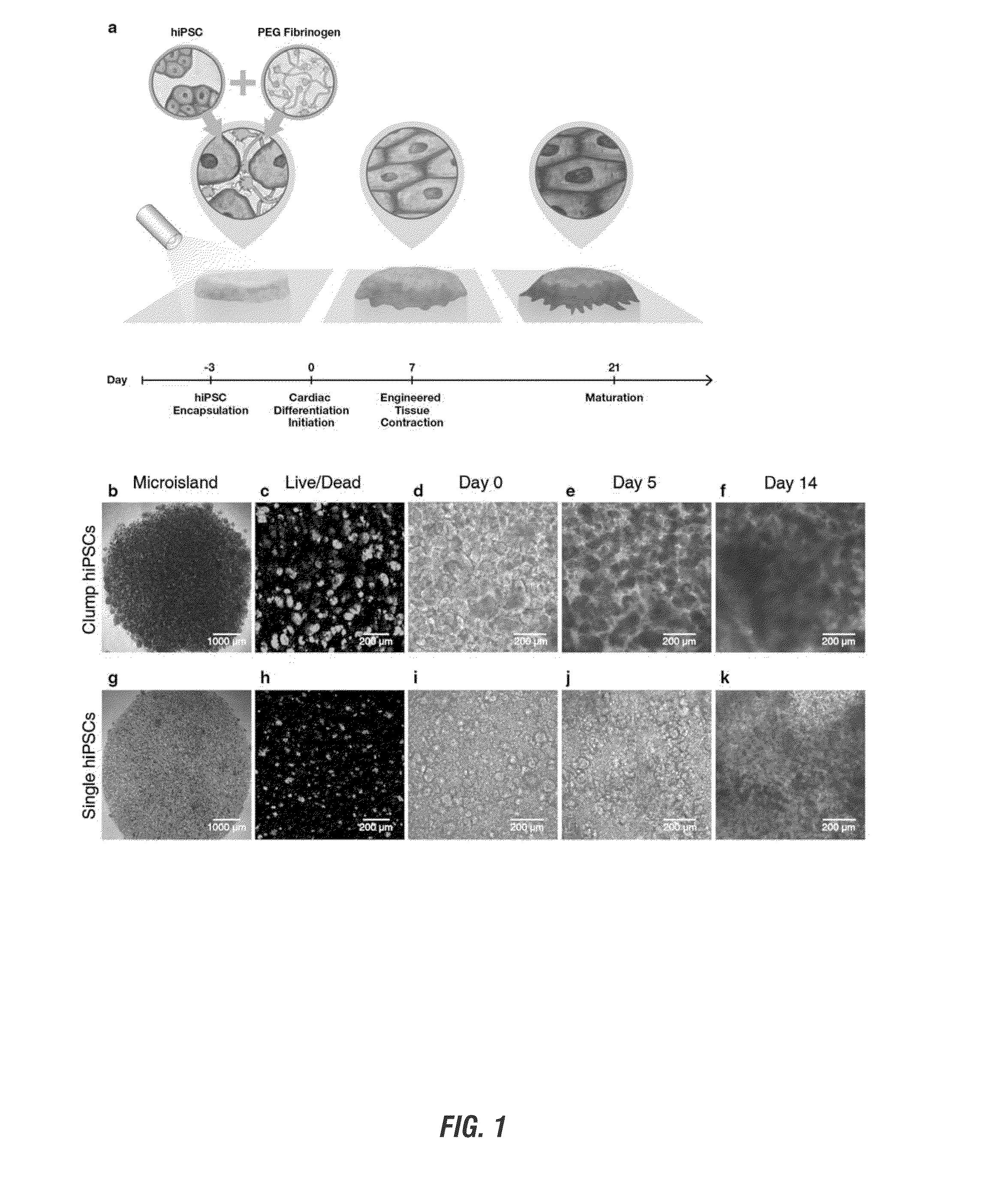
PEG-Fibrinogen Hydrogels Support Stem Cell Survival and Proliferation

[0104]HiPSC Expansion and Culture.

[0105]IMR-90 Clone 1 human induced pluripotent stem cells (hiPSCs) were purchased from WiCell and maintained at 37° C., 5% CO2, and 85% relative humidity. HiPSCs were cultured as colonies on hESC qualified Matrigel (BD Biosciences) using mTeSR-1 media (Stem Cell Technologies). HiPSCs were passaged using Versene (Life Technologies) and 5 μM ROCK inhibitor (Y 27632, R&D Systems) was added to the mTeSR-1 media for 24 h post-seeding.

[0106]HiPSC viability on day −2 (24 hours post-encapsulation) was assessed using a LIVE / DEAD® viability kit (Molecular Probes) following manufacturer's instructions. Z-stacks (step size=5 μm) throughout the entire tissue thickness at several randomly selected locations within microislands were taken (n=3 locations per microisland). The percentage of viable cells was quantified by manual counting at two depths (33% and 75% height) per acquired stack using NI...

example 2

3D Encapsulated hiPSCs Form Synchronously Contracting, Functional Cardiac Tissues

[0123]One-step hiPSC encapsulation and cardiac differentiation process successfully resulted in synchronously contracting cardiac tissues with first areas of contraction by day 7 of differentiation for all performed hiPSC encapsulations. Microislands formed using both clump and single hiPSCs were all observed to contract (n>40 separate differentiations) with initiation of contraction occurring in localized areas between day 7 and day 11 depending on dissociation technique. Spontaneously contracting areas increased in number and synchronicity of contraction in both clump and single hiPSC encapsulated microislands over time, resulting in uniform contracting cardiac tissues by day 14.

[0124]Video recordings of spontaneously contracting cardiac tissues were acquired on a phase contrast microscope (Ti Eclipse, Nikon) using a high speed camera (Andor Luca S) to determine frequency of contraction. Number of con...

example 3

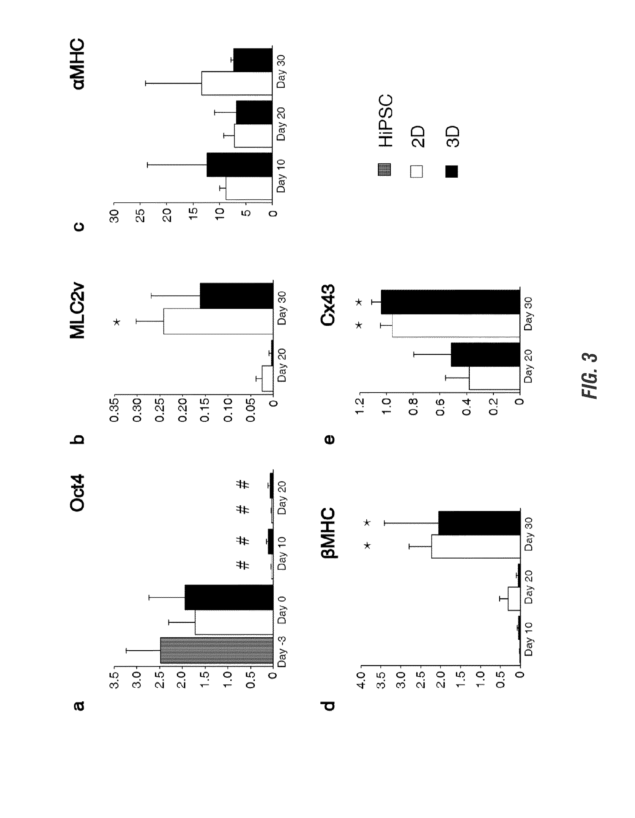
Developing Cardiac Tissues Express Cardiac Genes and Cardiac Efficiency Similar to Control 2D Monolayers

[0125]Tracking the ability to successfully and efficiently differentiate encapsulated hiPSCs into CMs, differentiation efficiency using flow cytometry and assessment of pluripotency and cardiac gene expression was shown to be similar to high efficiency 2D monolayer differentiation.

[0126]To determine the efficiency of cardiac differentiation in 3D, the expression of cTnT was evaluated by flow cytometry on day 20 microislands. On day 20, 2D sheets were washed with PBS and incubated in 0.25% trypsin (EDTA, Mediatech) at 37° C. for 5 min. Age-matched 3D tissues were washed with PBS followed by 2 h incubation at 37° C. on a rotator (Boekel Orbitron Rotator, Model 260250, Boekel Scientific) with collagenase Type 2 (1 mg / ml, Worthington) in 120 mM NaCl, 5.4 mM KCl, 5 mM MgSO4, 5 mM Na-pyruvate, 20 mM glucose, 20 mM taurine, and 10 mM HEPES (pH 6.9) supplemented with 30 μM CaCl2, followed...


PUM


Abstract

The present invention relates to the production of cell cultures and tissues from undifferentiated pluripotent stem cells using three-dimensional biomimetic materials. The resultant cell cultures or tissues can be used in any of a number of protocols including testing chemicals, compounds, and drugs. Further, the methods and compositions of the present invention further provide viable cell sources and novel cell delivery platforms that allow for replacement of diseased tissue and engraftment of new cardiomyocytes from a readily available in vitro source. The present invention includes novel methods required for the successful production of cell cultures and tissues, systems and components used for the same, and methods of using the resultant cell and tissue compositions.

Description

CROSS REFERENCE TO RELATED APPLICATION[0001]This application claims priority under 35 U.S.C. §119 to provisional application Ser. No. 61 / 902,453 filed Nov. 11, 2013, herein incorporated by reference in its entirety.GRANT REFERENCE[0002]This invention was made with government support under Contract No. CBET-1150854, awarded by the National Science Foundation. The government has certain rights in the invention.BACKGROUND OF THE INVENTION[0003]Stem cells are undifferentiated cells that possess two hallmark properties; self-renewal and the ability to differentiate into one or more different cell lineages. The process of self-renewal involves the self-replication of a stem cell to allow for propagation and expansion, wherein the stem cell remains in an undifferentiated state. Progenitor cells are also undifferentiated cells that have the ability to differentiate into one or more cell lineages, but have limited or no ability to self-renew. When maintained in culture, undifferentiated cell...

Claims


Application Information

Patent Timeline
IPC IPC(8): C12N5/077
CPCC12N5/0657C12N2506/45C12N2501/999C12N2537/10C12N2500/46C12N2500/50C12N5/0696C12N2513/00C12N2533/52C12N2533/54
Inventor LIPKE, ELIZABETH A.KERSCHER, PETRAHODGE, ALEXANDER J.
Owner AUBURN UNIV
Why Eureka
Try Eureka