Methods and Compositions for Preparing Biological Specimens for Microscopic Analysis

a technology of biological specimens and compositions, applied in the direction of liquid/fluent solid measurements, fluid pressure measurements, peptides, etc., can solve the problems of prohibitively inefficient and damaging mechanical sectioning and reconstruction, and is not compatible with immunostaining/molecular phenotyping, so as to increase the optical clarity of the specimen

Active Publication Date: 2015-05-28
THE BOARD OF TRUSTEES OF THE LELAND STANFORD JUNIOR UNIV
View PDF0 Cites 57 Cited by
  • Summary
  • Abstract
  • Description
  • Claims
  • Application Information

AI Technical Summary

Benefits of technology

[0006]In some embodiments, clearing the hydrogel-embedded specimen comprises electrophoresing the specimen. In some embodiments, the specimen is electrophoresed using a buffer solution that comprises an ionic surfactant. In some embodiments, the ionic surfactant is sodium dodecyl sulfate (SDS). In some embodiments, the specimen is electrophoresed using a voltage ranging from about 10 to about 60 volts. In some embodiments, the specimen is electrophoresed for a period of time ranging from about 15 minutes up to about 10 days. In some embodiments, the methods further involve incubating the cleared specimen in a mounting medium that has a refractive index that matches that of the cleared tissue. In some embodiments, the mounting medium increases the optical clarity of the specimen. In some embodiments, the mounting medium comprises glycerol.

Problems solved by technology

Current methods, exemplified by array tomography or serial block-face scanning electron microscopy can provide sub-cellular fine details, but involve prohibitively inefficient and damaging mechanical sectioning and reconstruction.
While these methods can bypass laborious mechanical sectioning and reconstruction processes, they are not compatible with immunostaining / molecular phenotyping.

Method used


Image

  • Methods and Compositions for Preparing Biological Specimens for Microscopic Analysis
  • Methods and Compositions for Preparing Biological Specimens for Microscopic Analysis
  • Methods and Compositions for Preparing Biological Specimens for Microscopic Analysis

Examples


example 1

Process for Imaging Whole Tissue

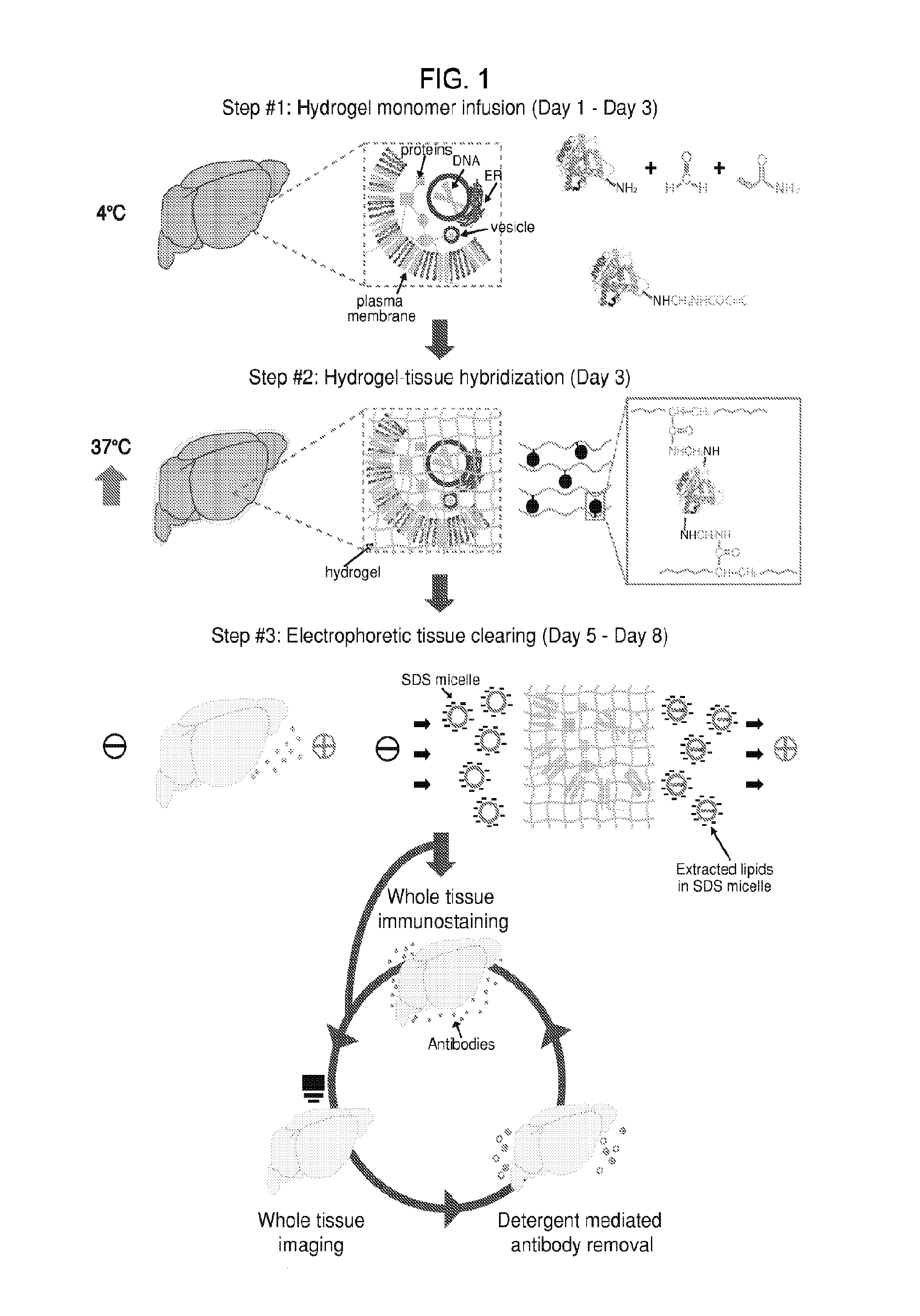
[0158]A process was developed to image whole tissue (including whole organs / organisms) for precise three-dimensional (3-D) imaging, immunohistology, clinical diagnostics, and functionalization. By embedding a tissue in nano-porous hydrogel that physically supports the ultrastructure of the tissue and then electrophoretically removing only membrane lipids, the whole tissue becomes highly transparent and permeable to macro-molecules. This technology preserves proteins, small peptides, small molecules, and nucleic acids in the tissue in their three-dimensional distribution, secured by the fine hydrogel mesh. By bypassing destructive sectioning of the tissue, subcellular structures may be analyzed. In addition, tissue can be iteratively stained, unstained, and restained with other reagents for comprehensive analysis. An illustration of the process, termed “CLARITY,” is provided in FIG. 1. Panel a: tissue is crosslinked using formaldehyde (red) in the pres...

example 2

Devices and Systems for CLARITY Process

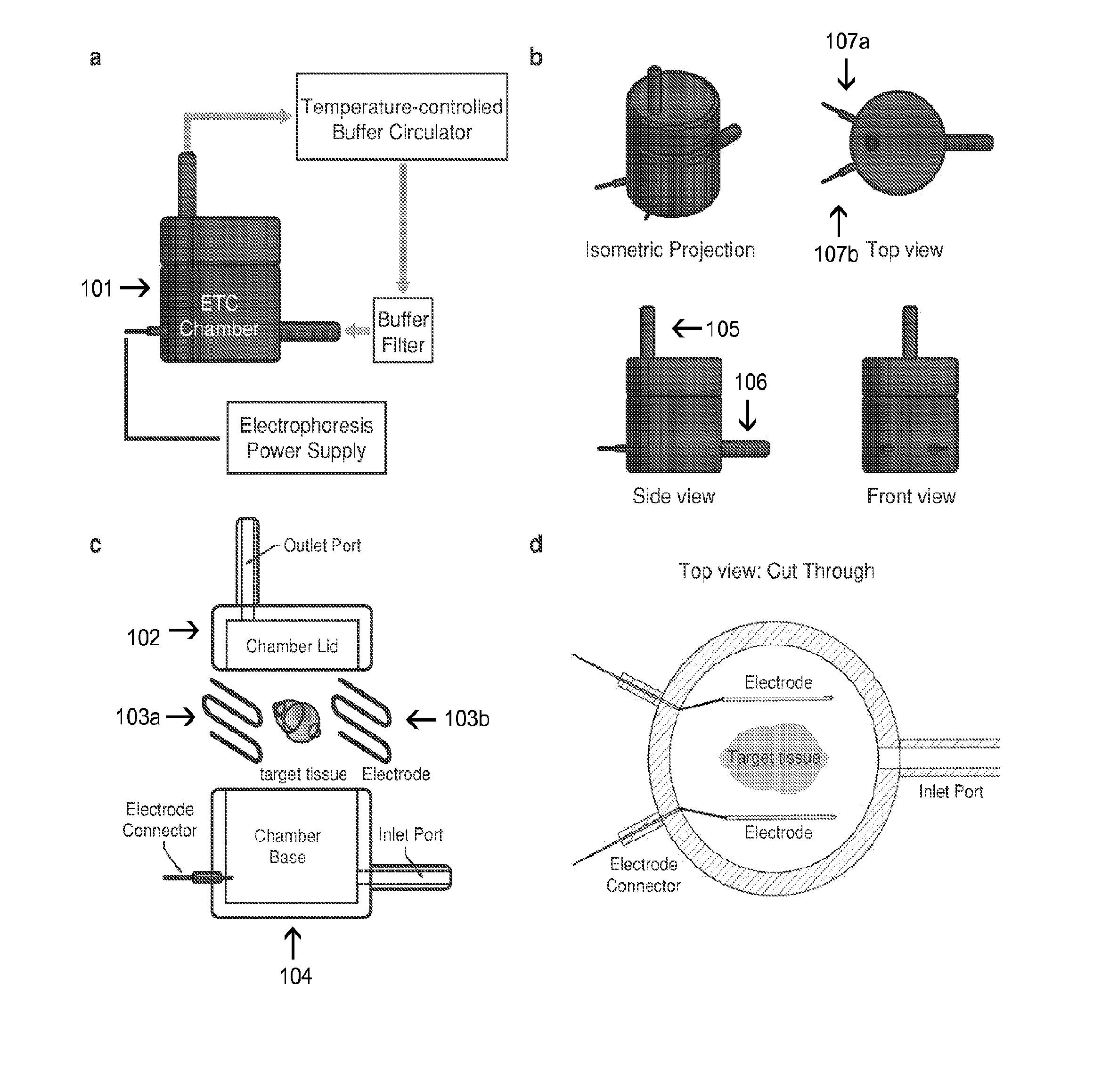
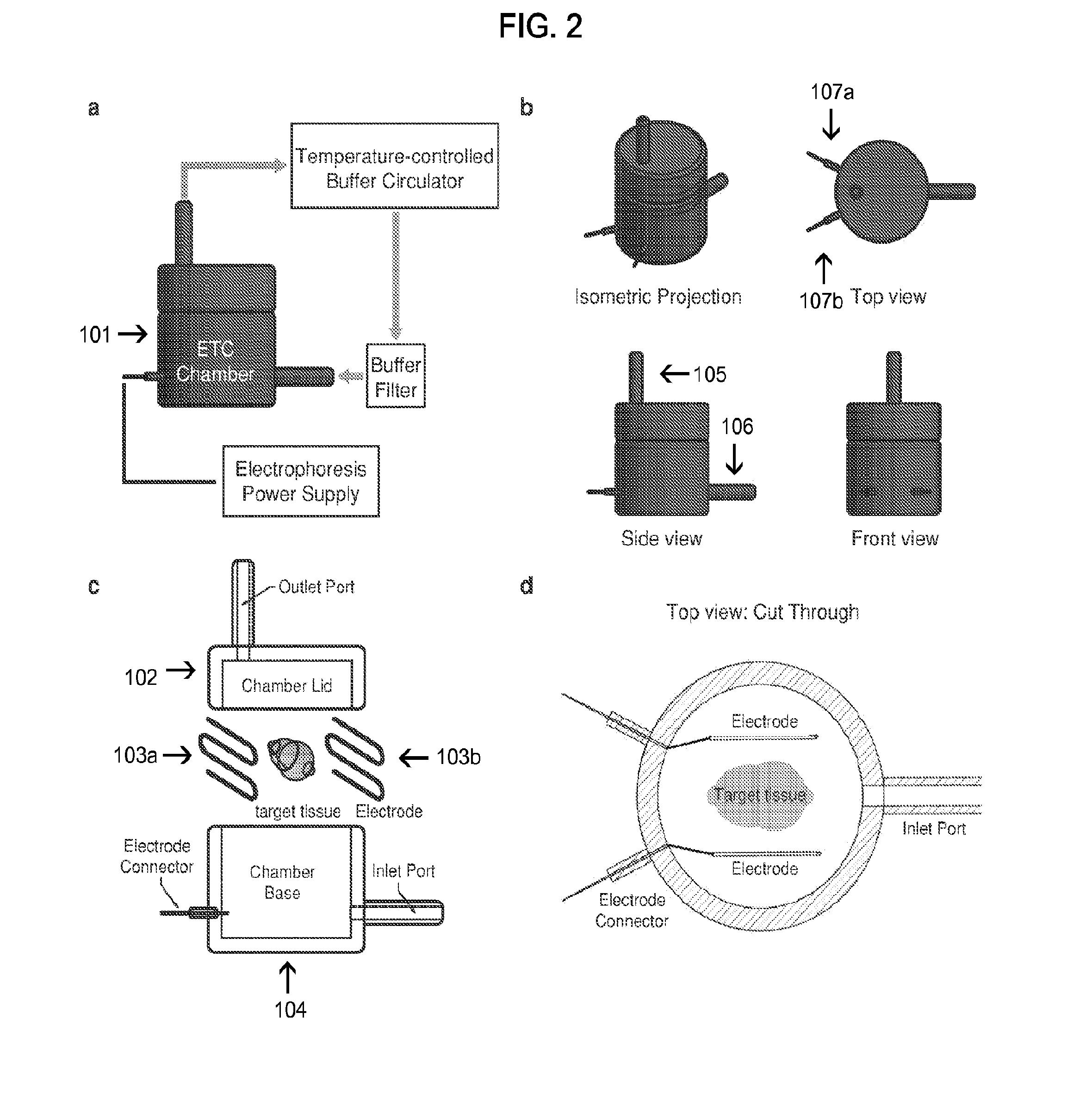
[0159]An electrophoretic tissue clearing (ETC) chamber and associated components (including a temperature-controlled buffer circulator, buffer filter, and electrophoresis power supply) were developed to carry out portions of the CLARITY process described in Example 1. Illustrations of the devices and systems are provided in FIG. 2. Panel a: ETC setup consists of the custom ETC chamber, a temperature-controlled buffer circulator (RE415, Lauda, Germany), a buffer filter (Cat. No. 4422K61, McMaster, Robbinsville, N.J.), and a power supply (Cat. no. 164-5052, Biorad, Hercules, Calif.). The sample is electrophoresed by applying 20-60V to the electrodes. Buffer solution is circulated through the chamber to maintain temperature and the composition of the buffer solution constant throughout the clearing process. Panels b-d: ETC chamber design. Panel b shows various views of a representative device showing a cylindrical plastic housing, inlet / outlet por...

example 3

Imaging of Intact Adult Mouse Brain Samples

[0160]Intact adult mouse brain samples were subjected to the CLARITY process and imaged. FIG. 3 shows the results. Panels a-d: images of whole mouse brains (three months old) with a Ramon y Cajal quote in the background. Panel a: before CLARITY. Panel b: after CLARITY: Thy1-EYFP H line brain after hydrogel-tissue hybridization, ETC, and refractive index (RI) matching with FocusClear™. Panel c: fluorescent image of the same brain shown in Panel b. Panel d: mounting of the whole cleared mouse brain for imaging. The brain is enclosed between two coverglasses. The dorsal half of the brain is first imaged using single photon (1P) excitation microscopy, and then the brain is inverted and the ventral half is imaged. Panel e: 3D reconstruction of the intact CLARITY-processed mouse brain (three months old) that was imaged using a 10× water immersion objective (numerical aperture (NA)=0.3, working distance=3.6 mm) and 514 nm excitation. Left, reconst...


PUM

PropertyMeasurementUnit
voltageaaaaaaaaaa
timeaaaaaaaaaa
depthsaaaaaaaaaa

Abstract

Methods and compositions are provided for preparing a biological specimen for microscopic analysis. These methods find many uses, for example in medicine and research, e.g., to diagnose or monitor disease or graft transplantation, to study healthy or diseased tissue, to screen candidate agents for toxicity and efficacy in disease modification. Also provided are reagents, devices, kits and systems thereof that find use in practicing the subject methods.

Description

CROSS-REFERENCE TO RELATED APPLICATIONS[0001]This application claims priority benefit to the filing date of U.S. Provisional Patent Application Ser. No. 61 / 681,551, filed on Aug. 9, 2012, the disclosure of which application is herein incorporated by reference in its entirety.FIELD OF THE INVENTION[0002]This invention pertains to preparing biological specimens for microscopic analysis.BACKGROUND OF THE INVENTION[0003]To study complex organs and tissues, such as brain and tumor, it is necessary to understand its integrated 3-D structure and fine molecular details throughout the whole tissue. Current methods, exemplified by array tomography or serial block-face scanning electron microscopy can provide sub-cellular fine details, but involve prohibitively inefficient and damaging mechanical sectioning and reconstruction. Optical sectioning techniques combined with tissue clearing methods have been developed, in which light-scattering is reduced to increase the depth at which tissue can b...

Claims


Application Information

Patent Timeline
Patent Type & Authority Applications(United States)
IPC IPC(8): G01N1/30G01N1/31
CPCG01N1/31G01N1/30G02B21/34G01N33/4833G01N2001/4038G01N33/549G01N1/40G01N27/44743G01N27/44747
Inventor DEISSEROTH, KARL A.CHUNG, KWANGHUN
Owner THE BOARD OF TRUSTEES OF THE LELAND STANFORD JUNIOR UNIV
Why Eureka
Try Eureka