Optoacoustic-Ultrasonic System for Coregistered Functional and Morphological Imaging of Placentas

a technology of optical and morphological imaging and co-registration, applied in the field of biomedical imaging and obstetrics, can solve the problems of inability to estimate local placental perfusion, inability to measure tissue oxygenation, and limitations of those approaches

Active Publication Date: 2015-06-04
TOMOWAVE LAB INC
View PDF2 Cites 13 Cited by
  • Summary
  • Abstract
  • Description
  • Claims
  • Application Information

AI Technical Summary

Benefits of technology

[0009]The present invention is directed to a dual modality imaging system for evaluating functional parameters of placental blood within displayed anatomical structures of placenta. The dual modality imaging system comprises means for delivering near infrared light to placental vasculature and blood diffused within tissue, means for generating and delivering ultrasonic pulses to placental tissue and means for detecting ultrasonic pulses either generated within the tissue or reflected within the tissue and for producing electronic signals. The means of generating ultrasound pulses can utiliz

Problems solved by technology

However, oxygenation of the tissue is not measurable by ultrasound and so it is not usable to estimate local placental perfusion.
Magnetic resonance imaging (MRI) and near infrared spectroscopy (NIRS) have been used experimentally to evaluate oxygenation, however, there are limitations in those approaches (6-8).
Due to cost, logistics, and lack of r

Method used


Image

  • Optoacoustic-Ultrasonic System for Coregistered Functional and Morphological Imaging of Placentas
  • Optoacoustic-Ultrasonic System for Coregistered Functional and Morphological Imaging of Placentas
  • Optoacoustic-Ultrasonic System for Coregistered Functional and Morphological Imaging of Placentas

Examples


example 1

LOUIS Imaging System

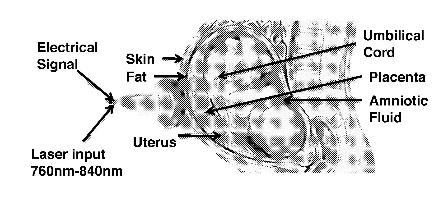
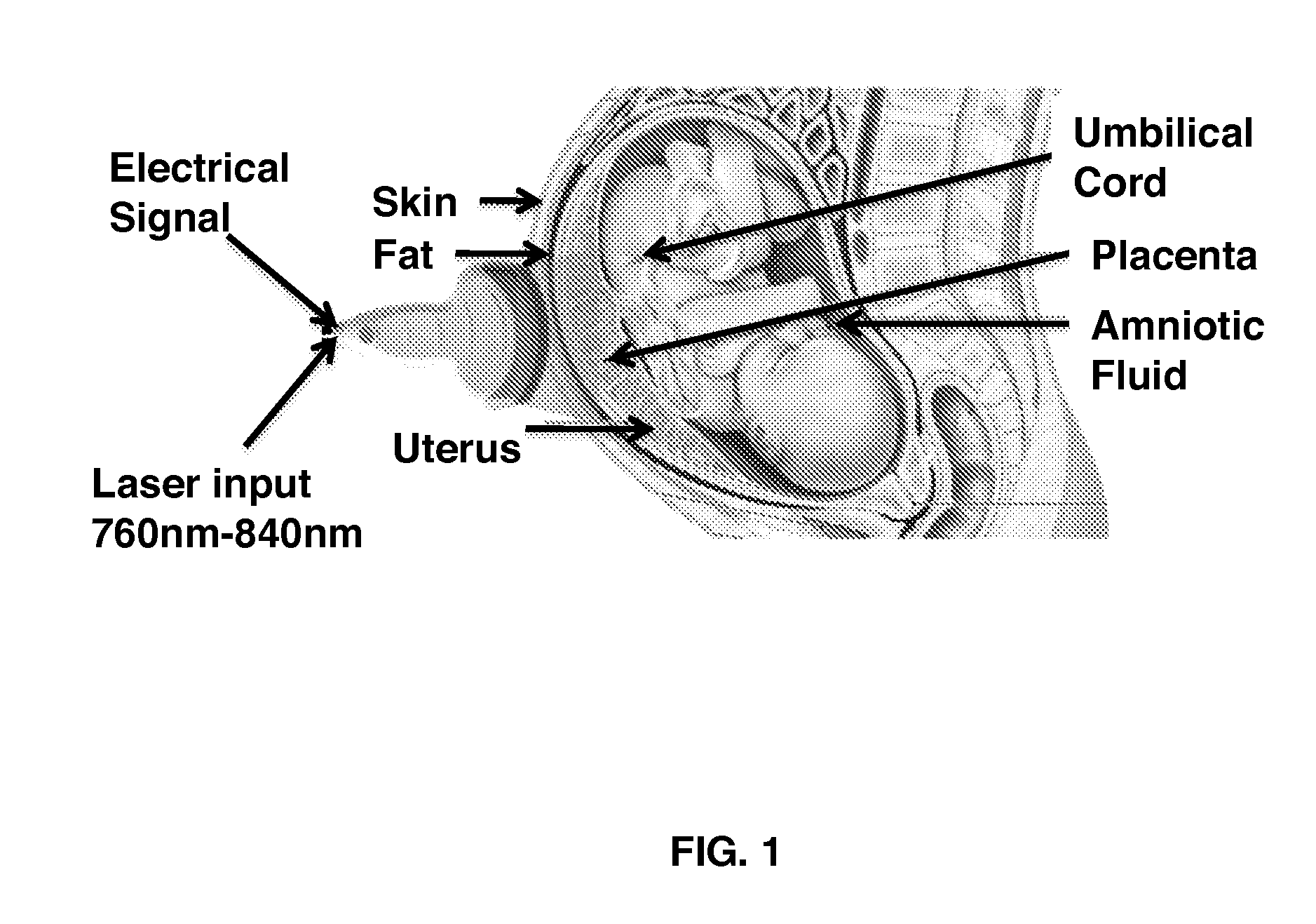
[0045]FIG. 1 illustrates the LOUIS imaging system which is configured or enabled for high resolution real-time optoacoustic imaging of intra- and extraplacental blood perfusion, for high resolution real-time optoacoustic imaging of placental blood and oxygenation and for mapping the functional information of perfusion and oxygenation onto the morphological structures that is visualized concurrently using transabdominal or transvaginal ultrasound. More particularly, the figure illustrates:

[0046]1) an ultrasonic probe upgraded with light delivery system;

[0047]2) OAT Data Acquisition Board (DAB) and software that works either in “sequence” mode or in a “toggling” acquisition mode with ultrasound; and

[0048]3) an additional laser unit that provides Q-switched fiber optically coupled output in a near infrared optical range or spectral window of about 750-840 nm.

example 2

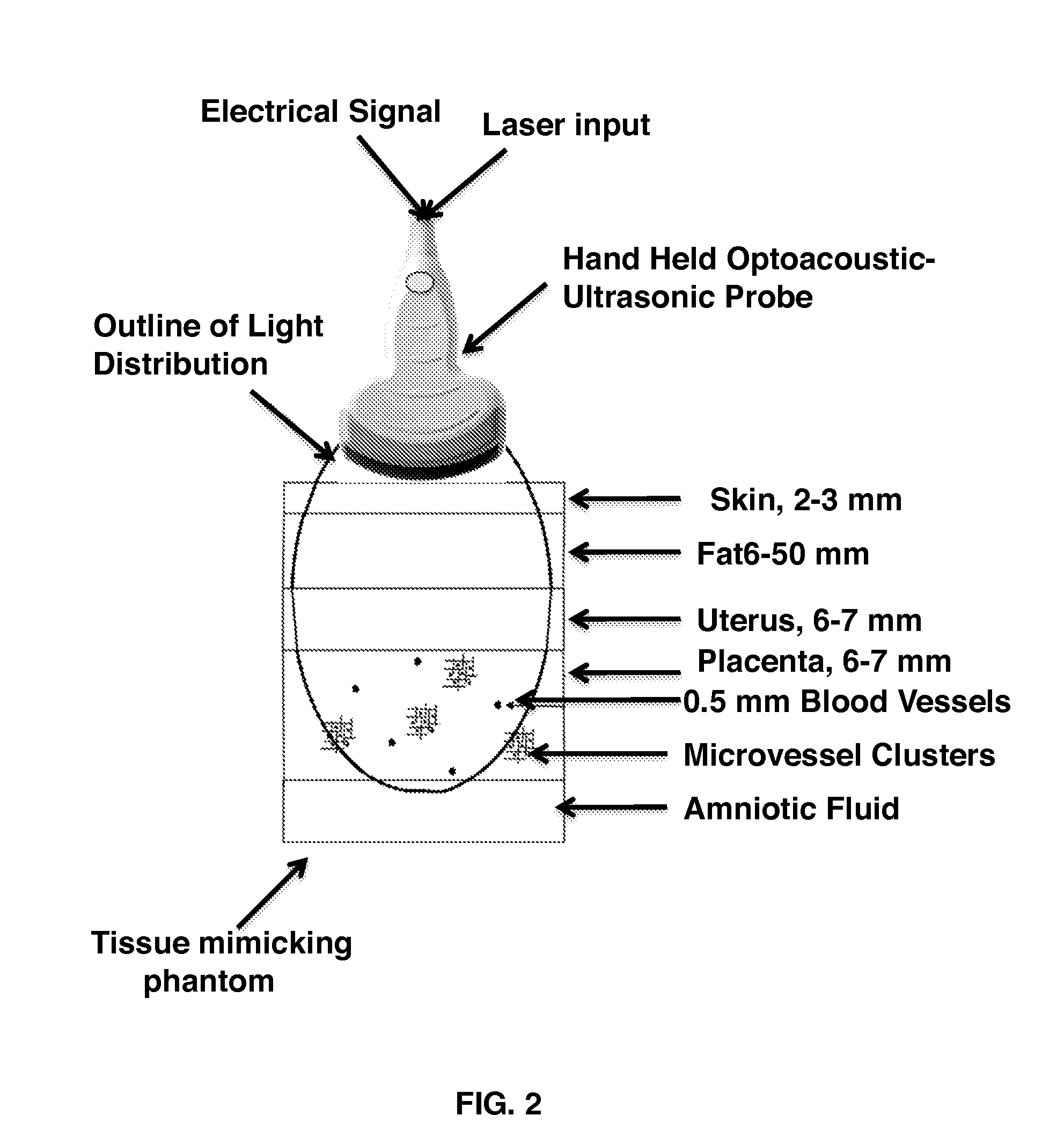
Digital Phantoms for Computer Simulations of Transabdominal Optoacoustic Imaging of Placenta

[0049]Digital phantoms are utilized for Monte Carlo based simulations of OAT imaging. A typical digital OAT phantom is a 3D map of optical properties which simulates a portion of the body affected by illumination during an OAT imaging procedure. Special care is taken with respect to boundary conditions by accounting for optical continuity of deep tissues and by modeling physical contact of the skin and optoacoustic probe. The digital phantoms utilized herein have a stratified geometry with individual layers representing skin, adipose tissue, uterus, placenta, and amniotic fluid (FIG. 2). The thickness of each layer is either obtained from published reference data or is estimated from at least 10 transabdominal ultrasonic images acquired during routine clinical examinations of the placenta at various gestational ages.

[0050]Each layer, except for the placenta, is represented as a homogeneous me...

example 3

Optimization of Shape, Size, and Arrangement of the Optical Fibers within Standard Transabdominal Ultrasound Probe Using Monte Carlo Computer Simulation of Optoacoustic Imaging and Digital Phantom

[0052]Optical illumination is critical for good quality 2D or 3D optoacoustic imaging. Conventionally it is achieved using two fiber optic bundles shaped at the output termini in a rectangular pattern and attached on both sides of the ultrasound probe (FIG. 3). Two illumination modes, dark field and bright field, have been employed currently for 2D OAT imaging. Dark field is formed when light bars are significantly separated from the imaging plane of the probe. In that case OAT image is formed by backscattered light. Such an approach significantly limits the depth of view and is not practical for imaging tissue that is deeper than 1 cm. The bright field mode is formed by forward scattered photons. It is traditionally used in clinical applications for 2D OAT imaging of breast and prostate (1...


PUM


Abstract

Provided herein are dual modality imaging systems and methods within displayed anatomical structures of placenta in real time. The imaging system comprises a dual modality laser optoacoustic and ultrasonic platform with a plurality of subsystems for delivering near infrared light, optoacoustic and ultrasonic pulses to the placenta and/or associated tissue and deep anatomic structures, for detecting ultrasonic pulses generated or reflected within the tissue using a multi-channel optoacoustic-ultrasound probe and associated transducers. The dual modality imaging system displays the results obtained as quantitative functional images of the parameters coregistered with anatomic tissue images. A multichannel electronic system comprising a computer tangibly storing software enables processor-executable instructions to calculate quantitative functional parameters of the placental blood within specific anatomical tissue structures and display quantitative functional optoacoustic images of the functional parameters within specific anatomical structures in the tissue that are visualized by ultrasound.

Description

CROSS-REFERENCE TO RELATED APPLICATIONS[0001]This nonprovisional application claims benefit of priority of provisional application U.S. Ser. No. 61 / 910,154, filed Nov. 29, 2013, now abandoned, the entirety of which is hereby incorporated by reference.BACKGROUND OF THE INVENTION[0002]1. Field of the Invention[0003]The present invention generally relates to the fields of biomedical imaging and obstetrics. Particularly the present invention discloses a dual modality laser optoacoustic-ultrasound imaging system (LOUIS) on a single platform that can provide medically relevant information about the developing placenta and associated tissues.[0004]2. Description of the Related Art[0005]The placenta and umbilical cord are vital for proper growth and transfer of nutrients and gases to and from the fetus. Several conditions are related to abnormal function of placenta, including abnormal fetal growth, stillbirth, preeclampsia, and preterm birth (1-2). Current in vivo methods of fetal surveill...

Claims


Application Information

Patent Timeline
IPC IPC(8): A61B5/00A61B8/08A61B8/00A61B5/145A61B5/1464A61B8/14A61B5/1455
CPCA61B5/0035A61B5/7425A61B8/14A61B5/0095A61B8/5261A61B5/14552A61B5/4848A61B5/1464A61B8/0866A61B5/7278A61B5/4362A61B8/4444A61B5/14546A61B8/4416
Inventor ORAEVSKY, ALEXANDER A.ERMILOV, SERGEY A.
Owner TOMOWAVE LAB INC
Why Eureka
Try Eureka