Automated histological diagnosis of bacterial infection using image analysis

an image analysis and histological analysis technology, applied in the field of image processing, can solve the problems of difficult to extract cell regions and bacteria from histological slides, high false negative rate, and exhausting procedure, and achieve the effect of improving performan

Inactive Publication Date: 2015-07-30
PANGEA DIAGNOSTICS
View PDF8 Cites 53 Cited by
  • Summary
  • Abstract
  • Description
  • Claims
  • Application Information

AI Technical Summary

Benefits of technology

[0019]The invention includes an initial module comprising template matching methods. This initial classifier identifies objects in images that have image characteristics of bacteria, in particular of HP. The output of this module is a set of candidates of such image objects. A candidate object component is a part of an image, which may be as small as a pixel, which is considered more likely to be within HP. These are fed to a subsequent classifier that further processes the candidates by algorithms of pattern recognition, such as machine learning and CNN, and confirms or rejects their identity as the target object. The information about confirmation or rejection by each pathologist may then be communicated back to a central processor. The data thus obtained may be evaluated and maintained on a central server(s) such that the data can be reintroduced into the CNN. This feedback information is accumulated from multiple, possibly independent users of the system who are located in diverse and distant locations. It is used to further calibrate and fine-tune the CNN for improved performance.

Problems solved by technology

This procedure is exhausting and prone to a high false negative rate due to the tedium and fatigue suffered by the pathologist.
Some attempts in the field of automated image segmentation and classification of histological specimens have been made, based on morphometric and color features, but have encountered difficulties due to the unique characteristics of histology imaging and the complex structures associated with cells and tissue architecture.
Furthermore, the cell architecture and bacteria may be presented in various 3D orientations due to the method of slicing that exacerbate the challenge of image analysis.
Those complexities have made histological slides difficult to extract cell regions and bacteria by traditional image segmentation approaches.
When attempting to segment and recognize bacteria in tissue all the above challenges exist and are made significantly more complicated because of the small size of bacteria in histological images both in absolute measure and by their much smaller size relative to the surrounding tissue features.
Identification of the boundaries of a bacterium is especially difficult on such background.
However, a convolutional neural network (“CNN”) or similar machine learning algorithm has not previously been applied to the identification of one or more bacteria in a histological specimen, mainly because the small size of bacteria coupled with the background “noise” in the image impedes automated identification.

Method used


Image

  • Automated histological diagnosis of bacterial infection using image analysis
  • Automated histological diagnosis of bacterial infection using image analysis
  • Automated histological diagnosis of bacterial infection using image analysis

Examples


Embodiment Construction

[0031]In the following detailed description, numerous specific details are set forth in order to provide a thorough understanding of the invention. However, it will be understood by those skilled in the art that the present invention may be practiced without these specific details. In other instances, well-known methods, procedures, and components have not been described in detail so as not to obscure the present invention.

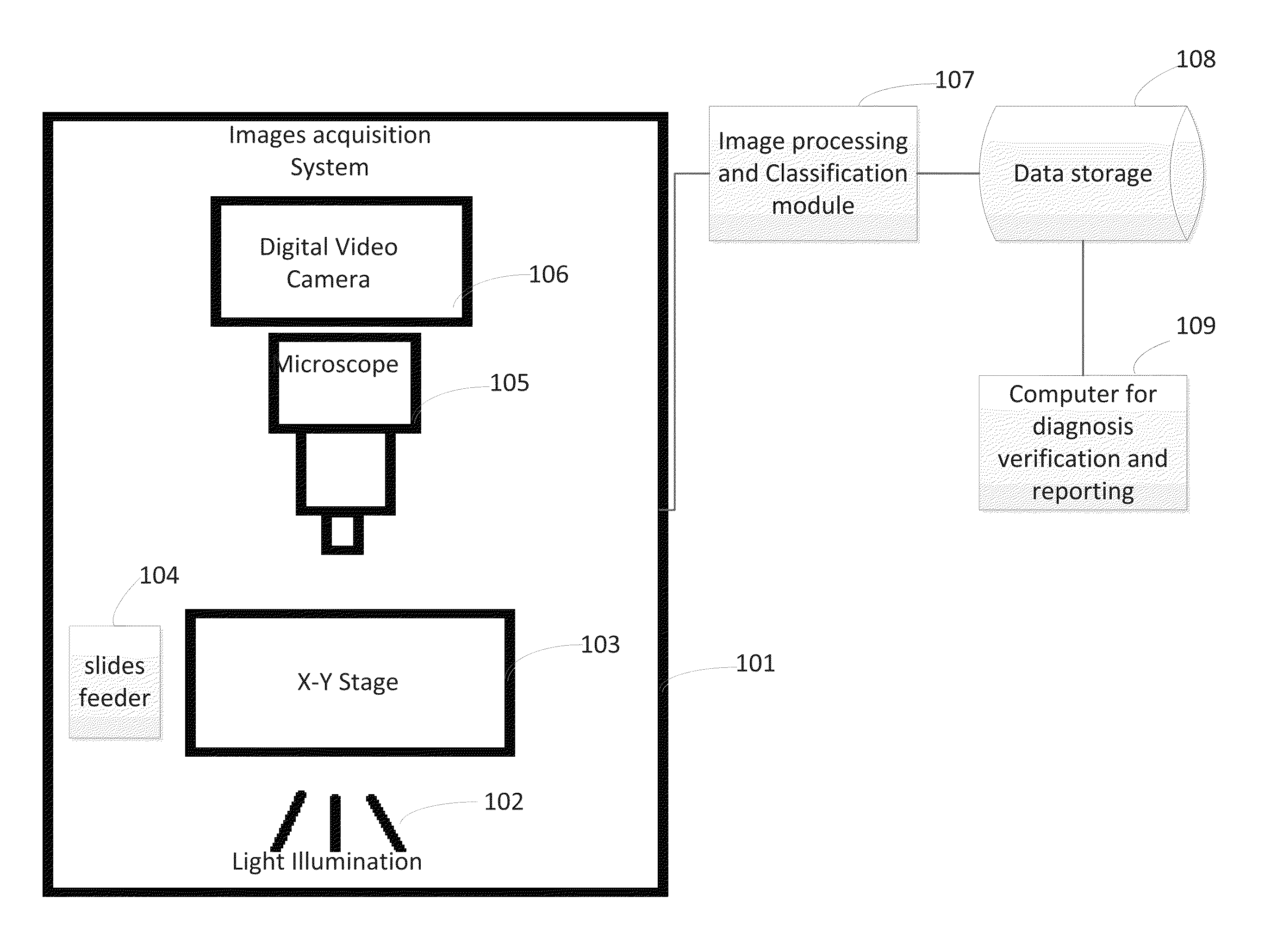
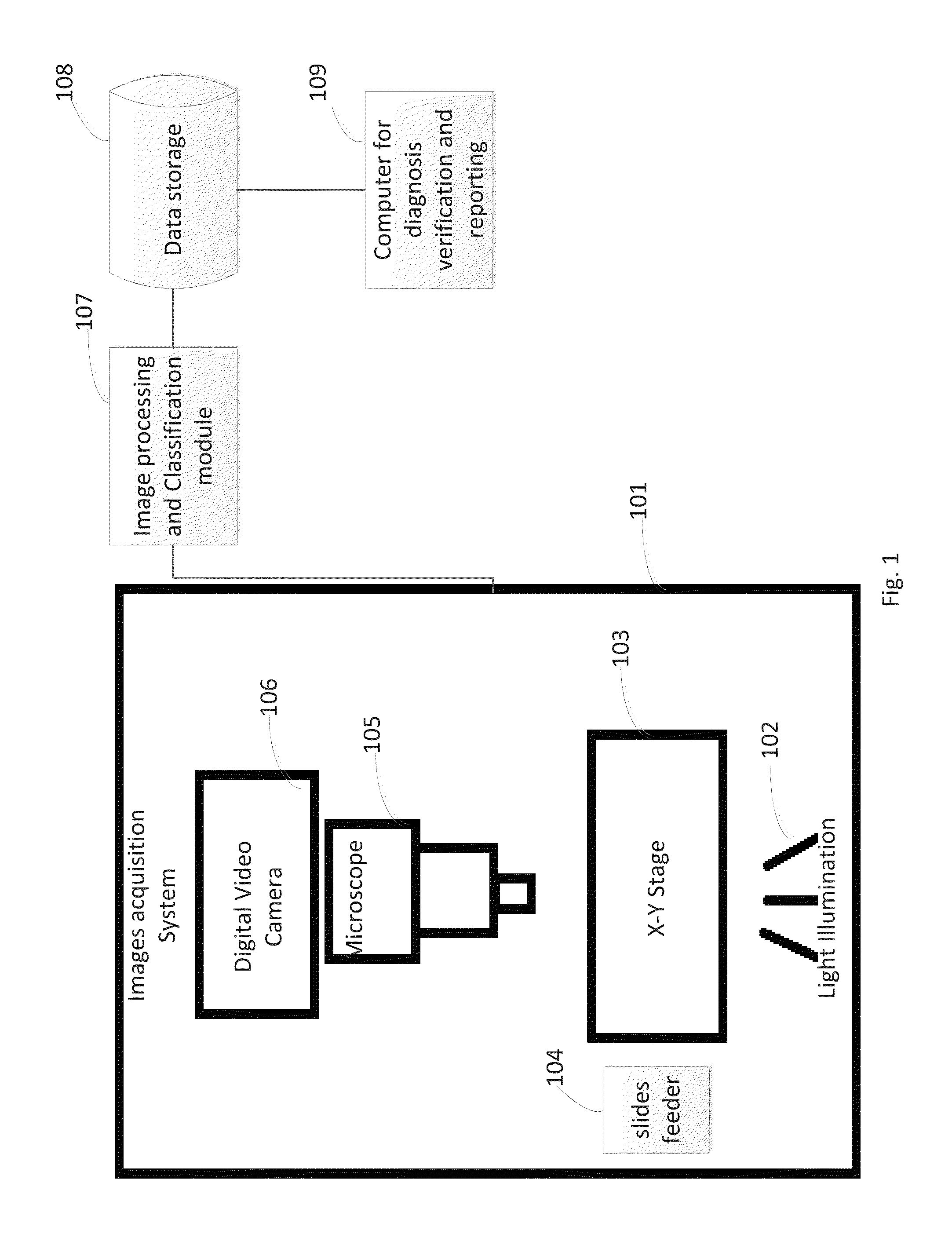
[0032]FIG. 1 illustrates a schematic representation of an automated screening apparatus and system according to an embodiment of the invention. Image acquisition system 101 includes visible light illumination 102, X-Y automated stage 103 equipped with a motor controller for stage movement, slide feeder 104, microscope 105, and a color RGB CCD or CMOS camera 106 to acquire digital images. The images obtained from image acquisition system 101 are transferred to image processing module 107 for automated classification decision. Slide data is sent to the data storage ...


PUM


Abstract

The invention relates to an automated decision support system, method, and apparatus for analysis and detection of bacteria in histological sections from tissue biopsies in general, and more specifically, of Helicobacter pylori (HP) in histological sections from gastric biopsies. The method includes image acquisition apparatus, data processing and support system conclusions, methods of transferring and storing the slide data, and pathologist diagnosis by reviewing and approving the images classified as containing bacteria, and more specifically HP findings.

Description

CROSS REFERENCE TO RELATED APPLICATIONS[0001]This application claims the benefit of U.S. Provisional Application No. 61 / 931,572, filed on Jan. 25, 2014 and entitled AUTOMATED HISTOLOGICAL DIAGNOSIS OF BACTERIAL INFECTION USING IMAGE ANALYSIS, which is incorporated in its entirety herein by reference.BACKGROUND OF THE INVENTION[0002]1. Field of the Invention[0003]The invention relates generally to the field of image processing, and more particularly relates to automated optical inspection, template matching segmentation, classification, features extraction, pattern recognition techniques and machine learning of digitized microscope slides.[0004]The invention relates also to histology analyses, and in particular, to decision support systems for diagnostic use by pathology professionals for the determination of the presence of bacteria in tissue biopsies, in general, and presence of Helicobacter pylori (“HP”) in histological specimens of the stomach, in particular.[0005]2. Description ...

Claims


Application Information

Patent Timeline
Patent Type & Authority Applications(United States)
IPC IPC(8): G06T7/00G06K9/52G06F17/30G06K9/62G06V10/26G06V10/42G06V10/44G06V10/764
CPCG06T7/0012G06K9/6201G06K9/627G06K9/52G06F17/3028G06K9/6267G06T2207/10056G06T2207/20084G06T2207/30024G06V20/695G06V20/698G06V10/26G06V10/44G06V10/454G06V10/42G06V10/751G06V2201/03G06V10/764G06F18/2413G06F16/51G06T2207/30092
Inventor BUZAGLO, ISAAC DANIELHANDZEL, AMIR AHARON
Owner PANGEA DIAGNOSTICS
Why Eureka
Try Eureka