
Novel cancer marker and utilization thereof

Inactive Publication Date: 2015-09-17
NARA MEDICAL UNIVERSITY
View PDF0 Cites 0 Cited by
  • Summary
  • Abstract
  • Description
  • Claims
  • Application Information

AI Technical Summary

Benefits of technology

The present invention relates to a new method for diagnosing urothelial cancer by detecting ubiquilin 2 in a urine sample from the patient. The method is non-invasive and accurate, making it a painless and effective way to diagnose the cancer. Ubiquilin 2 has also been found to be associated with squamous cancer, such as esophageal cancer and cervical cancer. The invention also provides a method for treating urothelial and squamous cancer by targeting ubiquilin 2. This innovation is based on the discovery that suppressing ubiquilin 2 expression can induce cancer cell death and enhance the effectiveness of therapy.

Problems solved by technology

However, the former, radiographic diagnostic imaging, has difficulty in detection of a non-invasive tumor and has difficulty in differentiating between a calculus or an inflammatory lesion and a cancer, although it is a non-invasive diagnostic method.
Further, since it is necessary to find diseased tissue with the endoscope and collect the diseased tissue, manipulation of the endoscope and determination of the diseased tissue require much skill.
Furthermore, even if the endoscope has successfully been inserted into the body, an accurate diagnosis cannot be made unless diseased tissue is finally found.
Further, ready availability of a sample makes it possible to perform an examination any number of times. However, urine cytodiagnosis has limitations in a case where the number of cells in a sample is small and in morphological determination of poorly atypical cancer cells, and requires much skill for determination of cancer cells.
For this reason, in a case where the number of cells in a sample is small or in a case where the cells are not in a form sufficient to be judged as malignant, there is a risk that a diagnosis of cancer may not be made when it should be made.
For this reason, there is a risk that non-cancer cells may be diagnosed as cancer cells.
Therefore, the existing urine cytodiagnosis is said to be fatally low in diagnostic accuracy (sensitivity), although it is high in specificity (i.e. it has a low probability of diagnosing non-cancer cells as cancer cells).
Furthermore, a urine specimen that is used as a sample in urine cytodiagnosis is easily denatured, and depending on handling of the urine specimen, an accurate diagnosis may not be made.
It is said that when ubiquilin 2 does not function normally, damaged proteins accumulate in nerve cells to severely destroy the nerve cells.

Method used


Image

  • Novel cancer marker and utilization thereof
  • Novel cancer marker and utilization thereof
  • Novel cancer marker and utilization thereof

Examples


example 1

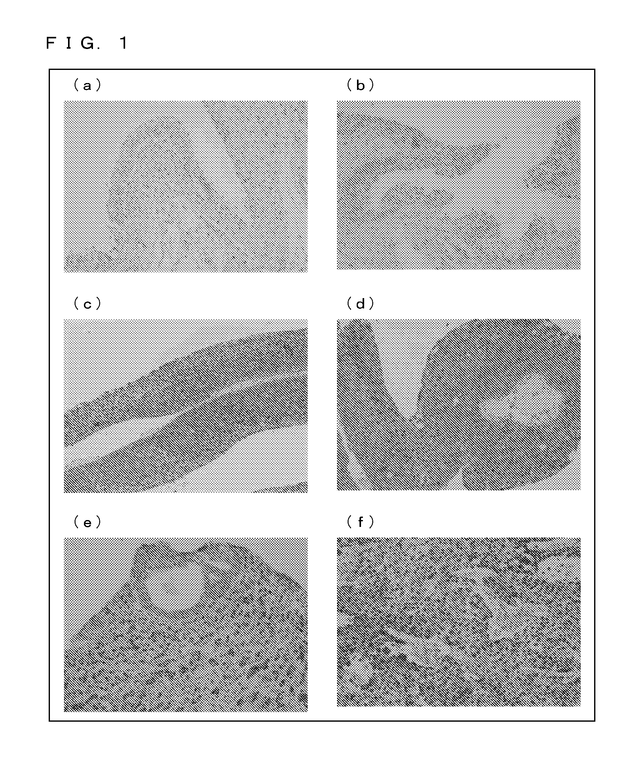
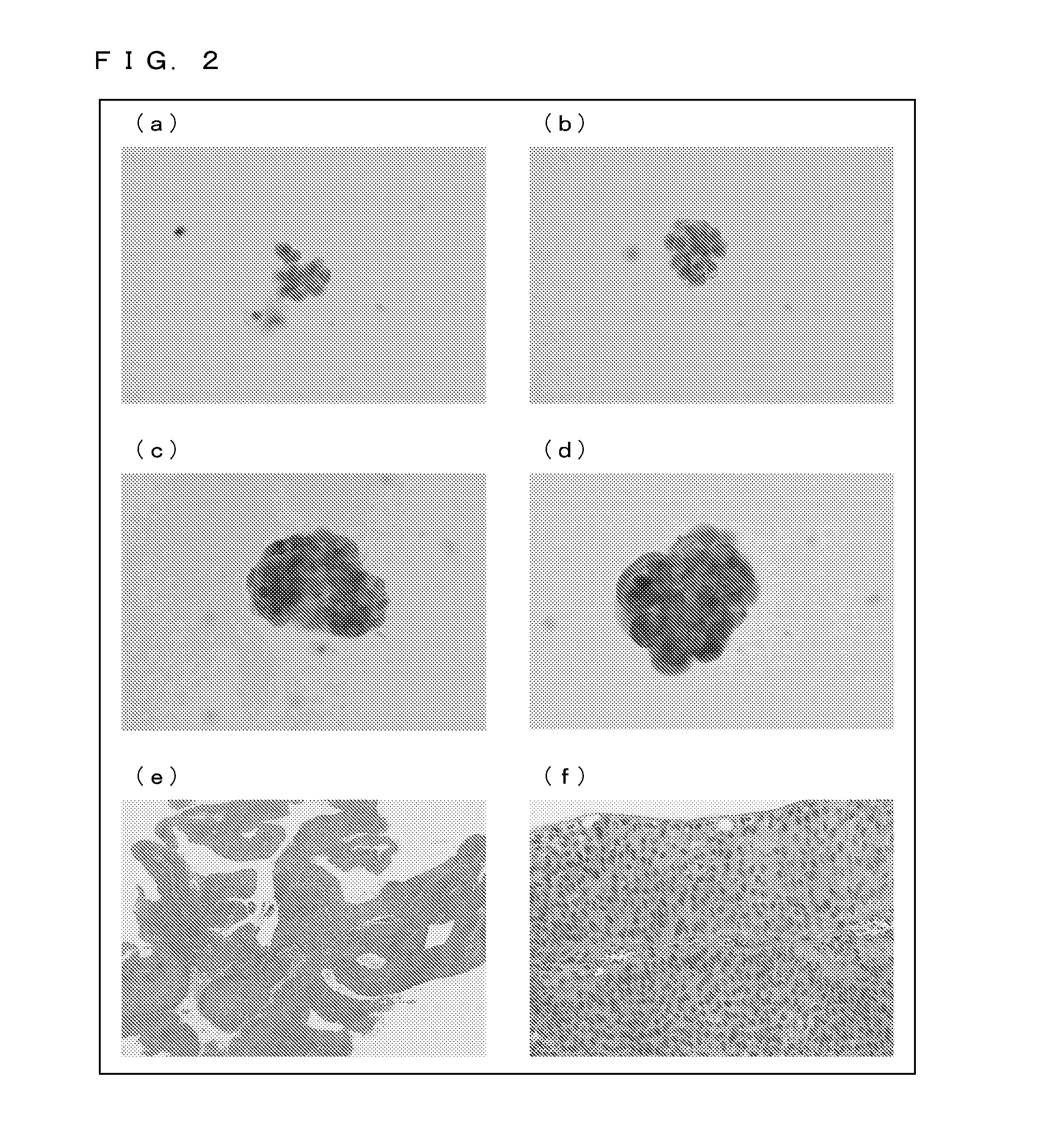
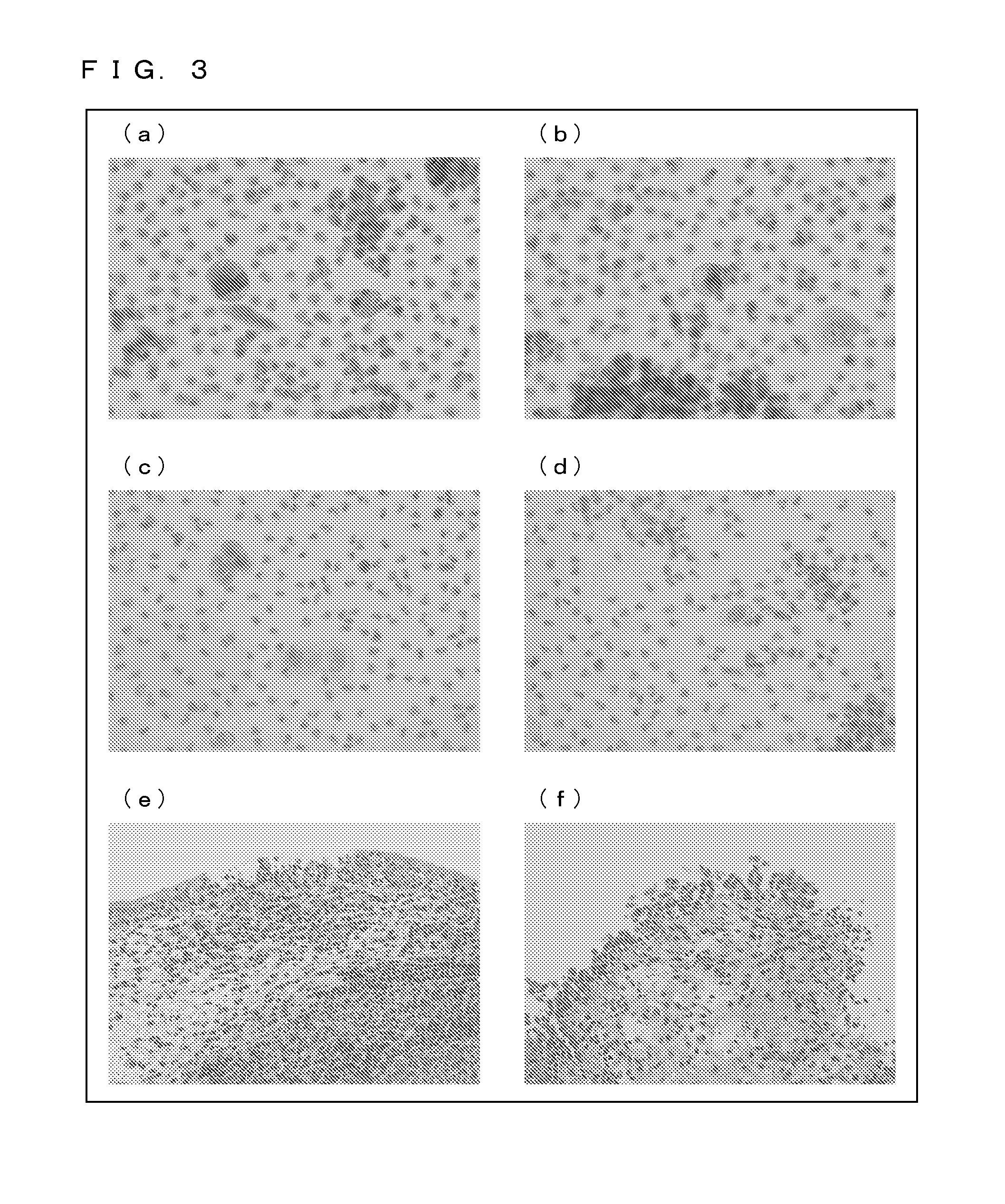
Immunohistological Staining and Immunocytological Staining with Use of a Ubiquilin 2 Specific Antibody

[0175](1) Experimental Methodology

[0176]

[0177]The ubiquilin 1 specific antibody, the ubiquilin 2 specific antibody, and the ubiquilin 2 specific antibody used were those purchased from Santa Cruz, Inc. Each of the antibodies was 500-fold diluted for use in experiment.

[0178]

[0179]A patient specimen (i.e. a surgically-resected tissue sample) was formalin-fixed, and was then embedded in paraffin. A section was prepared, deparaffinized, rehydrated, and brought into reaction with a primary antibody (ubiquilin 2 specific antibody) (4° C., 18 hours). Then, after a peroxidase-labeled secondary antibody (Nichirei Corporation) was applied and an enzyme-specific substrate was added, the specimen wan stained with a coloring reagent.

[0180]

[0181]Stained samples were prepared with urine specimens collected from patients (preparation of liquefied cytological specimens). Each urine specimen was fixe...

example 2

Examination 1 of the Effects of Suppression of Ubiquilin 2 Gene Expression

[0215](1) Methodology

[0216]Control RNA (QIAGEN) or ubiquilin 2 siRNA was introduced into human urothelial cancer cell strain KU7 (donated by the School of Urology at the Department of Medicine in Keio University). The ubiquilin 2 siRNA used was siRNA synthesized with 5′-TCCCATAAAGAGACCCTAATA-3′ (SEQ ID NO. 3) of the ubiquilin 2 gene as a Target sequence. The introduction of each RNA was performed by a Lipofection technique with a Lipofectamine™ RNAimax (Life Technologies Japan Ltd.).

[0217]After 72 hours of culture of the cells transfected with the RNA, TUNEL positive cells (i.e. cells having undergone apoptosis) were detected under a fluorescence microscope by the TUNEL (Terminal deoxynucleotidyl transferase dUTP nick end labeling) technique, and the percentage of the TUNEL positive cells to the total number of cells was calculated.

[0218](2) Results

[0219](a) of FIG. 15 shows a result of DAPI staining of cells ...

example 3

Examination 2 of the Effects of Suppression of Ubiquilin 2 Gene Expression

[0223](1) Methodology

[0224]To a culture solution prepared by culturing, for 24 hours, cells transfected with RNA in the same manner as in Example 2, thapsigargin (Sigma Corporation), which is an endoplasmic reticulum stress agent, was added so that predetermined concentrations (0.1 μM, 0.5 μM, 1 μM, and 2.5 μM) were achieved.

[0225]Seventy-two hours after the addition of thapsigargin, a cell survival assay (MTS assay: with use of a kit from Promega KK) was performed to obtain the number of cells that survived, and the ratio (cell survival rate) of the number of cells that survived in the presence of thapsigargin to the number of cells that survived in the absence of thapsigargin was calculated.

[0226](2) Results

[0227]FIG. 16 shows cell survival rates as observed 72 hours after the addition of thapsigargin. In a case where the ubiquilin 2 gene is not disrupted (knocked down), thapsigargin hardly exhibits cytotoxi...


PUM

PropertyMeasurementUnit
Luminescenceaaaaaaaaaa

Abstract

The present invention provides a novel cancer marker that is useful in the diagnosis of urothelial cancer. The present invention uses ubiquilin 2 as a cancer marker for urothelial cancer (renal pelvis cancer, ureteral cancer, and bladder cancer). Detection of ubiquilin 2 in a urine sample allows easy and accurate diagnosis of a possibility of urothelial cancer. The present invention is also applicable to the diagnosis of squamous cancer (esophageal cancer, cervical cancer, etc.).

Description

TECHNICAL FIELD[0001]The present invention relates to a novel cancer marker that is useful in the diagnosis of urothelial cancer (renal pelvis cancer, ureteral cancer, and bladder cancer) and representative examples of uses thereof. The novel cancer marker according to the present invention is also applicable to the diagnosis (detection) of squamous cancer (esophageal cancer, cervical cancer, etc.).BACKGROUND ART[0002]The diagnosis of urothelial cancer (renal pelvis cancer, ureteral cancer, and bladder cancer) depends greatly on radiographic diagnostic imaging and histopathological diagnosis of a lesion collected with a ureteroscope or a cystoscope. However, the former, radiographic diagnostic imaging, has difficulty in detection of a non-invasive tumor and has difficulty in differentiating between a calculus or an inflammatory lesion and a cancer, although it is a non-invasive diagnostic method. On the other hand, the latter, histopathological diagnosis, is an invasive diagnostic m...

Claims


Application Information

Patent Timeline
IPC IPC(8): G01N33/574C12Q1/68
CPCG01N33/57488C12Q1/6886G01N2333/4703C12Q2600/118C12Q2600/158A61K31/713A61K45/06A61P1/00A61P13/02A61P15/00A61P35/00A61P43/00C12Q2600/136G01N33/57407G01N33/57411G01N33/574
Inventor KONISHI, NOBORUSHIMADA, KEIJI
Owner NARA MEDICAL UNIVERSITY
Learn More