
Novel luciferase sequences utilizing infrared-emitting substrates to produce enhanced luminescence

a luciferase and infrared-emitting substrate technology, applied in the field of infrared-emitting substrates for infrared-emitting substrates, can solve the problems of limited utility of whole-animal optical imaging, limited light penetration by the absorption coefficient of particular components in blood, and limited tissue penetration limitations

Active Publication Date: 2016-03-17
PROMEGA
View PDF3 Cites 5 Cited by
  • Summary
  • Abstract
  • Description
  • Claims
  • Application Information

AI Technical Summary

Benefits of technology

The present invention is about a new polynucleotide that encodes a click beetle red luciferase (CBR) variant polypeptide. This polypeptide has at least 80% amino acid sequence identity to a CBR polypeptide of SEQ ID NO: 1. The CBR variant polypeptide may have enhanced luminescence, altered light emission wavelength, altered substrate specificity, or a combination of these compared to the CBR polypeptide of SEQ ID NO: 1. The CBR variant polypeptide may also have at least one amino acid substitution at a position corresponding to position 351, 389, 444, and 251 of SEQ ID NO: 1. The CBR variant polypeptide may have at least 2 fold increase in luminescence compared to a CBR polypeptide of SEQ ID NO: 1 or at least 4 fold increase in luminescence compared to a CBR polypeptide of SEQ ID NO: 1. The CBR variant polypeptide may have altered light emission spectra compared to a CBR polypeptide of SEQ ID NO: 1. The CBR variant polypeptide may also have improved stability and / or enhanced activity compared to the CBR polypeptide of SEQ ID NO: 1.

Problems solved by technology

Many standard systems for optical imaging have limited utility in a whole-animal context due to the diminished transmission of light through biological samples.
Light penetration is limited by the absorption coefficients of particular components in blood.
This region of maximum light penetration is known as the whole animal “optical window.” Bioluminescent reporter systems have been used extensively in research animals, yet still suffer from the limitations of diminished tissue penetration.
While numerous bioluminescent systems have been modified to shift visible light emission toward the red, none have achieved a strong red emission to overlap significantly with the critical “optical window” of blood transmittance.
However, this approach is limited by the signal intensity for Renilla luciferase and by the relatively low aqueous solubility of the coelenterazine substrate.

Method used


Image

  • Novel luciferase sequences utilizing infrared-emitting substrates to produce enhanced luminescence
  • Novel luciferase sequences utilizing infrared-emitting substrates to produce enhanced luminescence
  • Novel luciferase sequences utilizing infrared-emitting substrates to produce enhanced luminescence

Examples


example 1

Characterization of Near-IR Substrates PBI-4739 and PBI-4813 with Ultra-Glo™ Luciferase, QuantiLum® Recombinant Luciferase, and Purified Click Beetle Red Luciferase (CBR)

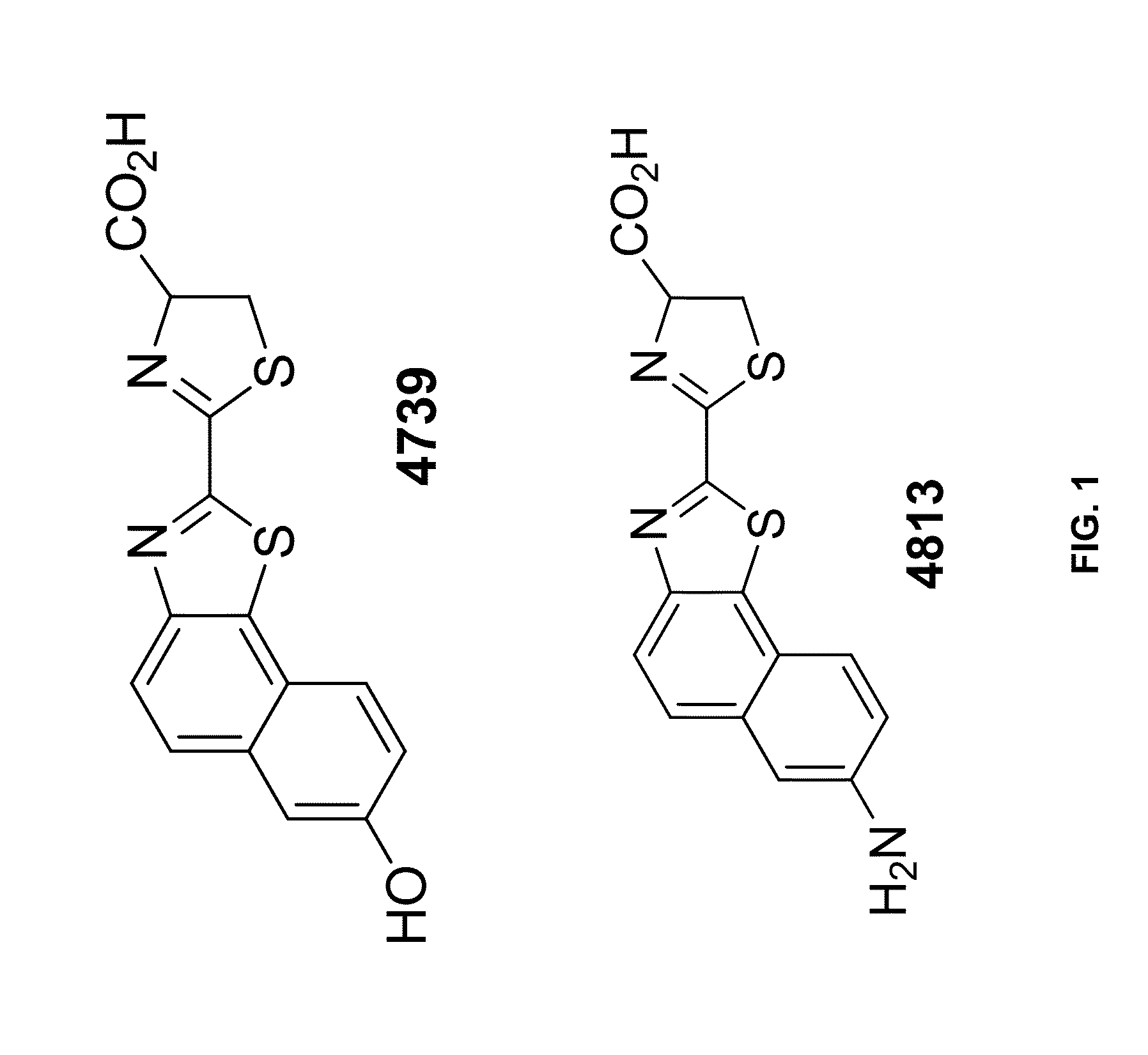
[0307]Materials: The following were used in the Examples: Ultra-Glo™ Luciferase (Promega Cat. No. E140); QuantiLum® Recombinant Luciferase (Promega Cat. No. E1701); Click Beetle Red Luciferase (CBR; 0.5 mg / mL purified; Promega); Bright-Glo™ assay buffer (Promega Cat. No. E264A); PBI-4739 (Promega—See FIG. 1); and PBI-4813 (Promega—See FIG. 1).

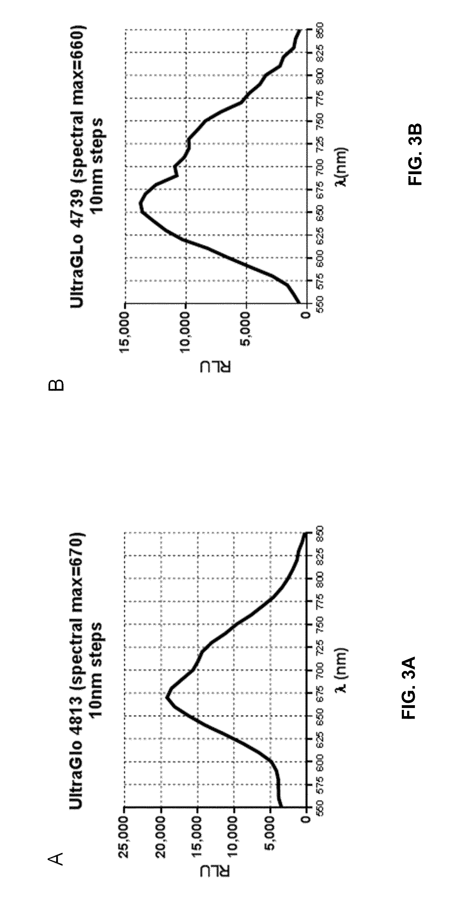
[0308]Experimental Details. The substrates, PBI-4813 and PBI-4739, were diluted to 1 mM (final concentration) in Bright-Glo™ Assay buffer+1 mM ATP. 50 μL of substrate solution was added to 50 μL of Click Beetle Red, UltraGlo® or QuantiLum® purified enzyme (0.5 mg / mL diluted in DMEM+0.1% Prionex) in triplicate, and then the samples assayed using a Tecan M-1000 plate reader in spectral scan mode.

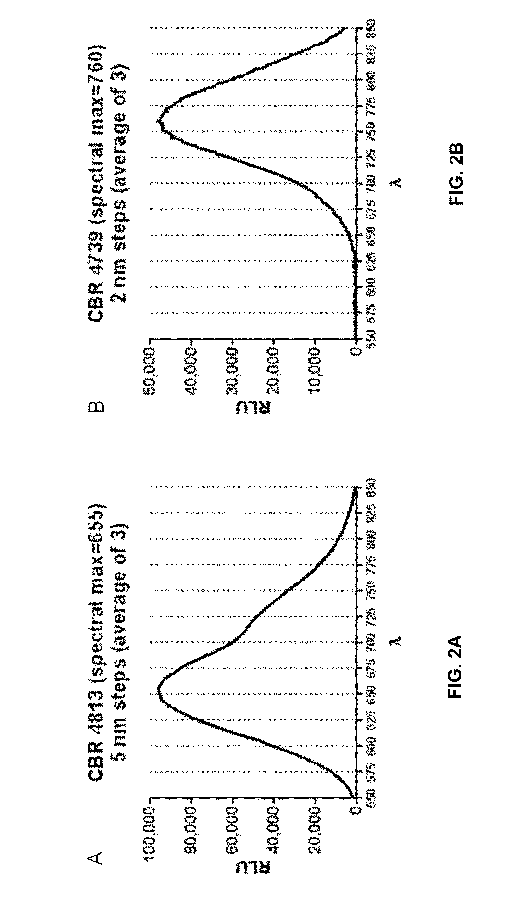
[0309]FIG. 2 provides the spectral data for Click Beetle Red Luciferase with PBI-4813...

example 2

Library Screening

[0310]A library of Click Beetle Red variants was prepared using the Diversify™ PCR Random Mutagenesis Kit (Clontech) according to the manufacturer's instructions using pF4Ag-HT7-CBR as a template, i.e., CBR-HALOTAG® fusion protein. The library of variant DNA was cloned into the pF4Ag-HT7 (Promega) and transformed into 50 μL KRX competent cells (Promega Corporation). The cells were grown overnight on LB-ampicillin plates at 37° C.

[0311]Colonies were picked and grown overnight at 37° C. in 200 μL of M9-minimal media (1× M9 salts, 0.1 mM CaCl2, 2 mM MgSO4, 1 mM Thiamine-HCL, 1% gelatin, 0.2% glycerol and 100 μg / mL Ampicillin) in wells of 96-well plates. 10 μL of the overnight culture was diluted into 190 μL M9-minimal media and grown overnight at 37° C. 10 μL of the second overnight culture was diluted into 190 μL M9-minimal induction media (M9 minimal media+0.05% glucose and 0.02% rhamnose) and grown overnight at 25° C.

[0312]The cells were assayed via a robot. Briefly...

example 3

Click Beetle Red Mutation Combinations

[0313]Some of the mutations identified in the library screen outlined in Example 2 were combined to identify beneficial combinations of mutations. The mutations were introduced into the Click Beetle Red (CBR) luciferase (SEQ ID NO: 1) using the QuikChange Multi Site-Directed Mutagenesis Kit (Agilent). Variants were cloned, expressed and screened as described in Example 2. Table 2 and FIG. 5 list the variants and their fold change in luminescence over CBR luciferase with the substrate PBI-4813 or PBI-4739.

TABLE 2Sample sequenceSamplePBI-4813PBI-4739I389F + S444RATG 10782.482.19I79V + I389F + I309T11G112.471.98G251SATG 10232.281.27I370T + I389F + I409T11A82.161.81I79V + I370T + I389F11C41.881.55I389FATG 10321.821.48S444RATG 10201.681.87I370T + I389F1G111.421.17WTWT1.061.05WTWT1.051.08WTWT1.041.02WTWT1.041.06


PUM

PropertyMeasurementUnit
bioluminescent light emission wavelengthsaaaaaaaaaa
temperatureaaaaaaaaaa

Abstract

Provided herein are isolated polynucleotide encoding modified click beetle luciferase polypeptides that have enhanced luminescence and longer wavelength near-infrared signals. The disclosure also relates to near-infrared bioluminescence systems that include said modified click beetle luciferase polypeptides and novel luciferin derivatives, as well as methods of using said modified click beetle luciferase polypeptides and bioluminescence systems.

Description

CROSS-REFERENCE TO RELATED APPLICATIONS[0001]This application claims priority to U.S. Provisional Application No. 62 / 049,150, filed Sep. 11, 2014, which is incorporated herein by reference in its entirety.FIELD OF THE INVENTION[0002]The present invention relates to an isolated polynucleotide encoding click beetle luciferase variant polypeptides that have enhanced luminescence and produce longer, near-infrared wavelength signals. The present invention also relates to near-infrared bioluminescence systems that include said click beetle luciferase variant polypeptides and infrared-emitting substrates and methods of using said click beetle luciferase variant polypeptides and near-infrared bioluminescence systems.BACKGROUND[0003]Bioluminescence with a longer-wavelength and lower-energy emission is of significant interest both for multiplexing applications with multiple emission colors and for in-depth tissue imaging where shorter wavelengths tend to be strongly absorbed. Many standard sy...

Claims


Application Information

Patent Timeline
Patent Type & Authority Applications(United States)
IPC IPC(8): C12Q1/66C12N9/02G01N21/64G01N33/68
CPCC12Q1/66C12N9/0069G01N33/6803G01N2021/6439G01N2333/90241C12Y113/12007G01N2021/6432G01N21/6428G01N33/542
Inventor ENCELL, LANCE P.HALL, MARY P.WOOD, KEITH V.WOOD, MONIKA G.
Owner PROMEGA
Learn More