Methods of preparing Anti-human papillomavirus antigen t cells

a technology of human papillomavirus and antigen t cells, which is applied in the field of preparation of antihuman papillomavirus antigen t cells, can solve the problems of poor prognosis of many cancers, including hpv-associated cancers

Inactive Publication Date: 2016-05-26
US DEPT OF HEALTH & HUMAN SERVICES
View PDF0 Cites 2 Cited by
  • Summary
  • Abstract
  • Description
  • Claims
  • Application Information

AI Technical Summary

Benefits of technology

[0003]An embodiment of the invention provides a method of preparing a population of HPV-specific T cells, the method comprising: dividing an HPV-positive tumor sample into multiple fragments; separately culturing the multiple fragments in the presence of only one cytokine; obtaining T cells from the cultured multiple fragments; tes...

Problems solved by technology

Despite advances in treatments such as chemotherapy, the progn...

Method used


Image

  • Methods of preparing Anti-human papillomavirus antigen t cells
  • Methods of preparing Anti-human papillomavirus antigen t cells
  • Methods of preparing Anti-human papillomavirus antigen t cells

Examples


example 1

[0085]This example demonstrates a method of preparing HPV-positive tumor-infiltrating lymphocytes (TIL) for adoptive cell therapy.

[0086]Patients were entered into clinical protocols and signed informed consents that were approved by the Institutional Review Board of the National Cancer Institute prior to tumor resection. Tumors were excised from patients. Tumors were tested for HPV 16 E6, HPV 16 E7, HPV 18 E6, and HPV 18 E7 expression using reverse transcriptase (RT) polymerase chain reaction (PCR) genotyping.

[0087]Multiple (24) independent cultures of HPV 16 E6 positive, HPV E7 positive, HPV 18 E6 positive, and HPV E7 positive TIL were set up using enzymatic digests and tumor fragments (1-2 mm3) procured by sharp dissection. TIL from tumor digests were generated by culturing single-cell suspensions (5×10⁵ / mL) obtained by overnight enzymatic digestion of tumor fragments in media containing collagenase, hyaluronidase, and DNAse. Cultures of tumor fragments and digests were initiated ...

example 2

[0091]This example demonstrates the reactivity of the TIL from Patient 1.

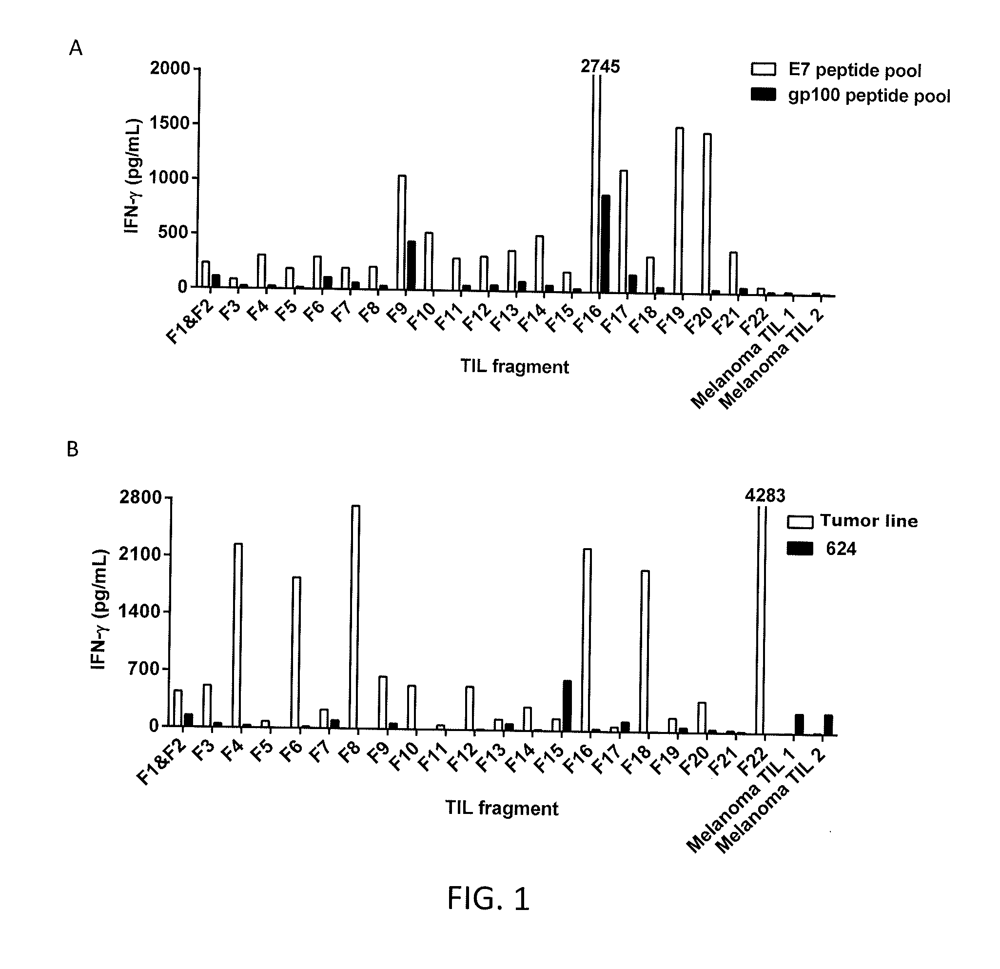
[0092]TIL were generated as described in Example 1 from 22 different tumor fragments (F1-F22) from Patient 1. The TIL from Patient 1 or melanoma TIL (control) were co-cultured with dendritic cells pulsed with the HPV 18 E7 peptide pool or a gp100 peptide pool (control) and IFN-γ was measured. The results are shown in FIGS. 1A-1B. As shown in FIGS. 1A-1B, the TIL from tumor fragment 22 of Patient 1 recognized an autologous tumor line but not HPV 18 E7 peptides.

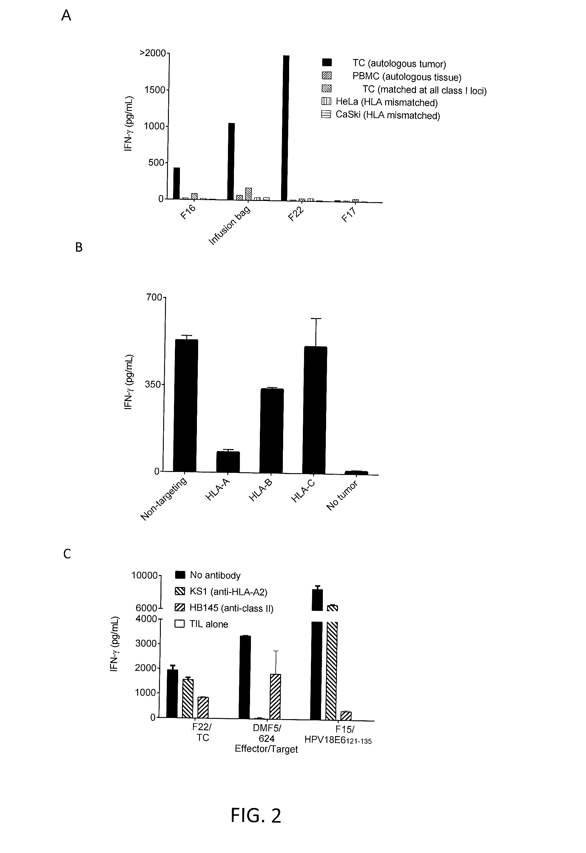
[0093]The TIL from tumor fragments F16, F17, or F22 of Patient 1 or cells given to the patient for treatment (“infusion bag”) were co-cultured with autologous tumor, peripheral blood mononuclear cells (PBMC) from autologous tissue, tumor cells matched at all class I loci, HeLa cells (HLA mismatched), or CaSki cells (HLA mismatched). IFN-γ was measured. The results are shown in FIG. 2A. As shown in FIG. 2A, TIL from tumor fragments F16 and F22 showed autolo...

example 3

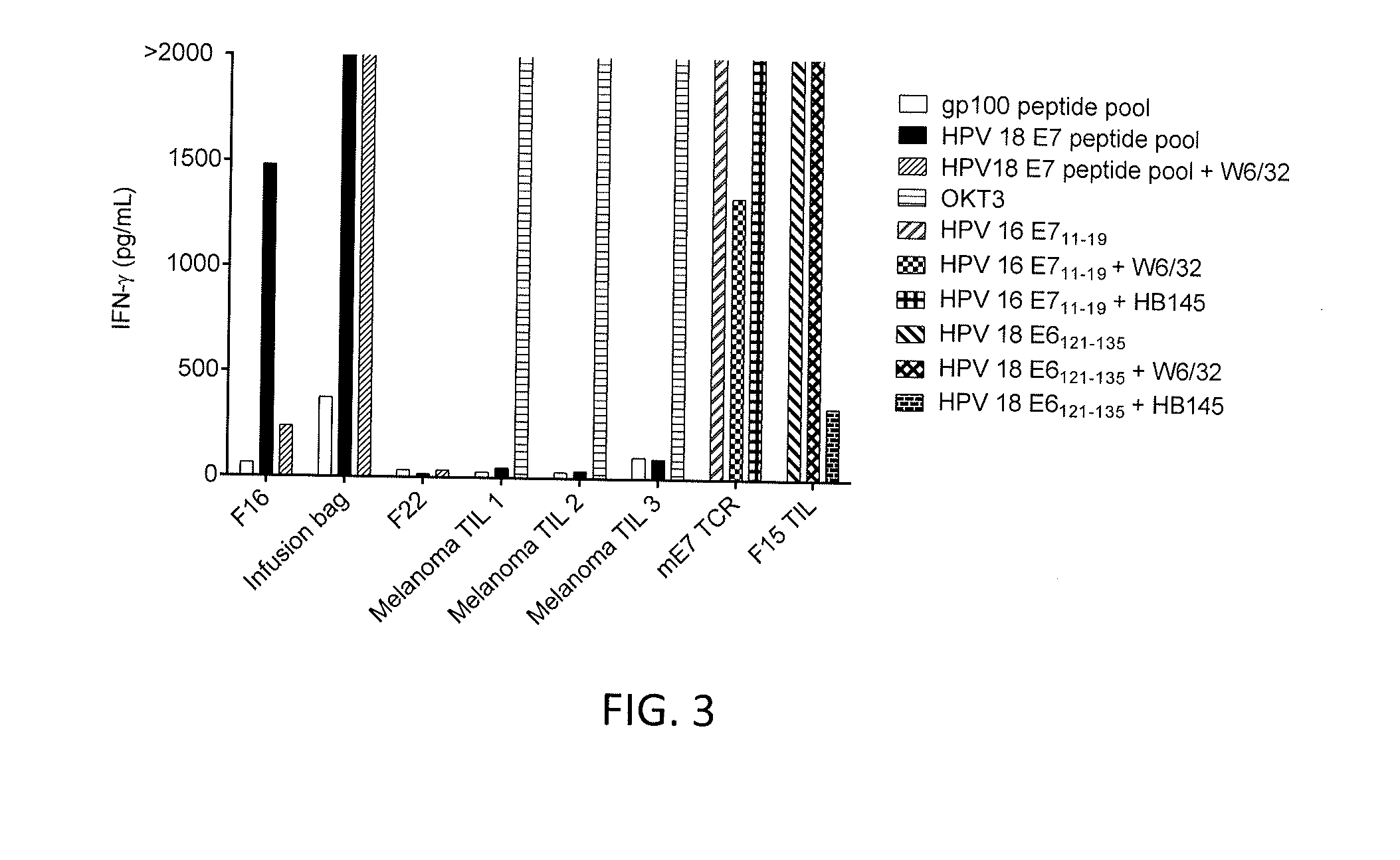
[0097]This example demonstrates the cloning of TIL from tumor fragment 16 of Patient 1 to isolate HPV 18 E7 reactive CD8 positive T cells.

[0098]DCs were loaded with HPV 18 E7 and co-cultured with TIL from tumor fragment 16 (F16) of Patient 1. The TIL were sorted for 4-1BB positive cells using fluorescence activated cell sorting (FACS). The sorted cells were cultured in 96-well plates with two cells per well. The clones were screened for tumor reactivity against a gp100 peptide pool or a HPV 18 E7 peptide pool. The results are shown in FIGS. 4A and 4B. As shown in FIGS. 4A and 4B, CD8 positive T cell cloning from tumor fragment F16 using 4-1BB-based FACS sorting resulted in the isolation of two clones (12 and 21) with E7 peptide pool reactivity.


PUM


Abstract

Disclosed are methods of preparing an isolated population of human papillomavirus (HPV)-specific T cells comprise dividing an HPV-positive tumor sample into multiple fragments; separately culturing the multiple fragments; obtaining T cells from the cultured multiple fragments; testing the T cells for specific autologous HPV-positive tumor recognition; selecting the T cells that exhibit specific autologous HPV-positive tumor recognition; and expanding the number of selected T cells to produce a population of HPV-specific T cells for adoptive cell therapy. Related methods of treating or preventing cancer using the T cells are also disclosed.

Description

CROSS-REFERENCE TO RELATED APPLICATION[0001]This patent application claims the benefit of U.S. Provisional Patent Application No. 61 / 846,161, filed Jul. 15, 2013, which is incorporated herein by reference in its entirety.BACKGROUND OF THE INVENTION[0002]The primary cause of some cancer types such as, for example, uterine cervical cancer, is human papillomavirus (HPV) infection. Despite advances in treatments such as chemotherapy, the prognosis for many cancers, including HPV-associated cancers, may be poor. Accordingly, there exists an unmet need for additional treatments for cancer, particularly HPV-associated cancers.BRIEF SUMMARY OF THE INVENTION[0003]An embodiment of the invention provides a method of preparing a population of HPV-specific T cells, the method comprising: dividing an HPV-positive tumor sample into multiple fragments; separately culturing the multiple fragments in the presence of only one cytokine; obtaining T cells from the cultured multiple fragments; testing th...

Claims


Application Information

Patent Timeline
IPC IPC(8): A61K39/12C12N7/00C12N5/0783
CPCA61K39/12C12N5/0638C12N7/00A61K2039/5158C12N2710/20011A61K2039/572C12N2501/2302C12N2502/30A61K2039/585A61K35/17C12N5/0636A61P1/04A61P11/04A61P15/00A61P15/02A61P35/00C12N7/025
Inventor HINRICHS, CHRISTIAN S.ROSENBERG, STEVEN A.
Owner US DEPT OF HEALTH & HUMAN SERVICES
Why Eureka
Try Eureka