
Corneal stromal keratocyte culture

a keratocyte and corneal stromal technology, applied in the field of cell culture, tissue culture and tissue engineering, can solve the problems of contamination source, difficult routine cell monitoring of growing cells in the opaque amniotic membrane stromal matrix, and inability to proliferate in serum-free medium

Inactive Publication Date: 2016-08-11
SINGAPORE HEALTH SERVICES PTE
View PDF2 Cites 2 Cited by
  • Summary
  • Abstract
  • Description
  • Claims
  • Application Information

AI Technical Summary

Benefits of technology

The present invention is about a method for culturing corneal stromal keratocytes (CSKs) using a dual culture medium protocol. The method involves contacting CSKs with two different culture media, A and B, which are supplemented with different proteins and growth factors. The first medium, A, is supplemented with serum and a liquid amnion extract, while the second medium, B, is supplemented with a liquid amnion extract or a minimum essential medium. This method allows for the production of a population of CSKs with improved growth and functionality. The invention also relates to the isolated population of CSKs obtained by this method and the use of the various supplements mentioned above. Overall, the invention provides a better understanding and control over the culture of CSKs which can be useful in the field of corneal regeneration.

Problems solved by technology

Unfortunately, they do not proliferate in serum-free medium.
Nonetheless, growing cells in the opaque amniotic membrane stromal matrix is difficult for routine cell monitoring, for example cell viewing to examine cell growth status.
Although they are sparsely located and should be destroyed by deep frozen storage, their remnants could affect keratocyte attachment and be a source of contaminants.
Lack of unique markers also makes the isolation of a homogenous and well-defined stem cell population difficult.
However, the induction efficiency and cell purity are yet to be optimized.

Method used


Image

  • Corneal stromal keratocyte culture
  • Corneal stromal keratocyte culture
  • Corneal stromal keratocyte culture

Examples


example 1

Materials and Methods

Corneal Stromal Tissue

[0071]Research grade human cadaveric cornea tissues were obtained from Lions Eye Institute for Transplant and Research Inc. (Tampa, Fla., US). In addition, transplant grade human cadaveric corneoscleral tissues after transplantation were obtained from Singapore Eye Bank, Singapore National Eye Centre (Singapore), with consent for research use. The human corneoscleral specimens with endothelial cell count greater than 2,000 cells per mm2 were procured, and preserved in Optisol-GS at 4° C. and transported to the culture laboratory within 14 days of preservation.

Isolation and Culture of Human Corneal Stromal Keratocytes

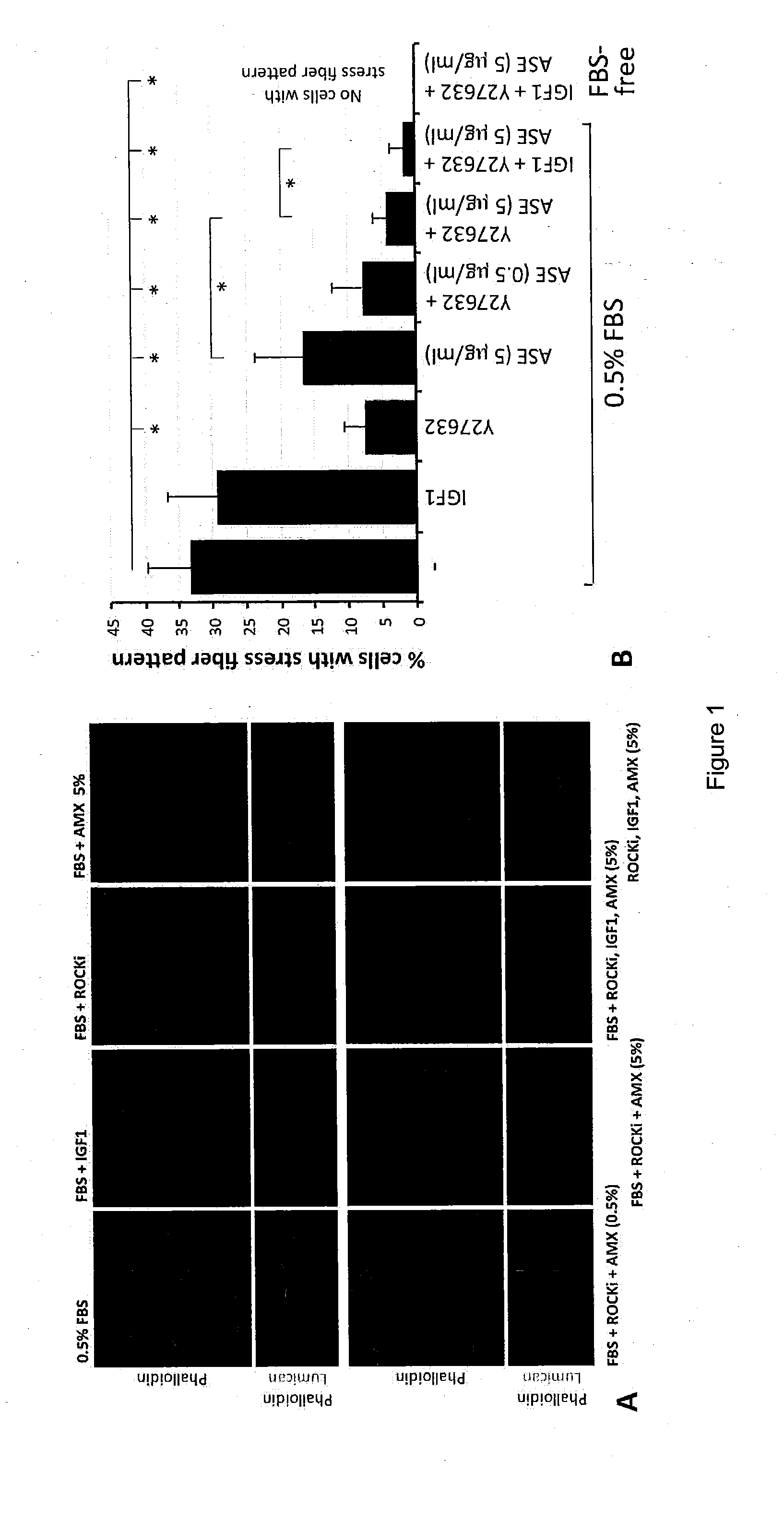
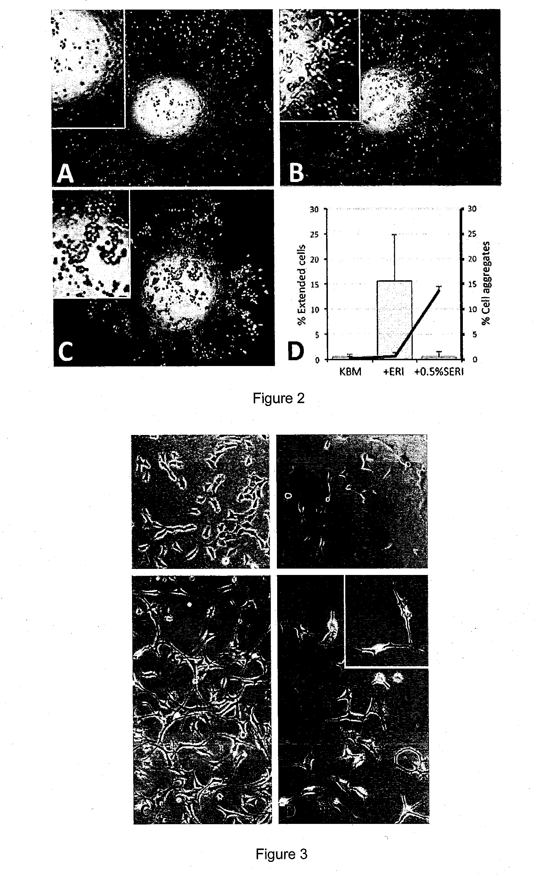
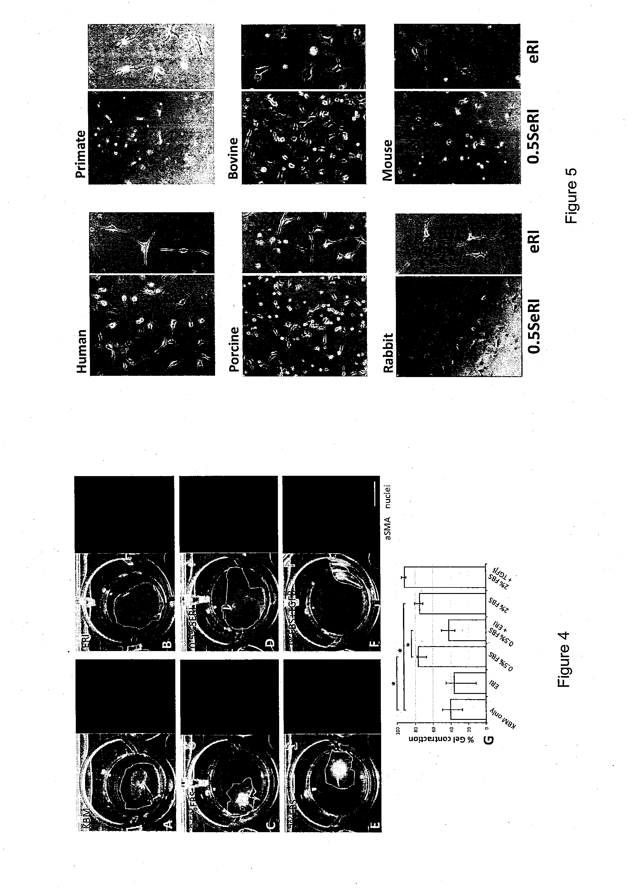
[0072]Cornea specimens were washed in sterile PBS (0.01 M, Invitrogen, Carlsbad, Calif., US) added with 3% antibiotics-antimycotes (penicillin S, streptomysin sulfate and amphoptericin B, Invitrogen). Central corneal buttons (1 mm from peripheral limbus) were trephined and treated with dispase II (20 mg / ml, Roche, Basal, Switzer...

example 2

Results and Discussion

LAE (ASE) and Protein Characterization

[0084]Soluble LAE (ASE) was prepared from frozen AM collected from the proximal one-fourth to the distal one-third to the placental disc. The devitalized AM was grounded to homogenate and extracted with ice-cold PBS. After removing debris by high-speed centrifugation, the clear supernatant was concentrated by spinning in UltraCel-3K. The protein profile of was successfully mapped with peptide homology ≧95% from the database of 178,828 proteins (Table 1). The candidate protein list was validated by choosing to measure TIMP1 level using ELISA and the concentration was 6.4±4.7 ng per μg protein. Furthermore, significant pathway analysis by MetaCore™ predicted that these proteins could participate in 12 major pathways (P−4 and False Discovery Rate FDR−3). They included TGFβ signaling, cytoskeleton and ECM remodeling, protein folding and maturation as well as immune responses (Table 2).

TABLE 1Protein list from LAE (ASE) by nanoL...


PUM


Abstract

The present invention relates to corneal stromal keratocytes (CSKs) culture. In particular the present invention relates to a method for culturing corneal stromal keratocytes ex vivo. The method utilises a dual culture medium protocol. In the presence of serum (in particular low serum concentration) in a first culture medium A, the “activated keratocytes” are expandable while specific CSK gene expression is maintained in serum-free ERI condition in a second culture medium B. The invention also relates to culture medium A and B which is supplemented with liquid amnion extract or other additional supplements. This protocol also applied to CSK from other species. Additionally, the present invention provides method for CSK cultivation to provide sufficient quantity of genuine CSKs for corneal tissue engineering without a risk of fibroblastic changes.

Description

FIELD OF THE INVENTION[0001]The present invention relates to the field of cell culture, tissue culture and tissue engineering. In particular, the invention relates to methods and systems for culturing cells ex vivo or in vitro through the use of various conditions and agents, to control the growth and / or development of the cells; maintain cellular morphology and / or phenotype; and / or prevent changes (e.g. physical changes) to the cells.BACKGROUND OF THE INVENTION[0002]The cornea is a transparent, avascular structure in the anterior part of the eye. Besides acting as a protective barrier, it provides 70% of refractive power to converge incoming light to the lens and retina. It contains 3 major layers: an outermost non-keratinized stratified epithelium, a middle collagen-rich stroma and an inner single cell-layered endothelium. The stroma spans about 90% of corneal thickness and consists of transparent extracellular matrix deposited by the resident corneal stromal keratocytes (CSKs). C...

Claims


Application Information

Patent Timeline
IPC IPC(8): C12N5/079
CPCC12N5/0621C12N2501/105C12N2533/54C12N2502/025C12N2500/84C12N2501/727
Inventor YAM, HIN-FAI GARYMEHTA, JODHBIR
Owner SINGAPORE HEALTH SERVICES PTE
Learn More