
Antibody Compositions and Immunoassay Methods to Detect Isoforms of Anti-Müllerian Hormone

an anti-mullerian hormone and immunoassay technology, which is applied in the field of anti-mullerian hormone antibody compositions and immunoassay methods to detect isoforms of the hormone, can solve the problems of inability to accurately measure amh in a given sample, limited assay, and inability to distinguish between monomeric and dimeric forms of the protein

Inactive Publication Date: 2016-09-22
ANSH LABS
View PDF1 Cites 8 Cited by
  • Summary
  • Abstract
  • Description
  • Claims
  • Application Information

AI Technical Summary

Benefits of technology

The present invention provides methods and compositions for accurately measuring AMH and its fragments in biological samples. The invention distinguishes between dimeric and monomeric forms of AMH, and measures the full-length AMH and its related polypeptides in a sandwich immunoassay. The invention also provides methods for measuring biologically-active AMH and evaluating its clinical potential. The invention can be used for assessing and diagnosing several clinical conditions involving expression, over-expression, under-expression, or absence of expression of AMH polypeptides in one or more selected cells, tissues, or biological fluids. The invention includes an isolated antibody that specifically binds to an epitope of human AMH. The antibody or its fragments can be used as a detection tool in various applications such as immunodetection-based assays.

Problems solved by technology

In addition, their report determined that the antibodies used in the Hudson assay did not recognize the mature region of the AMH protein.
While able to detect and quantify AMH in different biological samples (including mammalian samples such as human, monkey, mouse, rat, bovine and horse), the assay is limited in that in can only bind to the mature region of an AMH molecule.
Thus, this assay fails to accurately measure AMH in a given sample.
Unfortunately, none of the commercial assays presently available for AMH can distinguish between the monomeric and dimeric forms of the protein.
The antibodies of each assay all bind to both forms of the protein, making them unsuitable for distinguishing between the two forms of AMH either in vitro, or in situ.
(2011) elaborated on the particular weaknesses of the DSL assay and the IOT assays saying that they used different antibodies and different calibration materials, which resulted in substantially different results when the two assays were employed to quantify the protein under similar sample conditions.
The data suggested that AMH was not stable in some conditions, and such stability issues were more pronounced with the Gen II assay (Rustamov, et al., 2012).
This variability in results is extremely troubling.
According to the Stages of Reproductive Aging Workshop (STRAW) committee, “lack of standardized assays for [AMH] remains an important limitation in efforts to stage reproductive aging and to translate research findings to cost effective tools.

Method used


Image

  • Antibody Compositions and Immunoassay Methods to Detect Isoforms of Anti-Müllerian Hormone
  • Antibody Compositions and Immunoassay Methods to Detect Isoforms of Anti-Müllerian Hormone
  • Antibody Compositions and Immunoassay Methods to Detect Isoforms of Anti-Müllerian Hormone

Examples


example 1

Antibody Production

[0242]Male AMH knockout mice on a C57bl / 6 background were immunized with recombinant human AMH. An initial injection of 40 μg of recombinant human AMH was given to each mouse subcutaneously in Freund's Complete Adjuvant (MP Biomedical 0855828) and one booster injection of 40 μg in Freund's Incomplete Adjuvant (MP Biomedical 0855829) was given after one month. Tail bleeds were collected from the mice and titrations of the samples were screened on AMH coated plates with 10 μg / well. The highest-responding mouse was determined by color intensity (O.D. from 0-3, light yellow-dark yellow), and it was selected for final boosting. Final boosting was given one month after the booster injection date. For four consecutive days, the mouse received approximately 162 μg of recombinant human AMH, administered by intraperitoneal injection. On the fifth day, the mouse was sacrificed and the spleen was removed aseptically. Spleen cells were centrifuged, aliquoted and stored in liqu...

example 2

Antibody Screening

[0243]A fusion with SP2 / 0 cells was performed with one aliquot of the spleen cells. Fusions were initially screened on the immunogen, recombinant human AMH. Secondary screens were performed on the pro and mature fragments of AMH, to obtain more information on epitope location (FIG. 7). Positive clones were re-cloned, scaled up, and the secreted antibody purified on the GE AktaPrime Plus. After purification, the antibodies were paired and optimized based on the signal to noise ratio on full-length AMH, and the pro and mature fragments (See FIG. 9-FIG. 12).

[0244]Western blots were performed to determine which antibodies bound to the pro and mature region of AMH. Antibodies #9 and #10 cleanly and specifically detected the human AMH pro region in both human reduced (monomeric) and non-reduced (dimeric) forms (see FIG. 19 and FIG. 20). Antibodies #17 and #23 detect both monomeric and dimeric mature regions for both human and rat AMH (See FIG. 21 and FIG. 22).

example 3

Immunoassay

[0245]Coating of Microtiter Plates:

[0246]The monoclonal antibody used for coating was purified by Protein G (GE) affinity chromatography. Microtiter plates from Greiner Bio-One (Maybachstr. D-72636 Frickenhausen, Germany, cat: 705071, Greiner Bio-One North America. MONROE, N.C., USA) were coated with 100 μL / well of 10 μg / mL antibody in borate buffer overnight at room temperature. Excess antibody was removed by washing the plates once with 300 μL / well of neutral buffer. The plates were blocked with 200 μL / well of protein-based buffer and sucrose for 16-24 hours at room temperature (85% humidity). The blocking solution was aspirated, and the plates were dried in an environmental chamber at 34° C. for 4-5 hours. The plates were then packed in foil pouches with desiccant, labeled and stored at 2-8° C.

[0247]Biotinylation of Anti-AMH Monoclonal Antibody:

[0248]Purified monoclonal antibody was dialyzed in 0.1 M sodium borate buffer and biotinylated with NHS-Biotin. After two hour...


PUM

PropertyMeasurementUnit
Concentrationaaaaaaaaaa

Abstract

Disclosed are compositions and methods for detecting and quantifying human anti-Mtillerian hormone (AMH) in biological samples. In particular, the invention provides novel methods of measuring different forms of AMH in a biological sample, such as human plasma The anti-AMH antibody compositions disclosed herein enable reproducible measurement and quantitation of AMH, including dimeric forms of the AMH protein and fragments thereof. The antibody compositions disclosed herein find particular utility as diagnostic tools for single epitope sandwich-based AMH assays, which can be used to diagnose a variety of medical conditions.

Description

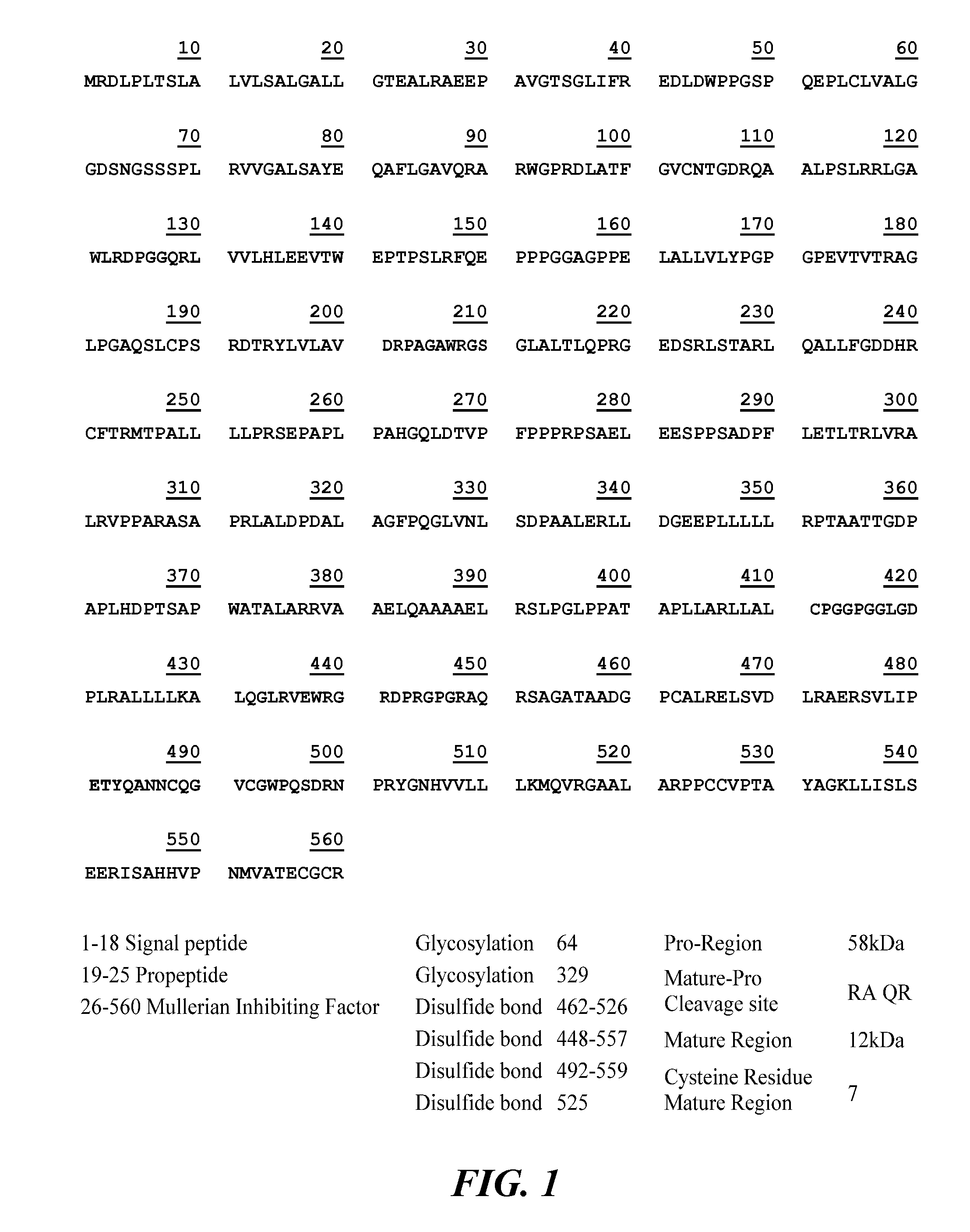
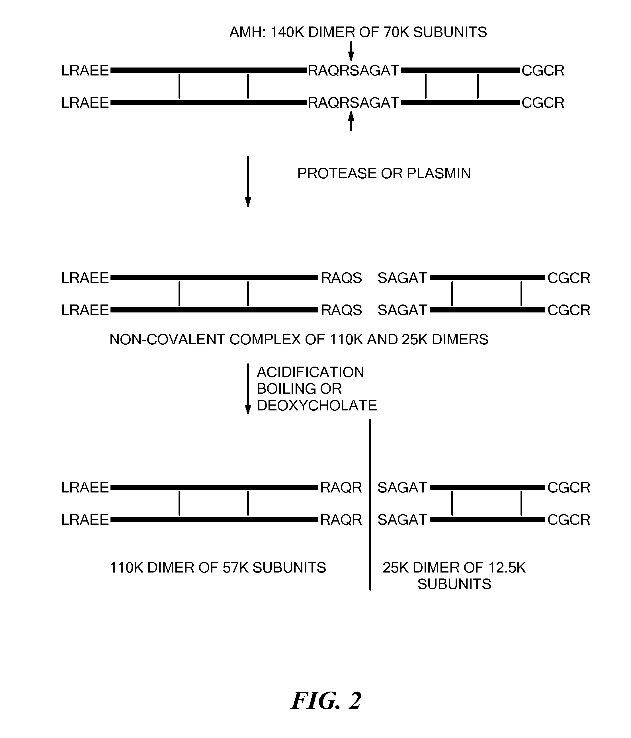
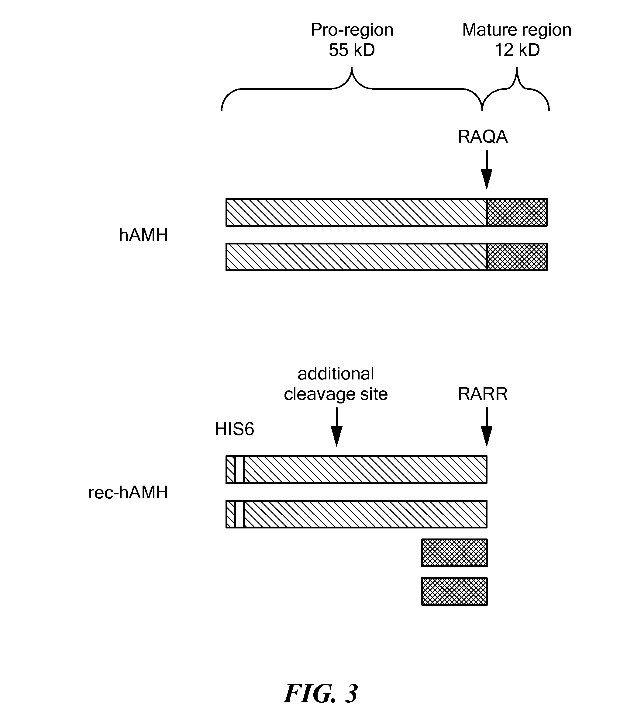
BACKGROUNDAnti-Müllerian Hormone[0001]Anti-Müllerian Hormone (AMH), also referred to as Müllerian inhibiting factor (MIF), Müllerian-inhibiting hormone (MIH), and Müllerian-inhibiting substance (MIS), is a 140-kilodalton (kDa) dimeric glycoprotein hormone, that is part of the transforming growth factor-β (TGF-β) superfamily (Zec et al., 2011). All members of this superfamily are dimeric glycoproteins that are variously involved in the regulation of tissue growth and / or cell differentiation. Like other members of the TGF-β superfamily, AMH protein is synthesized as a large precursor having a short 18 amino acid signal sequence, that is processed into a pre-prohormone that ultimately yields homodimers (Zec et al., 2011). Before secretion, the mature hormone undergoes glycosylation and dimerization resulting in a 140-kDa dimer of identical, 70-kDa, disulfide-linked, monomer subunits (Zec et al., 2011). Each monomer has an N-terminal domain (known as the “pro” region) and a C-terminal d...

Claims


Application Information

Patent Timeline
IPC IPC(8): G01N33/74C07K16/26
CPCG01N33/74G01N2333/575C07K16/26G01N33/6863
Inventor SAVJANI, GOPAL V.KUMAR, AJAY
Owner ANSH LABS
Learn More