Apparatus and methods for treating cardiac valve regurgitation

a cardiac valve and valve valve technology, applied in the field of apparatus and methods for treating cardiac valve regurgitation, can solve the problems of cardiac valves being rendered less effective, serious cardiovascular compromise or even death, valve dysfunction, etc., and achieve the effect of reducing cardiac valve regurgitation

Active Publication Date: 2016-11-10
SEGUIN JACQUES
View PDF2 Cites 61 Cited by
  • Summary
  • Abstract
  • Description
  • Claims
  • Application Information

AI Technical Summary

Benefits of technology

[0021]The present disclosure overcomes the drawbacks of previously-known systems by providing apparatus and methods for repairing a cardiac valve, e.g., a mitral valve, a tricuspid valve, an aortic valve, or a pulmonary valve. The apparatus may include an expandable frame defining a curved structure in the expanded deployed state and a membrane coupled to the expandable frame. The membrane may curve around a native leaflet, e.g., the posterior leaflet, in a first plane and curve around another leaflet, e.g., the anterior leaflet, in an orthogonal plane. The membrane may be adapted to be suspended in the flow path of the cardiac valve such a first surface of the membrane abuts the native leaflet during systole and a second surface of the membrane abuts the other native leaflet during systole, thereby reducing cardiac valve regurgitation.

Problems solved by technology

These heart valves may be rendered less effective by acute or chronic ischemic disease of the heart, congenital, inflammatory, or infectious conditions, or disease, all of which may lead to dysfunction of the valves over time.
Such degradation may result in serious cardiovascular compromise or even death.
Because the left ventricle drives systemic circulation, it generates higher pressures than the right ventricle, and accordingly the aortic and mitral valves are more susceptible to dysfunction, such as stenosis or regurgitation.
A stenotic mitral valve may impede blood flow into the heart, causing blood to back up and pressure to build in the lungs.
Consequently, the presence of a stenotic valve may make it difficult for the heart to increase the amount of blood pumped during exercise, producing shortness of breath under physical activity.
Regurgitation occurs when the mitral valve leaflets do not coapt correctly, thus causing blood to leak backwards into the left atrium and lungs each time the heart pumps.
Although the heart may compensate for this overload for months to years, provided the progression of the leakage comes gradually, the heart will eventually begin to fail, producing shortness of breath and fatigue.
Some forms of ischemic cardiac disease cause the papillary muscles to lose their muscle tone, resulting in a loosening of the chordae tendineae.
This loosening, in turn, allows the leaflets of the affected valve to prolapse, causing regurgitation.
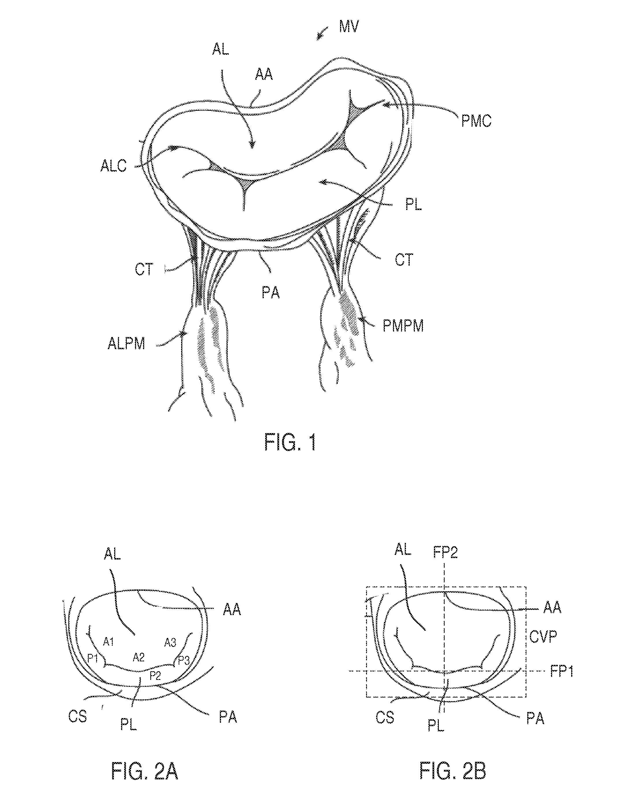
The mitral valve MV illustrated in FIG. 1 is defective as the mitral valve leaflets (AL and PL) do not coapt correctly, leaving one or more gaps between the leaflets, resulting in regurgitation.
Previously-known valve prostheses have some disadvantages, such as need for long-term maintenance with blood thinners, the risk of clot formation, limited durability, etc.
However, most dysfunctional valves are too diseased to be repaired using previously known methods and apparatus.
However, these surgeries are prone to many complications and long hospital stays for recuperation.
By expanding the prosthetic valve, the native valve leaflets are generally pushed aside and rendered ineffective.
Unfortunately, for a significant percentage of patients, mitral valve replacement is still necessary due to stenosis or anatomical limitations, and few less-invasive options are available for replacement procedures.
Such materials, while durable, are prone to blood clotting and thrombus formation, which in turn increases the risk of embolization and stroke or ischemia.
Anticoagulants may be taken to prevent blood clotting that may result in thromboembolic complications and catastrophic heart failure, however, such anti-clotting medication may complicate a patient's health due to the increased risk of hemorrhage.
A major disadvantage of tissue valves is they lack the long-term durability of mechanical valves.
Furthermore, valves are subject to stresses from constant mechanical operation within the body.
Such tension causes prosthetic valves to wear out over time, requiring replacement.
While iterative improvements have been made on surgical bioprosthetic valves over the last several decades, existing bioprosthetic valves still have drawbacks.
One drawback is the mismatch in size and mass between opposing surfaces of the stent-like supporting frame.
The mismatch is often due to the variability in the shapes and mechanical characteristics of the stent-like supporting frame.
For prosthetic valves with balloon-expandable stent-like supporting frames, the recoil of the supporting frames post-balloon-inflation may lead to perivalvular leaks around the circumference of the prosthetic valve and potential slippage and migration of the valve post-implantation.
Another risk associated with prosthetic valves having balloon-expandable supporting frames is potential damage to the leaflets of the prosthesis during implantation, when the leaflets may be compressed between the balloon and the supporting frame.
For prosthetic valves with self-expanding stent-like supporting frames, mismatch may arise due to the deformation / movement of the supporting frame, e.g., slight deformation of the frame into a less than circular shape during normal cardiac movement.
Such mismatch may lead to instability among components of a prosthetic valve, resulting in perivalvular leaks and uneven stress distribution in the valve leaflets, resulting in accelerated wear of the valve.
Another drawback in the construction of existing bio-prosthetic valves with self-expanding supporting frames is the potential for damage to the leaflet tissue arising from the spacing between the struts of the frame.
For example, when the stent-like supporting frame is deployed, the distance between struts during expansion of the frame may stretch both the surrounding tissue and the leaflet tissue further apart than designed, potentially resulting in damage to surrounding tissue and leaflet tissue.
With use of an oblong or circular radially self-expanding frame applied on the majority of the mitral valve, there is risk of left-ventricular outflow tract (LVOT) obstruction.
Because the clip transforms the mitral orifice into two orifices, the clip may significantly obstruct the flow of blood through the valve.
These techniques have the drawback of replacing a mitral valve that is itself generally normal or subnormal.
The native structures (mitral leaflets, chordae, papillary muscles, etc.) play an important role in left-ventricular function and therefore any valve replacement system that does not respect these elements may adversely impact the left-ventricular function.

Method used


Image

  • Apparatus and methods for treating cardiac valve regurgitation
  • Apparatus and methods for treating cardiac valve regurgitation
  • Apparatus and methods for treating cardiac valve regurgitation

Examples


Embodiment Construction

[0045]Apparatus and methods of the present disclosure are designed for repairing a defective cardiac valve, such as a mitral valve, a tricuspid valve, a pulmonary valve, or an aortic valve. In accordance with the principles of the present disclosure, the apparatus and methods may be optimized for use in treating cardiac valve regurgitation when the leaflets of the cardiac valve do not coapt correctly, thus causing blood to leak backwards through the valve as the heart pumps. Advantageously, apparatus of the present disclosure are configured for implantation at a cardiac valve within a blood flowpath such that the native leaflets abut the apparatus during the portion of the cardiac cycle when the cardiac valve attempts to close, thereby enhancing native leaflet coaptation and minimizing regurgitation.

[0046]Referring to FIG. 3A, an illustrative embodiment of a prosthetic device in accordance with the principles of the present disclosure is described. Illustratively, the prosthetic dev...


PUM


Abstract

Apparatus and methods for repairing a cardiac valve, e.g., a mitral valve, are provided. The apparatus may include an expandable frame defining a curved structure in the expanded deployed state and a membrane coupled to the expandable frame. The membrane may curve around a native leaflet, e.g., the posterior leaflet, in a first plane and curve around another leaflet, e.g., the anterior leaflet, in an orthogonal plane. The membrane may be adapted to be suspended in the flow path of the cardiac valve such a first surface of the membrane abuts the native leaflet during systole and a second surface of the membrane abuts the other native leaflet during systole, thereby reducing cardiac valve regurgitation.

Description

I. FIELD OF THE INVENTION[0001]This application generally relates to apparatus and methods for performing transcatheter or minimally invasive repair of a defective cardiac valve, such as the mitral, tricuspid, pulmonary, and aortic valves.II. BACKGROUND OF THE INVENTION[0002]The human heart has four major valves which moderate and direct blood flow in the cardiovascular system. These valves serve critical functions in assuring a unidirectional flow of an adequate blood supply through the cardiovascular system. The mitral valve and aortic valve control the flow of oxygen-rich blood from the lungs to the body. The mitral valve lies between the left atrium and left ventricle, while the aortic valve is situated between the left ventricle and the aorta. Together, the mitral and aortic valves ensure that oxygen-rich blood received from the lungs is ejected into systemic circulation. The tricuspid and pulmonary valves control the flow of oxygen-depleted blood from the body to the lungs. Th...

Claims


Application Information

Patent Timeline
Patent Type & Authority Applications(United States)
IPC IPC(8): A61F2/24
CPCA61F2/2454A61F2/2427A61F2230/0095A61F2/246A61F2220/0016A61F2/2409A61F2/2412
Inventor NGUYEN, THANSEGUIN, JACQUES
Owner SEGUIN JACQUES
Why Eureka
Try Eureka