Methods of primary tissue culture and drug screening using autologous serum and fluids

a primary tissue culture and autologous technology, applied in the field of cell biology and medicine, can solve the problems of death or biological alterations, complex selection of a cancer therapy for an individual, and inability to closely represent the conditions of the patient's artificial microenvironment, so as to improve the effectiveness of individualized cancer therapy, improve the success rate of primary culture, and improve the effect of drug sensitivity testing

Active Publication Date: 2016-12-22
TANG YAO
View PDF2 Cites 9 Cited by
  • Summary
  • Abstract
  • Description
  • Claims
  • Application Information

AI Technical Summary

Benefits of technology

[0015]The invention is based on the discovery of protocols for cell culturing which utilize a subject's own serum or body fluid and which closely mimic the cellular or tissue microenvironment. In some embodiments, a cancer patient's own serum is used to grow his / her cancer cells in a 3D culture condition. Other cells that surround the tumor or co-exist in the cancerous fluid can also survive in this system. With the methods of the invention, tumor cells remain in a microenvironment that is virtually identical to the one they experienced inside of the patient, with similar nutrients and biochemical supplies. The present invention provides for more accurate drug sensitivity testing to improve the effectiveness of individualized cancer therapy. In some embodiments, the methods use the patient's body fluid to culture their own tumors in vitro, including solid tumors, malignant serous tumors, peripheral blood mononuclear cells (PBMCs) and circulating tumor cells (CTCs), as well as stromal cells associated with the tumors. This greatly increases the success rate of primary culture for human cancers. In some embodiments, one difference from traditional cell cultures is that, in the present culture system, tumor cells and their autologous surrounding tissue cells survive and grow together, and are able to reconstruct the tumor's original histopathology structures. Because of the similarity in the in vivo and ex vivo microenvironments, no change in immunophenotypes was found in tumor tissues before and after cultures. In addition, human PBMCs also survived well for at least 4-5 days and CTCs in the blood circulation can be multiplied in this culture system. This culture technique can have many applications, including, but not limited to: (1) the study of cancer as a complete tissue instead of isolated cancer cells; (2) chemo-sensitivity testing with improved accuracy; (3) evaluation of the toxicity of a drug to PBMCs, (4) in vitro multiplication of CTCs in a patient's blood and; (5) provision of a new platform for other pre-clinical targeted therapies (e.g. gene-therapy or immunotherapy) for cancerous or non-cancerous diseases, to improve the accuracy and efficacy of individualized / personalized treatment.

Problems solved by technology

If the ‘soil’ is not well-suited to the growth of the tumor cells, death or biological alterations occur.
Although many efforts have been made in this field, a challenge remains regarding the creation of in vitro environments that are suitable for all types of tumors, from different individuals (See Edmondson R et al., Assay &Drug Development Technologies.
But because the source of these stroma cells is either biologically different from the tumor cells or has lost the function to communicate with tumor cells (after irradiation), these artificial microenvironments do not closely represent conditions in the patient.
All of these in vivo factors make the selection of a cancer therapy for an individual much more complicated and challenging.
However, implanting a human tumor into an animal results in an abrupt change in the microenvironment of the tumor, forcing the tumor cells to attempt to communicate with stroma cells from a different species.
In addition, the high financial cost of animal experimentation mitigates against its widespread clinical use.
However, because these cells occur in very low numbers and circulate through the body, isolating CTCs from the blood of cancer patients has been a technical challenge (See Castle J et al., The Breast.
Accordingly, the results of testing with this culture system may not accurately predict the response of the tumor when the drug is administered to the patient.

Method used


Image

  • Methods of primary tissue culture and drug screening using autologous serum and fluids
  • Methods of primary tissue culture and drug screening using autologous serum and fluids
  • Methods of primary tissue culture and drug screening using autologous serum and fluids

Examples


example 1

Materials and Methods

Clinical Samples Information:

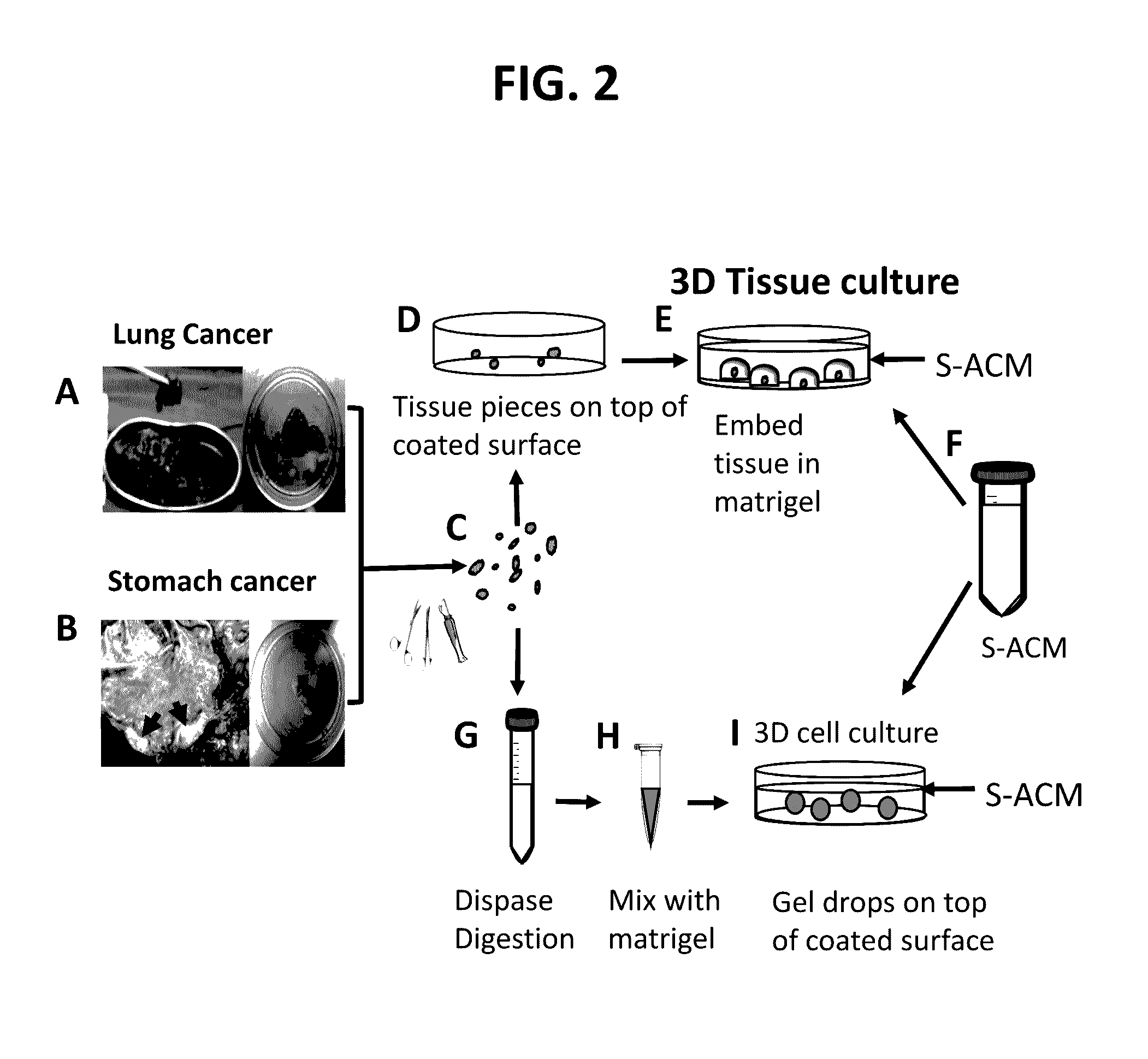
[0215]All clinical materials were collected from Dalian Central Hospital, Dalian, China, during 2014 and 2015. This study was approved and monitored under the hospital Humane Society and Research Committee (YN2014-023-01). Primary cultures were performed for fourteen solid tumors and thirteen serous tumors (malignant ascites and pleural effusions). The solid tumors were from seven male and seven female patients whose ages ranged from 40 to 80, with an average of 61. These patients were diagnosed clinically as stomach cancers (n=6), lung cancers (n=7), and lymph node metastatic cancer from a stomach cancer patient (n=1). For the lymph node metastatic cancer from a stomach cancer patient an enlarged lymph node (LN; 2.5×1×1 cm3) was used. Pathology confirmed that the enlargement of this LN was caused by cancer metastasis. All these cases were confirmed as cancers by histopathology (Table 1). For serous tumors, samples were collected fro...


PUM

PropertyMeasurementUnit
v/vaaaaaaaaaa
thicknessaaaaaaaaaa
timeaaaaaaaaaa

Abstract

The present invention provides methods for culturing primary cells and tissues from a subject in the presence of the subject's own serum, ascites or pleural effusion fluid. Methods of treating cancer, and screening for the effectiveness or toxicity of drugs are also provided herein.

Description

CROSS REFERENCE TO RELATED APPLICATIONS[0001]This application claims the benefit of U.S. Provisional Appl. No. 62 / 137,218, filed Mar. 23, 2015. The content of the aforesaid application is relied upon and incorporated by reference in their entireties.FIELD OF THE INVENTION[0002]The field of the invention relates to cell biology and medicine.BACKGROUND OF THE INVENTION[0003]Scientists have long sought to improve culturing systems to support medical research in general, and treatment, including cancer treatment, in particular. However, cancer patients are highly individualized in their response to chemotherapies or other anti-cancer regimens. The success of individualized therapy requires accurate pre-testing to determine drug-sensitivity, no matter if the target is DNA, protein, enzyme, hormone or cytoskeleton. In turn, accurate pre-testing requires a tumor sample growing in a microenvironment that is maximally similar to its original condition. Tumor cells grow as ‘seeds’ in their mi...

Claims


Application Information

Patent Timeline
Patent Type & Authority Applications(United States)
IPC IPC(8): C12N5/09
CPCC12N5/0694C12N5/0693C12N2503/02C12N2509/00C12N2500/84C12N2513/00C12N2509/10C12N2533/52C12N2533/54C12N2533/90
Inventor TANG, YAO
Owner TANG YAO
Why Eureka
Try Eureka