Visualization of bacterial colonization and biofilm formation on orthopaedic trauma explants

Active Publication Date: 2016-12-22
RHODE ISLAND HOSPITAL
View PDF0 Cites 0 Cited by
  • Summary
  • Abstract
  • Description
  • Claims
  • Application Information

AI Technical Summary

Benefits of technology

[0015]Therefore, there remains a need in the prior art for a reliable, rapid diagnostic tool that allows for real-time detection of bacterial presence on potentially infected explants, tissue surfaces, bone cements etc. Preferably, the systems and methods would take advantage of existing equipment and materials that are already typically present or available in the hospital setting. The systems and methods also can and accurately detect and characterize bacterial biofilm formation on potentially infected explant and

Problems solved by technology

Microbial infection and biofilm formation on orthopaedic trauma implants is one of the most serious problems in orthopaedic surgery.
In many cases, the heterogeneity of the tissue surrounding the complex surface of the implant may complicate the identification of microbial colonization or infection.
Subsequent analysis to determine the bacterial colonization, adhesion, and species on the explant may take several days or weeks, negatively impacting the efficacy and efficiency of possible treatment plans.
In many cases, current detection methods may yield false-negative findings, which further exacerbate the problem.
Furthermore, this infection rate dramatically increases in traumatic extremity injuries sustained during combat and in developing countries.
These one- or two-step revision surgeries mean delayed healing time, increased medical costs, and less successful post-operative outcomes.
One of the greatest ongoing challenges in the field of orthopaedic trauma remains decreasing the rates and consequent ill effects of bacterial infection.
One of the principal factors challenging effective infection treatment is the formation of bacterial biofilm, a coating of bacterial film that adheres to the infected tissue and/or orthopaedic implant.
Biofilms are difficult to eradicate because most antibiotics are unable to penetrate their surface, weakening the primary line of attack.
Despite advances in infection treatment, one of the most comprehensive problems in

Method used


Image

  • Visualization of bacterial colonization and biofilm formation on orthopaedic trauma explants
  • Visualization of bacterial colonization and biofilm formation on orthopaedic trauma explants
  • Visualization of bacterial colonization and biofilm formation on orthopaedic trauma explants

Examples


Embodiment Construction

[0058]The subject technology overcomes many of the prior art problems associated with quantifying and characterizing microbial colonization and biofilm formation on explants. The advantages, and other features of the system disclosed herein, will become more readily apparent to those having ordinary skill in the art from the following detailed description of certain preferred embodiments taken in conjunction with the drawings which set forth representative embodiments of the present invention and wherein like reference numerals identify similar structural elements. It is understood that references to the figures such as up, down, upward, downward, left, and right are with respect to the figures and not meant in a limiting sense.

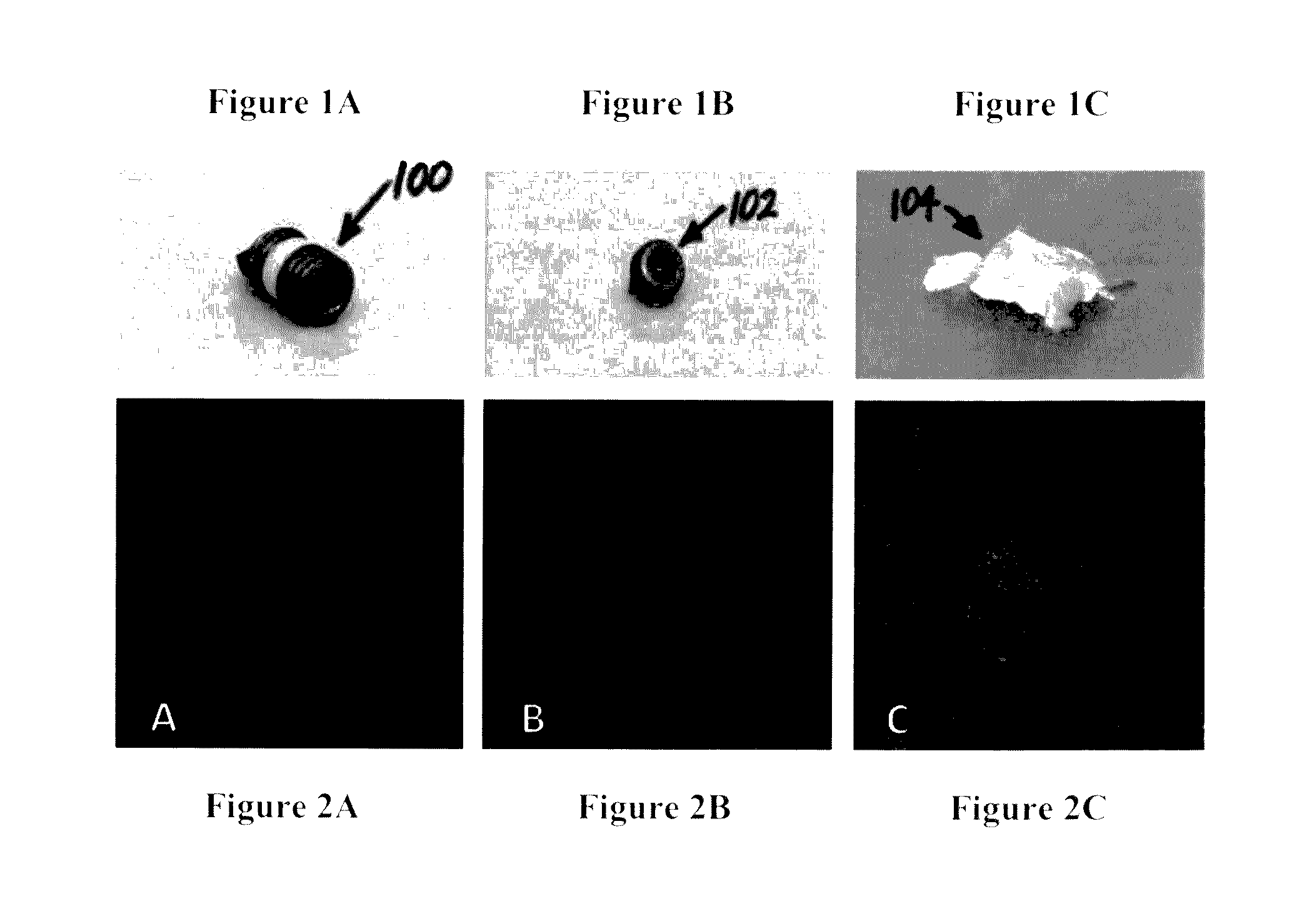
[0059]Referring now to FIGS. 1A-C, typical explants are shown. FIG. 1A is a top portion of a screw 100. FIG. 1B is a screwhead 102. FIG. 1C is a polymethyl methacrylate bone cement fragment 104. Referring now to FIGS. 2A-C, each is a confocal laser scanning m...


PUM


Abstract

Detection and quantification of microbial colonization and biofilm formation on orthopaedic explants would be important for deciding treatment interventions at the time of surgery. Methods and Systems for such treatments are shown that utilize various steps and kits to quickly generate confocal laser-scanning microscopy (CLSM) images to allow for the detection and quantification. The explants are prepared by applying antibodies of both gram-positive and grain-negative for at least one period of time.

Description

BACKGROUND OF THE INVENTION[0001]1. Field of the Invention[0002]The subject disclosure relates to systems and methods for rapid quantification and characterization of microbial colonization and biofilm formation on implants at the time of removal.[0003]2. Background of the Related Art[0004]Microbial infection and biofilm formation on orthopaedic trauma implants is one of the most serious problems in orthopaedic surgery. The formation of biofilms increases resistance to antibiotic treatment, and typically an orthopaedic device that becomes infected must be removed. In many cases, the heterogeneity of the tissue surrounding the complex surface of the implant may complicate the identification of microbial colonization or infection. Subsequent analysis to determine the bacterial colonization, adhesion, and species on the explant may take several days or weeks, negatively impacting the efficacy and efficiency of possible treatment plans. In many cases, current detection methods may yield...

Claims


Application Information

Patent Timeline
IPC IPC(8): G01N33/569G02B21/00
CPCG01N33/56911G02B21/0028G02B21/0076G01N2333/22G01N2333/31G01N2333/195G02B21/0056G02B21/16G02B21/365G01N21/6428G01N21/6458G01N2021/6439
Inventor GARCIA, DIOSCARISBORN, CHRISTOPHER T.JARRELL, JOHN
Owner RHODE ISLAND HOSPITAL
Why Eureka
Try Eureka