High-throughput cellular analysis using microbubble arrays

Inactive Publication Date: 2017-03-09
UNIVERSITY OF ROCHESTER
View PDF2 Cites 0 Cited by
  • Summary
  • Abstract
  • Description
  • Claims
  • Application Information

AI Technical Summary

Benefits of technology

[0025]According to an embodiment, the cells can be stem cells, cancer cells, mouse hybridoma cells, CHO cells, or B cells derived from human

Problems solved by technology

Normally, cells in MB wells are not easily displaced by fluid flow and, in fact,

Method used


Image

  • High-throughput cellular analysis using microbubble arrays
  • High-throughput cellular analysis using microbubble arrays
  • High-throughput cellular analysis using microbubble arrays

Examples


example 1

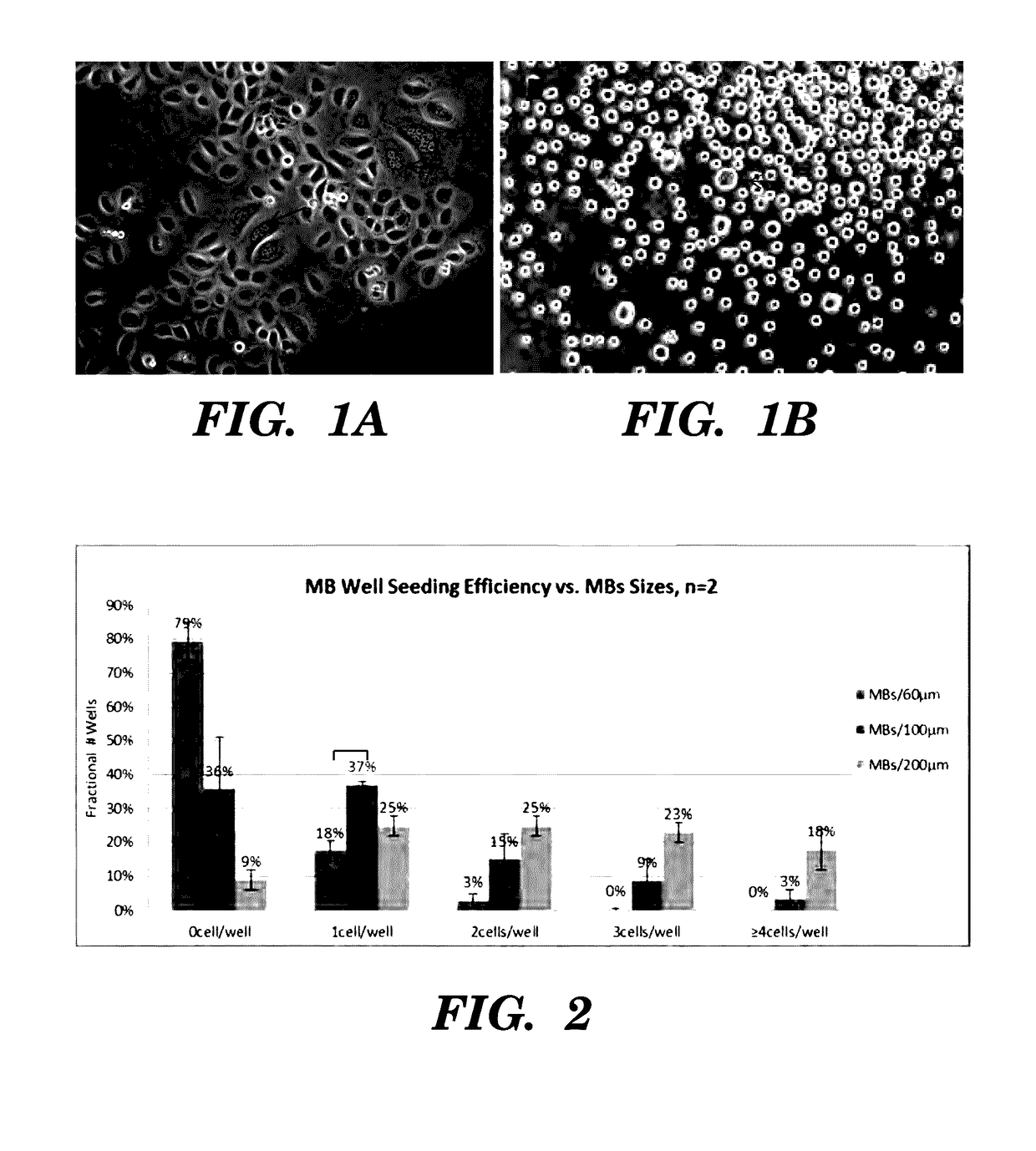
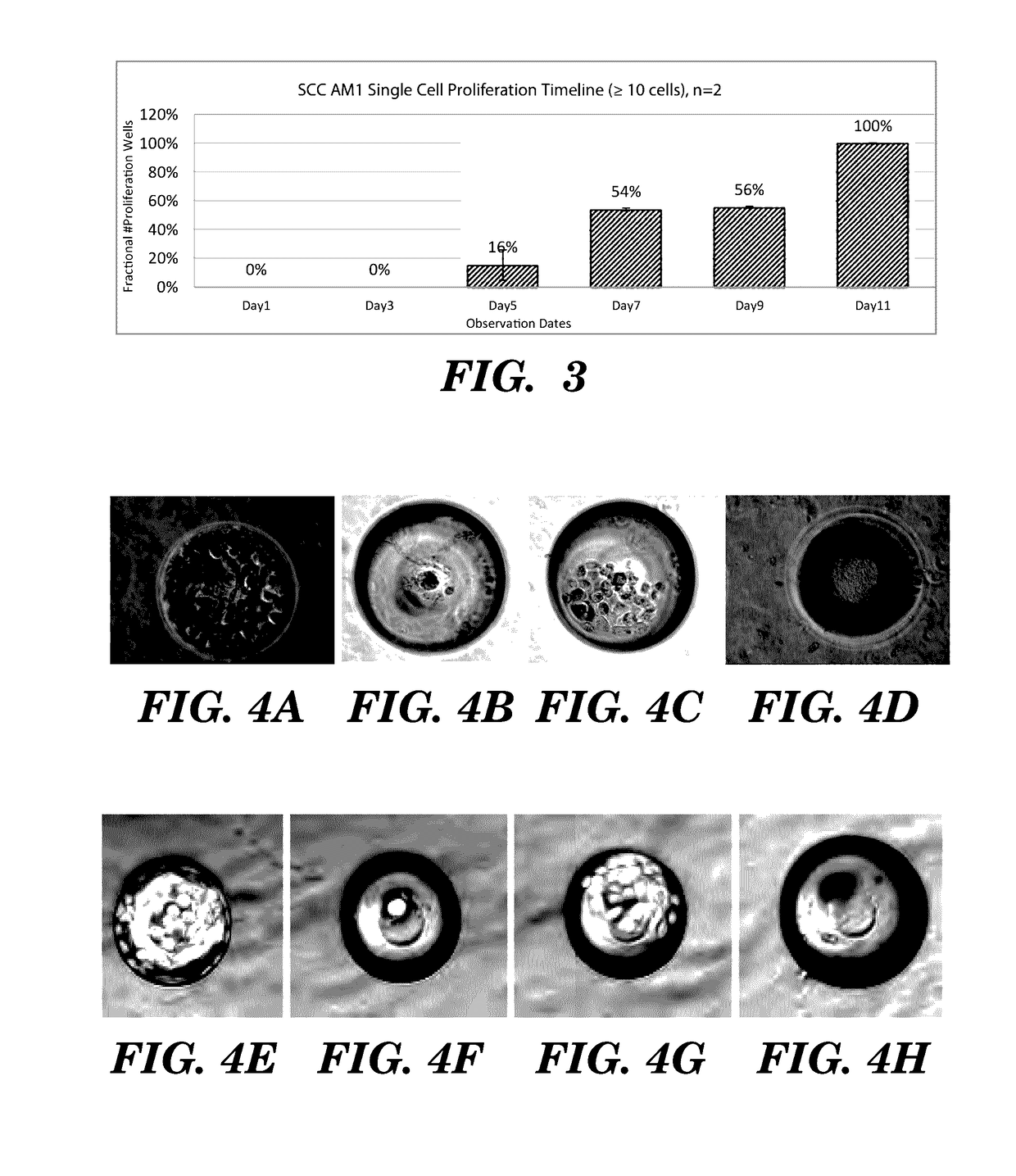
Sorting Tumor Initiating Cells by Clonal Proliferation and Morphology

[0085]Skin cancers comprise a large public health concern and an increasing burden on the health care system. Among 2 million diagnosed skin cancers annually in the U.S., ˜ 35% are squamous cell carcinomas (SCCs). SCC metastases account for the majority of non-melanoma skin cancer deaths. Tumor size and morphology are considered to be prognostic factors for recurrence, but there are no genetic mutations or predictive biomarkers that can be reliably use to identify tumor aggressiveness at an early stage. The characterization and quantification of cells with tumor initiating capability is essential for developing prognostic biomarkers and for advancing the basic understanding of metastases. Current methods of identifying tumor-initiating cells (TICs) are using flow cytometry and serial in vivo xenotransplantation models. However, the cell surface markers derived from these methods failed to identify consistent expres...

example 2

SCC Cytokeratin Gene Expression

[0096]Stem cells are immature cells and have the ability to develop into different cell types. To test whether SCC cells cultured in MBs express cytokeratin 5, validating a basal proliferative SCC phenotype, the expression of cytokeratin 5 and 10 was investigated. Briefly, a MB chip array was placed in 24-well TCP and then transferred into wells containing 1× PBS. It was then transferred into wells containing 10% formalin fixative with 0.1% Triton for 20 min at room temperature. The chip was rinsed twice with 1× PBS, and blocked with 2% BSA for 30 min to prevent any non-specific adsorption of antibodies. A mixture of primary keratin 5 and keratin 10 antibodies (500 μl of 1:500 dilution) in 2% BSA was pipetted onto the top of the chip. The TCP was placed in 4° C. fridge overnight in the dark. Then, the excess antibody solution was removed the next day and the chip was rinsed twice with 1× PBS. The TCP was placed on a shaker for 5 min. A mixture of secon...

example 3

Identification of Tumor Initiating Cells by Drug Resistance

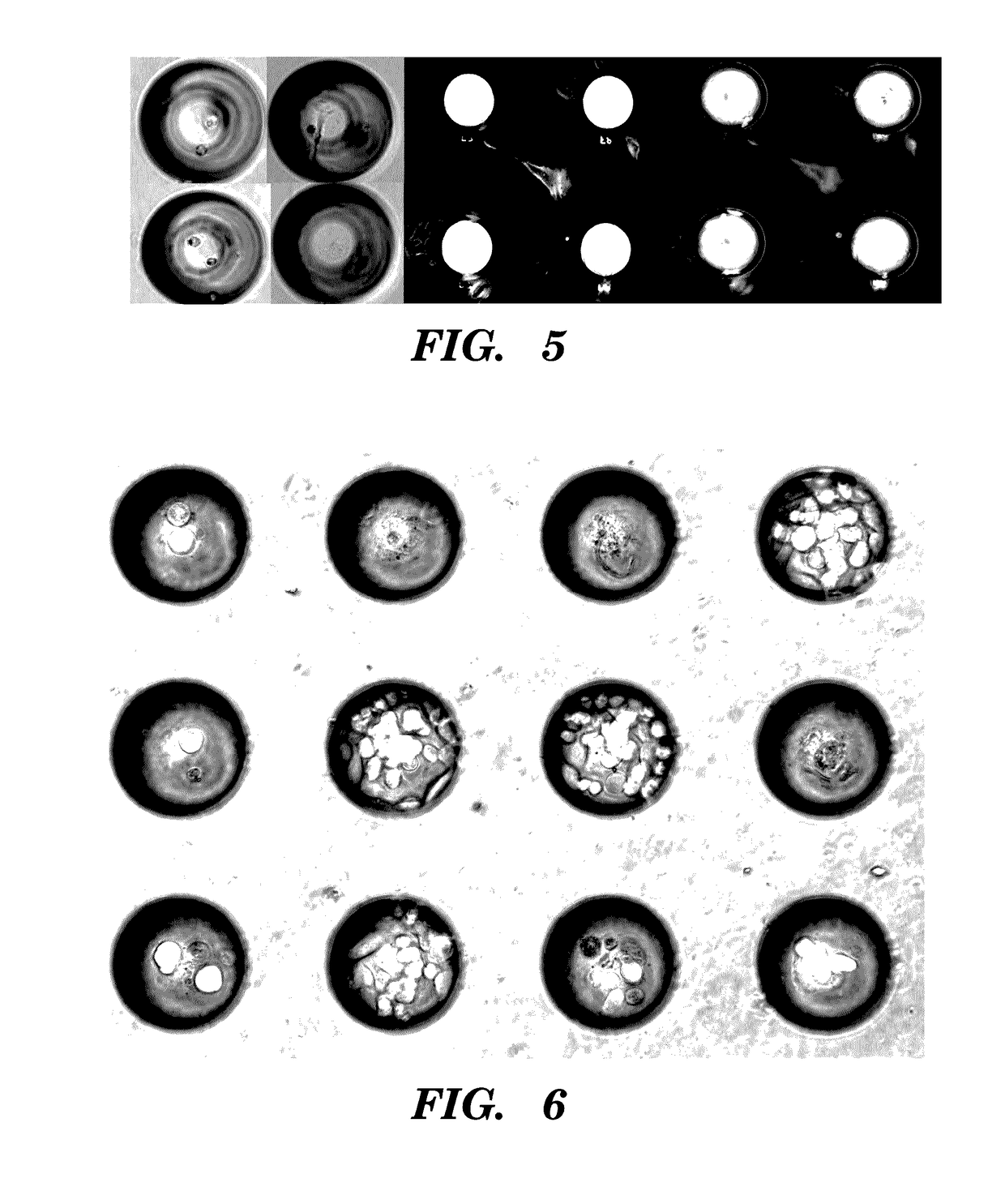
[0098]According to an embodiment is a single-cell screening method using microbubble well arrays where morphologically distinct clone can be sorted by drug resistance. Genetic mutations in cancer cells give rise to their aggressive phenotype and their ability to proliferate in a nonadherent fashion with reduced drug sensitivity, which are hallmarks cancer stem cells. Seeding and establishing clonally pure cancer cell colonies in microbubble well arrays is a means to screen for drug resistance as shown in FIGS. 13A-C, 14A-B, and 15. It is shown that the spheroid cell populations are drug resistant and may be representative of tumor initiating cells or cancer stem cells. To enrich for the sphere cells, established colonies were cultured in the presence of a chemotherapeutic agent (e.g. Cisplatin). Traditional chemotherapeutic agents work by killing rapidly dividing cells, one of the main properties of most cancer cells. This m...


PUM


Abstract

A microfabricated device and method having a substrate with an array of curvilinear cavities that is used for high throughput single cell screening. The substrate of the device is preferably fabricated in a low elastic modulus polymer such as polydimethylsiloxane. The architecture of the cavity forms a small volume micro-niche that seeded cells can rapidly condition to promote survival and proliferation which can be monitored for hours to days to weeks. The cavity architecture allows independent assays to be conducted with minimal influence from nearest neighbor cavities. Methods are disclosed to use the device to, for example, screen single cells by clonal proliferation, clonal morphology, secreted factors, secretion rate, surface markers, and cell functional characteristics including but not limited to migration, drug resistance, the ability to block or promote signaling pathways, or to enhance opsonization.

Description

[0001]This application claims the priority benefit of U.S. Provisional Patent Application Ser. No. 62 / 215,348, filed Sep. 8, 2015, which is hereby incorporated by reference in its entirety.STATEMENT REGARDING FEDERALLY SPONSORED RESEARCH OR DEVELOPMENT[0002]This invention was made with government support under Contract Nos. P30 AI078498, TL1 TR000096, KL2 TR000095, and UL1 TR000042, awarded by National Institutes of Health. The Government has certain rights in the invention.FIELD OF THE INVENTION[0003]The present disclosure is directed generally to methods and systems for high throughput single cell screening using a substrate comprising an array of curvilinear cavities.BACKGROUND[0004]The ability to sort cells from heterogeneous population and to study them at the single cell level provides unique opportunities for drug discovery, identification of tumor initiating cancer stem cells, and for understanding signaling pathways in disease, among other opportunities. This capability is ...

Claims


Application Information

Patent Timeline
IPC IPC(8): G01N33/483B01L3/00
CPCG01N33/4833B01L2300/0893B01L2200/0652B01L3/502761B01L3/5085
Inventor DELOUISE, LISA A.KOBIE, JAMES J.
Owner UNIVERSITY OF ROCHESTER
Why Eureka
Try Eureka