
Device for detecting target biomolecules

a biomolecule and target technology, applied in the field of target biomolecule detection devices, can solve the problems of reducing the sensitivity of the system, affecting the accuracy of the system,

Active Publication Date: 2017-05-18
STELLENBOSCH UNIVERSITY
View PDF3 Cites 5 Cited by
  • Summary
  • Abstract
  • Description
  • Claims
  • Application Information

AI Technical Summary

Benefits of technology

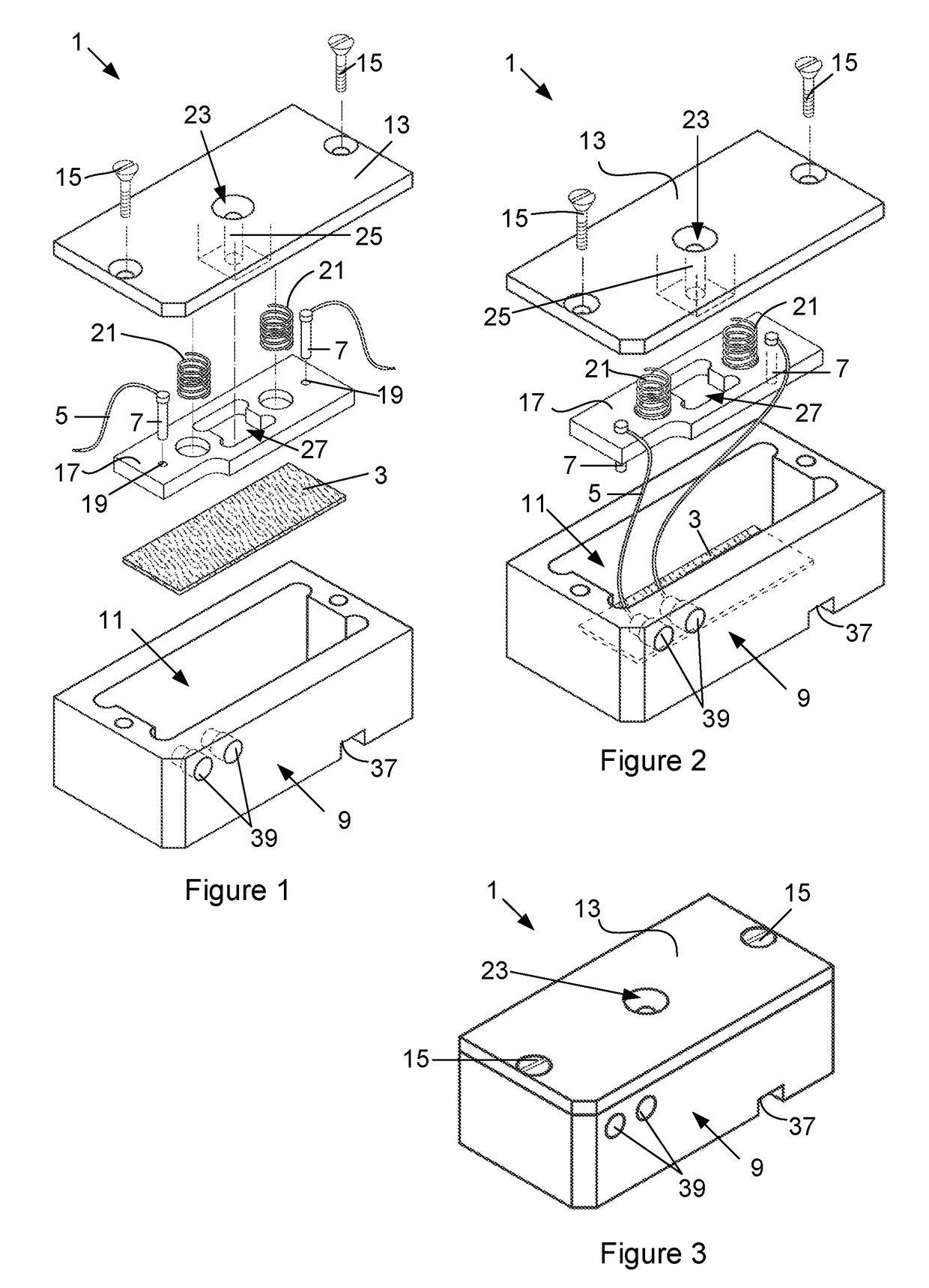
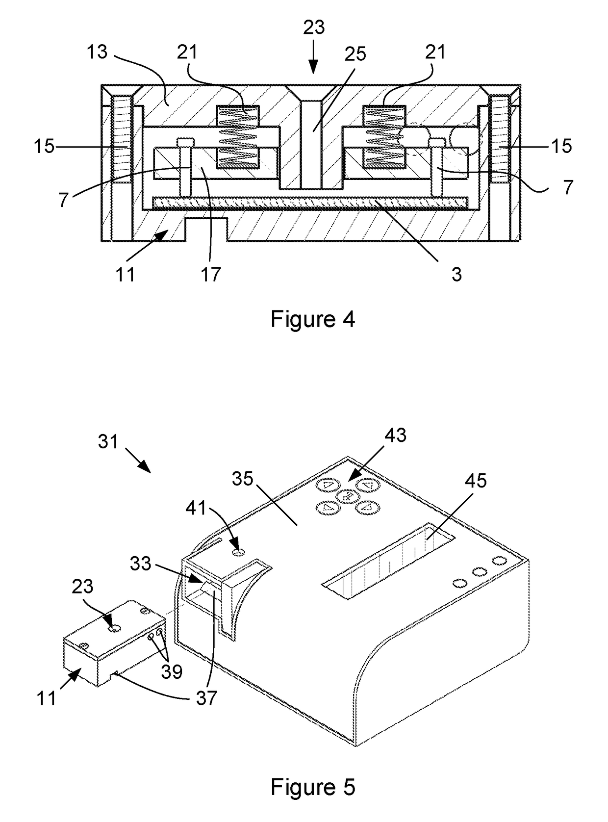
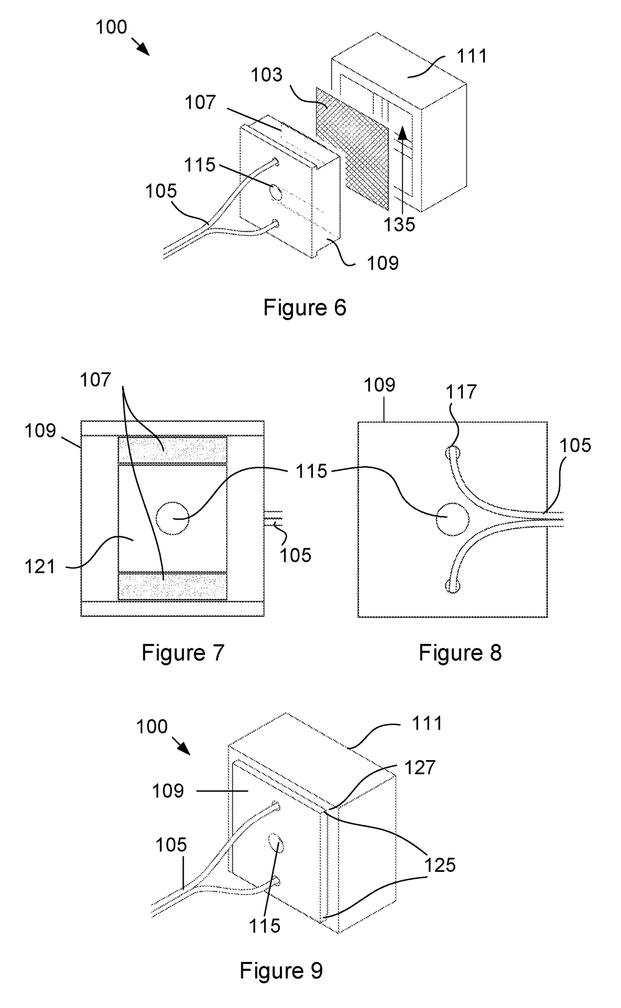
The invention provides a device for detecting target biomolecules by measuring the resistance of a membrane as fluid samples containing the target molecule are applied. The device includes an electroconductive membrane with a biological recognition component for binding the target molecule, connected to an electric circuit with voltage source. The membrane is supported on a non-conducting support and a selected volume of fluid sample delivered onto the membrane results in a change in resistance. The resistance monitoring device detects the target molecule and the level of contamination or infection in the fluid sample by measuring the change in resistance. The device is portable and battery-operated. The method allows for sensitive and specific detection of target molecules.

Problems solved by technology

Traditional pathogen detection methods such as polymerase chain reaction, immunology-based methods and culture and colony counting generally take a long time to provide accurate results and can be expensive and inaccessible in remote locations.
However, the submerged electrodes participate in electrochemical reactions with the salts in the solution, producing a substantial background effects during the detection process which reduces the sensitivity of the system.
As a result of being submerged in a saline solution, the electrodes are also susceptible to degradation over time caused by redox reactions on the surface of the electrodes.
The system also needs a relatively large volume of buffer solution, which may not be readily available outside of a laboratory environment.
In general, existing systems that utilize electrotextiles for the detection of biomolecules are complex, laboratory-scale systems that may not be suitable for use in the field or re-use and may not be sensitive enough to be used with very small fluid samples.

Method used


Image

  • Device for detecting target biomolecules
  • Device for detecting target biomolecules
  • Device for detecting target biomolecules

Examples


example 1

[0069]The ability of the device to detect the presence of anti-lysozyme antibody was tested with a membrane on which lysozyme is immobilized as the biological recognition component.

[0070]Methods

[0071]1. Preparation of the Conductive Membrane

[0072]A sheet of nonwoven, spun bound and spot melted polypropylene microfibers with a thickness of 50 g / m2 was cut into a 90×90 mm square sheet. The microfibers of the sheet were provided with a conductive coating comprising doped polypyrrole copolymers. The pyrrole monomer was copolymerized with carboxylic acid functional 3-thiopheneacetic acid (3TAA). A 3TAA solution with a concentration of 10 mg / ml was mixed with 10% pyrrole and the microfiber sheet was submerged in the solution for a few seconds and then removed. Thereafter the microfiber sheet was placed in a glass reaction vessel containing 30 ml of 0.1 M iron(III) chloride (FeCl3) in deionised water. A volume of 1 ml of 0.1 M 5-sulfosalicylic acid (5SSA), acting as a dopant, was added for...

example 2

[0134]The ability of the device to detect Escherichia coli (E. coli) bacteria was tested. The conductive membrane was prepared according to the steps in Example 1 above. However, instead of lysozyme, Anti-E. coli antibody ab25823 was immobilized on the membrane.

[0135]Methods

[0136]The immobilization of Anti-E. coli antibody ab25823 onto the membrane was achieved by firstly washing the conductive membrane with 0.01M PBS and drying it for 10 minutes. The membrane was then incubated in 2.5 mM glutaraldehyde for 1 hour at 4° C. Thereafter, the membrane was washed again with 0.01M PBS and dried for 10 minutes. The membrane was incubated in 600 mL Anti-E. coli antibody (6.67 ug / mL) at 37° C. for 1 hour and thereafter washed with 0.01M PBS and dried for 10 minutes. Thereafter 5% BSA Blocker™ was added to the membrane and it was left to react for 1 hour at room temperature. Finally the membrane was washed three times using 0.01M PBS.

[0137]Different concentrations of E. coli solutions of 3.4×...

example 3

[0141]In this example a paper-based conductive membrane was used as part of the device to demonstrate its ability to detect anti-lysozyme antibodies. Lysozyme protein was immobilized on the paper-based conductive membrane and the resistance of the membrane was monitored and recorded. The resistance measurements were processed and demonstrate that the paper-based conductive membrane is capable of resistive sensing of biomolecules.

[0142]Methods

[0143]Whatmann Grade 50 (97 g / m2) and Whatmann Grade 1 (87 g / m2) Filter Papers were chosen and acquired from Sigma Aldrich due to their wet strength. Whatmann Filter Papers (Grades 1,50) were cut into a standard size of 180×280 mm to be used for testing.

[0144]1. Conductive Coating

[0145]The cellulose fibers of the filter paper were made conductive through coating with a doped polypyrrole solution. The 180×280 mm sheets were first immersed in the pyrrole monomer and left to soak for 5 minutes, this ensured that the paper was fully saturated with t...


PUM


Abstract

A device for detecting target biomolecules is provided. The device consists of an electrically conductive membrane having a biological recognition component configured to bind a target biomolecule immobilized thereon. The membrane is connected to an electric circuit by means of electrodes. A voltage source applies a voltage to the membrane and a resistance monitoring device monitors the resistance of the membrane as a selected volume of fluid sample suspected of containing the target biomolecule is delivered onto the membrane.

Description

CROSS-REFERENCE TO RELATED APPLICATIONS[0001]This application claims priority from South African provisional patent application number 2015 / 08499 filed on 18 Nov. 2015, which is incorporated by reference herein.FIELD OF THE INVENTION[0002]This invention relates to a device for detecting target biomolecules. In particular, it relates to a device for detecting target biomolecules by measuring a change in conductivity of an electrically conductive membrane to which a biological recognition component configured to bind the target biomolecule is attached.BACKGROUND TO THE INVENTION[0003]The early diagnosis of pathogenic infections may help reduce the number of incidents of disease and fatalities caused by pathogenic infections in humans. Infections may be further reduced by the detection of pathogenic agents in water or food before consumption. Traditional pathogen detection methods such as polymerase chain reaction, immunology-based methods and culture and colony counting generally take...

Claims


Application Information

Patent Timeline
IPC IPC(8): G01N33/543G01N33/573G01N27/12
CPCG01N33/5438G01N27/125G01N2469/20G01N2333/936G01N33/573G01N27/04G01N27/041G01N33/553
Inventor VIVIERS, CHRISTIAAN GUNTER ALWYNPEROLD, WILLEM JACOBUSDICKS, LEON MILNER THEODOREMAYBERY, GILES HUBERT COYLE
Owner STELLENBOSCH UNIVERSITY
Learn More