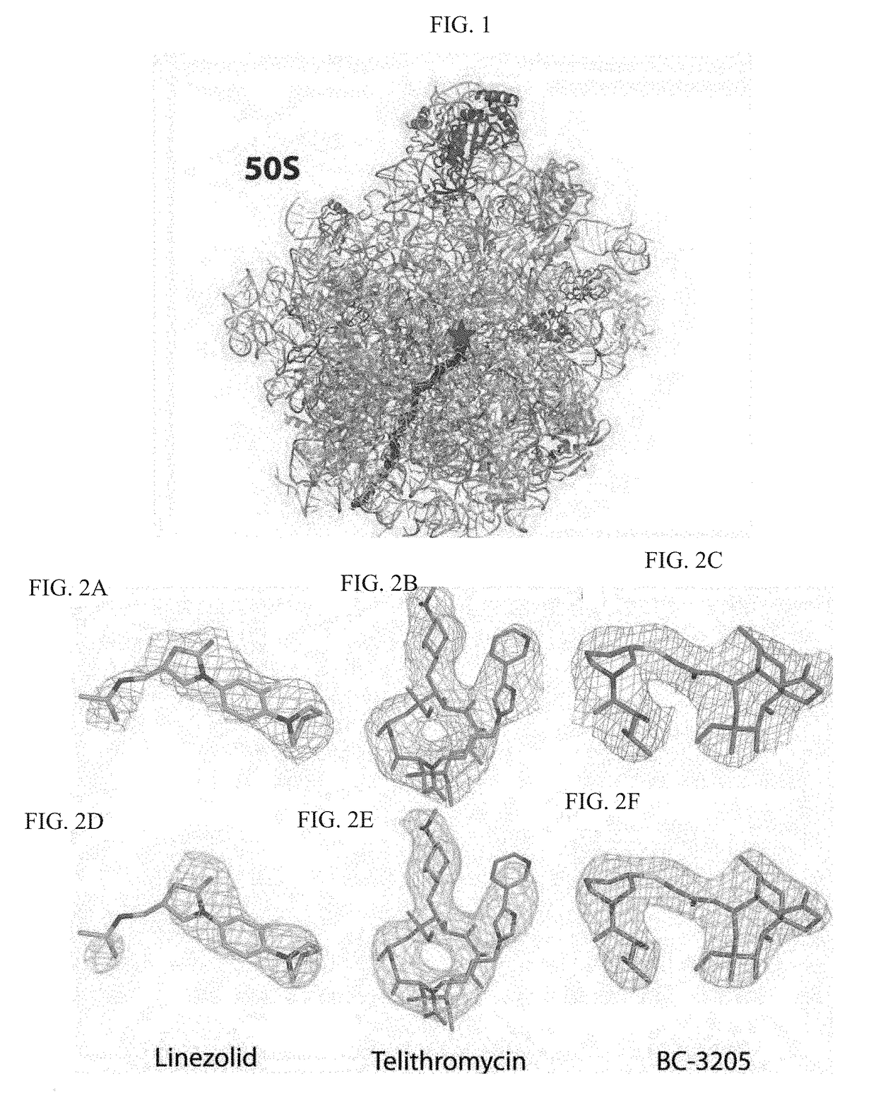
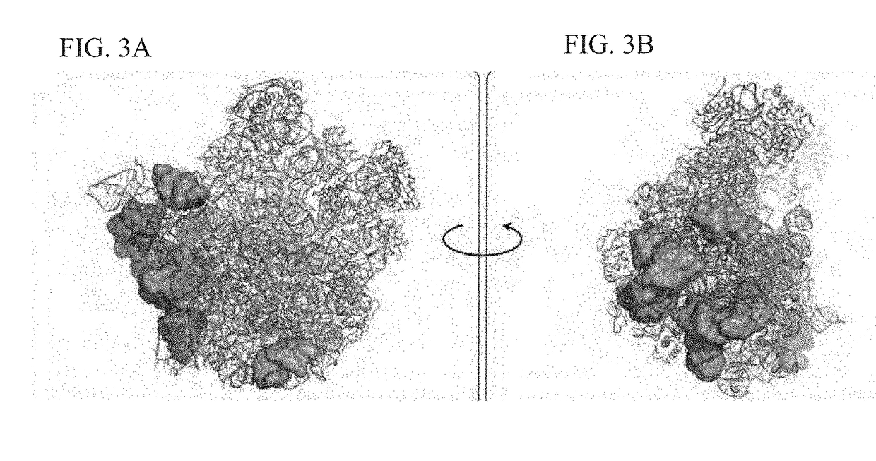
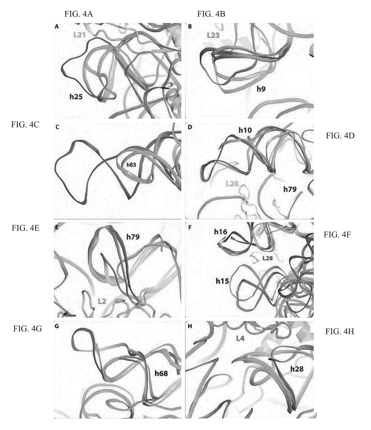
Crystal structure of the large ribosomal subunit from s. aureus
a ribosomal subunit and crystal structure technology, applied in the field of crystal structure and structure-based drug design, can solve the problems of bacterial resistance to antibiotics threatening to return to the pre-antibiotic era, emergence of resistance to this drug would occur rather slowly, and the use of currently available antibiotics is becoming ever more limited
- Summary
- Abstract
- Description
- Claims
- Application Information
AI Technical Summary
Benefits of technology
Problems solved by technology
Method used
Image
Examples
example 1
Preparation of 50S Subunit Sample from SA
[0471]SA Growth and Cell Wall Disruption:
[0472]Following Iordanescu and Surdeanu [Journal of General Microbiology, 1976, 96(2), p. 277-81]Staphylococcus aureus (SA) strain RN4220 (ATCC 35556) was grown overnight at 37° C., and cells were harvested at OD600 nm of about 1.5. The bacterial culture was centrifuged twice in a table top centrifuge for 10 minutes at 4000 rpm at 4° C. The supernatants were discarded and the wet cell pellets were weighed, resuspended in 10 mM Tris-Acetate buffer, pH=8.0, 14 mM Mg-Acetate, 1 M KCl, 1 mM DTT and 50 μg / ml lysostaphin (glycylglycine endopeptidase that breaks down the cell wall Staphylococci species), incubated at 37° C. for 1 hour and periodically inverted.
[0473]The lysates were centrifuged for 30 minutes at 36,000 rpm, at 4° C. for removing cell debris. The supernatants were incubated in 670 mM Tris-Acetate buffer pH=8.0, 20 mM Mg-Acetate, 7 mM DTT, 7 mM Na3-phosphoenolpyruvate, 5.5 mM ATP, 70 mM from ea...
example 2
Crystallization of 50S Subunit from SA
[0480]Native SA50S Crystals:
[0481]Crystals of the 50S ribosomal subunit extracted from Staphylococcus aureus (SA50S) were obtained at 20° C. by the hanging-drop vapor diffusion technique. The crystallization solution contained 0.166% 2-methyl-2,4-pentanediol (MPD), 0.333% EtOH, 20 mM Hepes, 10 mM MgCl2, 60 mM NH4Cl and 15 mM KCl buffer set to pH range of 6.8-7.8, 5 mM spermidine, 0.5 mM MnCl2 and 1-1.6 mg / ml SA50S. The reservoir solution contained 15% of 1:2 ethanol-MPD and 110 mM Hepes, 10 mM MgCl2, 60 mM NH4Cl and 15 mM KCl buffer. The SA50S subunits were heat activated for 30 minutes at 37° C. before crystallization. These conditions usually yield about 60-300 μm hexagonal crystals, which appeared as hexagons. High resolution diffracting crystals were obtained by macro seeding, using crystals that were extracted from the crystallization drop, washed in 10 μl of 7.5% EtOH, 7.5% MPD, 110 mM Hepes, 10 mM MgCl2, 60 mM NH4Cl and 15 mM KCl buffer a...
example 3
Crystal Structure of 50s Subunit from SA
[0485]Data Collection and Processing:
[0486]Prior to exposure to X-ray, the crystals were immersed in a cryoprotectant solution containing 20% MPD, 15% EtOH, 110 mM Hepes, 10 mM MgCl2, 60 mM NH4Cl and 15 mM KCl buffer and 0.5 mM MnCl2. Crystallographic X-ray diffraction data were collected at the ID23-1, ID23-2 and ID-29 beamlines, at the European Synchrotron Radiation Facility (ESRF), Grenoble, France, from the hexagonal crystals at a temperature of 100° K. Up to 15 SA50S crystals were used for yielding a complete dataset using 0.1 degree oscillations. Data were processed with HKL-2000 [Otwinowski, Z. and Minor, W., Methods in Enzymology, Macromolecular Crystallography, part A, 1997, 276, p. 307-326, Carter, C. W. Jr. and Sweet, R. M., Eds., Academic Press (New York)] and CCP4 package suite [Winn, M. D. et al., Acta Crystallographica Section D: Biological Crystallography, 2011, 67, p. 235-242].
[0487]Electron Density Map Calculation, Model Buil...
PUM
| Property | Measurement | Unit |
|---|---|---|
| Fraction | aaaaa | aaaaa |
| Fraction | aaaaa | aaaaa |
| Time | aaaaa | aaaaa |
Abstract
Description
Claims
Application Information
 - R&D
- Intellectual Property
- Life Sciences
- Materials
- Tech Scout
- Unparalleled Data Quality
- Higher Quality Content
- 60% Fewer Hallucinations
Browse by: Latest US Patents, China's latest patents, Technical Efficacy Thesaurus, Application Domain, Technology Topic, Popular Technical Reports.
© 2025 PatSnap. All rights reserved.Legal|Privacy policy|Modern Slavery Act Transparency Statement|Sitemap|About US| Contact US: help@patsnap.com


