Cell membrane-derived nanovesicles and use thereof

a cell membrane and nano-vesicles technology, applied in the field of cell membrane-derived nano-vesicles, can solve the problems that stealth liposomes cannot be used to deliver drugs to specific types of cells or tissues, none of them have passed clinical trials, etc., to achieve the effect of improving therapeutic efficacy, preventing the occurrence of potential side effects, and reducing side effects of therapeutic substances such as drugs

Active Publication Date: 2018-02-08
POSTECH ACAD IND FOUND
View PDF3 Cites 11 Cited by
  • Summary
  • Abstract
  • Description
  • Claims
  • Application Information

AI Technical Summary

Benefits of technology

[0060]The cell membrane-derived nanovesicles according to the present invention can prevent the occurrence of potential side effects because intracellular or intraluminal materials (e.g., genetic materials and cytosolic proteins) unnecessary for delivery of therapeutic or diagnostic substances are removed from the extracellular vesicles and artificial nanovesicles. In addition, since the nanovesicles can be targeted to the specific types of cells or tissues, therapeutic or diagnostic substances may be predominantly delivered to the targeted cells or tissues while delivery of therapeutic or diagnostic substances to untarge

Problems solved by technology

However, since liposomes and stealth liposomes lack the ability to recognize specific types of cells, the liposomes and stealth liposomes may not be used for the purpose of delivering drugs to specific types of cells or tissues.
To enable liposomes to bind to specific targets, liposomes conjugated with antibodies specific to a single target have been d

Method used


Image

  • Cell membrane-derived nanovesicles and use thereof
  • Cell membrane-derived nanovesicles and use thereof
  • Cell membrane-derived nanovesicles and use thereof

Examples


example 1

Preparation of Nanovesicles by Alkaline Solution Treatment and Sonication

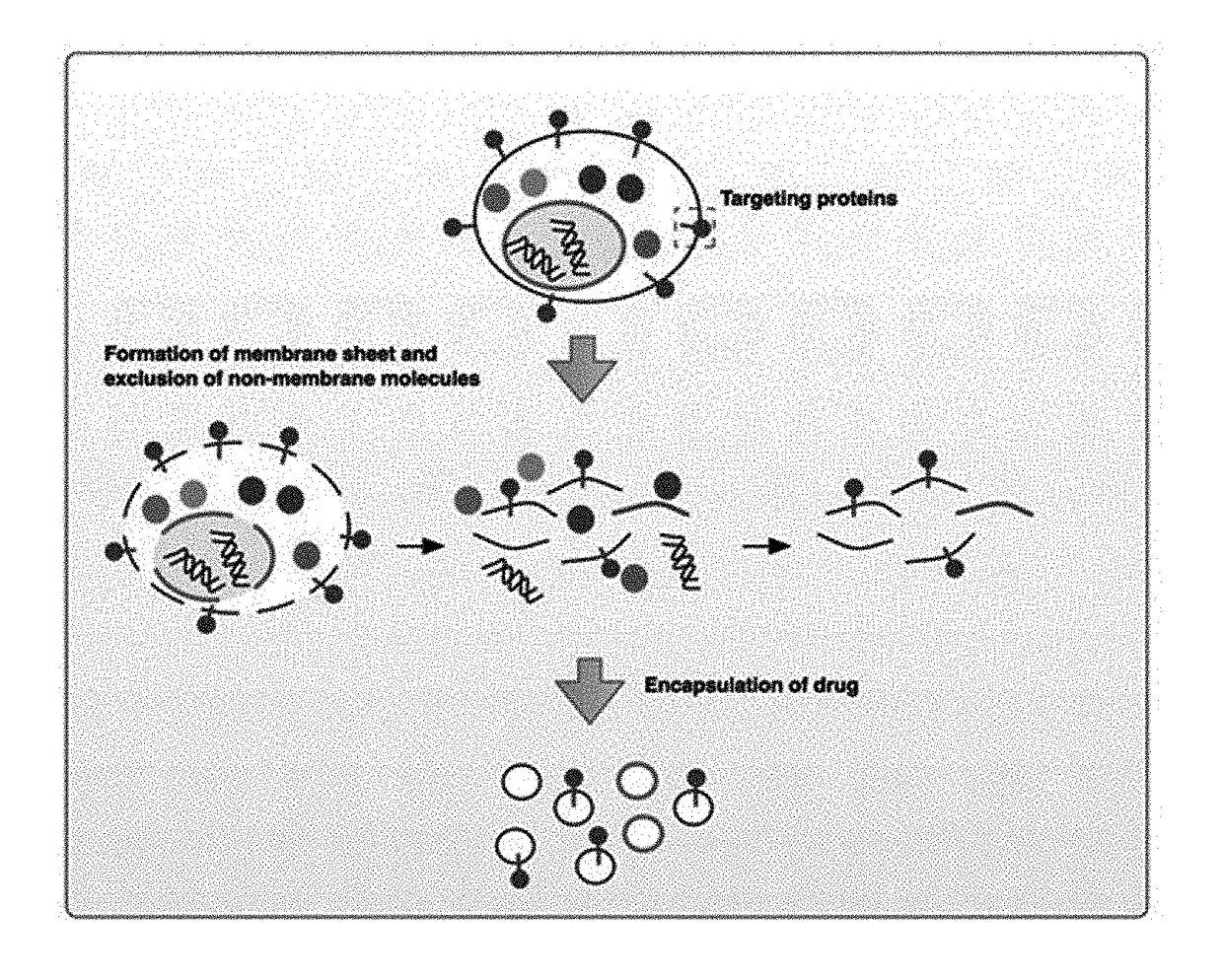
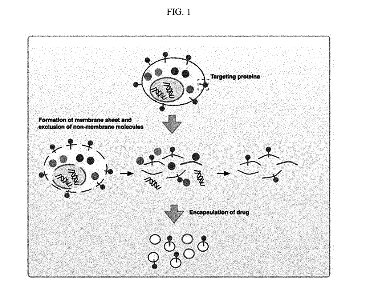
[0106]To prepare nanovesicles derived from nucleated mammalian cells or transformed nucleated mammalian cells from which intracellular components had been removed, monocytes or transformed cells were subjected to alkaline solution treatment, sonication, and a density gradient method, and a schematic diagram for preparing the nanovesicles is shown in FIG. 1.

[0107]8×10⁷ U937 cells (ATCC No. CRL-1593.2) were used as monocytes and 4×10⁷ HEK293 cells (ATCC No. CRL-1573) or 4×10⁷ HT1080 cells (ATCC No. CCL-121) were used as transformed cells. Each cell type was treated with 8 ml of an alkaline solution (200 mM Na2CO3, 1× phosphatase inhibitor, pH 11.5), the solution was sonicated 30 times with a sonicator under the conditions of cycle: 0.5 and amplitude: 50, and ultracentrifugation was performed at 100,000×g for 15 minutes to obtain a pellet. The pellet was resuspended in 8 ml of the alkaline solution, stored in a ro...

example 2

Characterization of Nanovesicles

2-1. Identification of Shape and Size of Nanovesicles

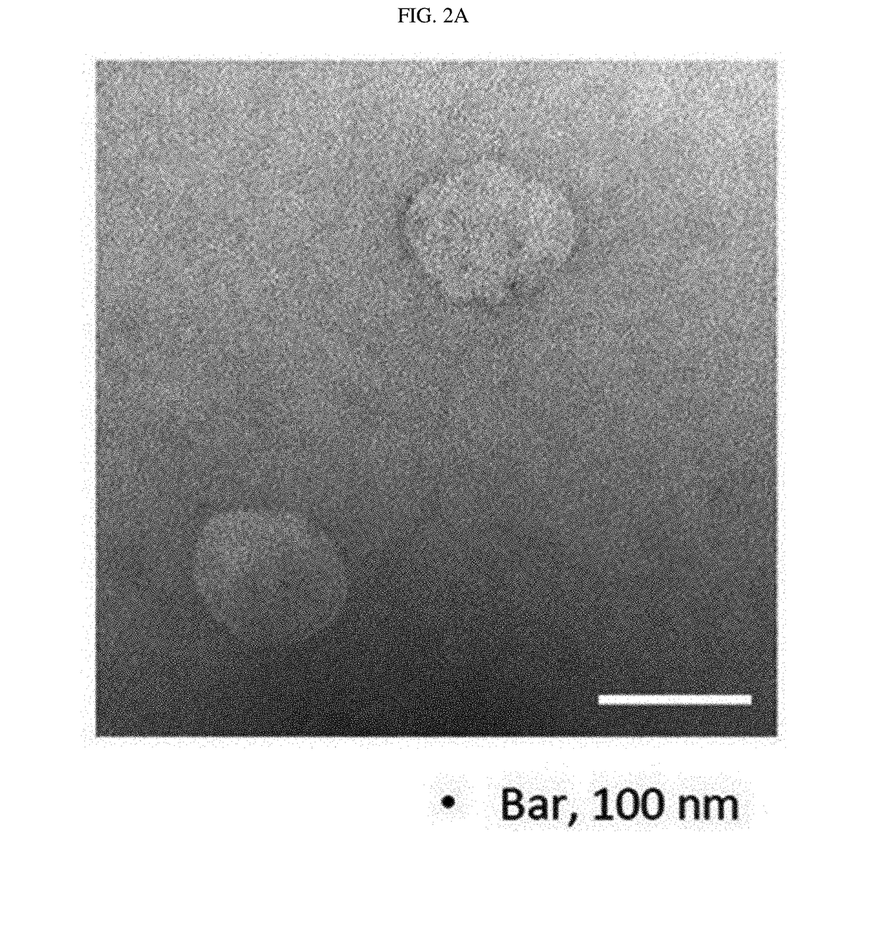
[0108]The shape and size of the monocyte- and transformed cell-derived nanovesicles prepared by the method of Example 1 were analyzed. First, the nanovesicles derived from monocytes were observed using a transmission electron microscope. The nanovesicles derived from monocytes were adsorbed in a glow-discharged carbon-coated copper grid for 3 minutes. The grid was washed with distilled water, stained with 2% uranyl acetate for 1 minute, and observed with JEM101 (Jeol, Japan), an electron microscope.

[0109]As a result, as shown in FIG. 2a, it was found that the nanovesicles prepared from monocytes using alkaline solution treatment were composed of lipid bilayers, had a size of 100 to 200 nm, and exhibited a generally spherical shape. In addition, the nanovesicles prepared from monocytes were diluted in 1 ml of HBS to a concentration of 0.5 μg / ml, 1 ml of the HBS solution containing the nanovesicles wa...

example 3

Preparation of Nanovesicles Containing Polyethylene Glycol

[0122]Nanovesicles containing polyethylene glycol were prepared by loading polyethylene glycol into the monocyte-derived nanovesicles prepared according to the method of Example 1.

[0123]After sonication, suspension was performed by adding HBS, and the solution was extruded through a 1 μm filter (two-stack). 0.5 ml of 50% Optiprep, 1 ml of 10% Optiprep, and 3ml of the suspension were sequentially added into an ultracentrifuge tube, and then μltracentrifugation was performed at 100,000×g for 2 hours to obtain nanovesicles present in the layer between the 50% Optiprep and 10% Optiprep. After ultracentrifugation, 5 μg / ml of cholesterol-polyethylene glycol-biotin was added and incubated at room temperature for 1 hour. Once again, 0.5 ml of 50% Optiprep, 1 ml of 10% Optiprep, and 3 ml of the suspension were sequentially added into a 5 ml ultracentrifuge tube, and then μltracentrifugation was performed at 100,000×g for 2 hours to ob...


PUM

PropertyMeasurementUnit
Acidityaaaaaaaaaa
Sizeaaaaaaaaaa
Adhesion strengthaaaaaaaaaa

Abstract

The present invention relates to cell membrane-derived nanovesicles, a method of preparing the same, and a pharmaceutical composition and a diagnostic kit using the nanovesicles. The cell membrane-derived nanovesicles according to the present invention may prevent the occurrence of potential side effects because intracellular materials (e.g., genetic materials and cytosolic proteins) unnecessary for delivery of therapeutic or diagnostic substances are removed from the nanovesicles. In addition, since the nanovesicles may be targeted to the specific types of cells or tissues, therapeutic or diagnostic substances may be predominantly delivered to the targeted cells or tissues while delivery of therapeutic or diagnostic substances to untargeted sites may be inhibited. Therefore, when the cell membrane-derived nanovesicles are applied to disease treatment, the side effects of therapeutic substances such as drugs may be reduced, so that suffering and inconvenience of patients may be alleviated during the course of treating diseases, and therapeutic efficacy may be improved. In addition, the cell membrane-derived nanovesicles of the present invention, in which substances for the treatment or diagnosis of diseases are loaded, and a method of preparing the nanovesicles may be used in vitro or in vivo for therapeutic or diagnostic purposes, or for experimental use.

Description

STATEMENT REGARDING GOVERNMENT RIGHTS[0001]The present invention was undertaken with the support of Studies on the mechanisms of bacterial exosome-mediated tumor metastasis No. 2015R1A2A1A10055961 grant funded by the Ministry of Science ICT and Future Planning.CROSS-REFERENCE TO RELATED APPLICATION[0002]This application claims priority to and the benefit of Korean Patent Application No. 10-2015-0014164, filed on Jan. 29, 2015 and International Patent Application No. PCT / KR2015 / 008801, filed on Aug. 24, 2015, the disclosure of which is incorporated herein by reference in its entirety.TECHNICAL FIELD[0003]The present invention relates to cell membrane-derived nanovesicles, a method of preparing the same, and a pharmaceutical composition and a diagnostic kit using the nanovesicles.BACKGROUND ART[0004]When a drug is administered to an individual for disease treatment, a drug delivery system (DDS) that controls the delivery and release of the administered drug into cells, tissues, and or...

Claims


Application Information

Patent Timeline
IPC IPC(8): A61K9/127A61K38/46A61K33/26A61K49/00A61K31/713A61K31/704
CPCA61K9/1273A61K31/713A61K31/704A61K33/26A61K49/0067A61K49/0086A61K38/465C12Y301/27005A61K9/127A61K48/00A61K49/00A61K47/50A61K9/5068A61K9/5089A61K9/5192A61K9/1271A61K9/1277A61K47/40A61K49/0084
Inventor GHO, YONG SONGCHOI, DONG-SICGO, GYEONGYUN
Owner POSTECH ACAD IND FOUND
Why Eureka
Try Eureka