
Novel biodegradable and non-biodegradable 3D printed implants as a drug delivery system

Inactive Publication Date: 2018-06-21
ROWAN UNIVERSITY
View PDF8 Cites 8 Cited by
  • Summary
  • Abstract
  • Description
  • Claims
  • Application Information

AI Technical Summary

Benefits of technology

The patent describes how to deliver a therapeutic substance to a patient through an implant. The substance can be placed in layers or reservoirs throughout the implant. It can also be dissolved or suspended in a safe substance and sprayed or coated onto polymer beads. The coating process can be repeated to add another layer of substance. This approach allows for controlled release of the therapeutic substance over time, which can help improve treatment outcomes and make the process more effective.

Problems solved by technology

Another contributing factor to the high cost burden relates to post-surgical infections associated with knee and hip surgeries.
However, the rate of antibiotic release for the cement blocks is not only subpar, but also uncontrollable, leading to substantial degree of clinical failure.
Unfortunately, even with this aggressive treatment regimen, re-infection rates are high.
In general, the use of PMMA as a drug delivery mechanism has the following problems: (a) when used in joint replacement setting, patients are left non-weight bearing on the extremity for 6-8 weeks, frequently placed in casts and have problems with cement dislodging and bone erosion; (b) drug elution properties of PMMA have been shown to be poor; (c) the implant is also susceptible to bacterial colonization and becomes a nidus for continued infection; finally (d) polymerization reaction for PMMA is highly exothermic limiting the choice of antibiotics that can be mixed into the cement.

Method used


Image

  • Novel biodegradable and non-biodegradable 3D printed implants as a drug delivery system
  • Novel biodegradable and non-biodegradable 3D printed implants as a drug delivery system
  • Novel biodegradable and non-biodegradable 3D printed implants as a drug delivery system

Examples


example 1

Coating PLA Beads with Antibiotics

[0096]In this example, bioactive 3D printing filaments using gentamicin sulfate, tobramycin, and nitrofurantoin antibiotics were created and a process was developed to coat PLA beads with 1%, 2.5%, and 5% coating of the antibiotics. The coated PLA beads were then extruded into filament usable for FDM 3D printing. The 3D printing material used in the additive coating study were commercially-available PLA beads. To perform the coating process, the antibiotics chosen for testing were gentamicin, tobramycin, and nitrofurantoin, with KJL 705 Silicone Oil as the chemical used to hold the antibiotics to the heads.

[0097]During the coating portion of the experiment, basic chemical measuring devices, test tubes, and sterilization chemicals were required. A vortex machine to ensure complete coating, as well as a mortar and pestle to crush the antibiotics into a uniform powder, were also used. For extrusion process, an Extrusionbot filament extruder turned the ...

example 2

Total Knee Replacement CAD Development

[0102]Using information gathered on total knee replacement design aspects, bone geometry data provided by Mimics software, and a total knee replacement sample, a total knee replacement three dimensional model was developed in Solidworks. FIG. 5 shows the model iteration. The total knee replacement 1200 includes a femoral component 1201, a tibial component 1203 and a spacer 1202 that is placed in between the femoral and tibial components. The femoral component possesses two lateral surfaces, one on each opposite side 1204 (opposite side not shown), each lateral surface having an outer edge 1205, and inner edge 1206. The outer edge 1205 may be of a shape of semi-wheel configured to be placed on top of the spacer 1202. The inner edge 1206 may be formed by a multiple concatenated straight lines at different lengths. The outer edge and inner edge define the area of the lateral surface.

example 3

Use of Microchannel and Reservoir Network Inside the Orthopedic Implant

[0103]In this example drug delivery methods were developed for manufacturing a microchannel / reservoir network inside the orthopedic implant, filled with antibiotics, which would then elute through the implant, primarily via diffusion, for the 6-8 week treatment period.

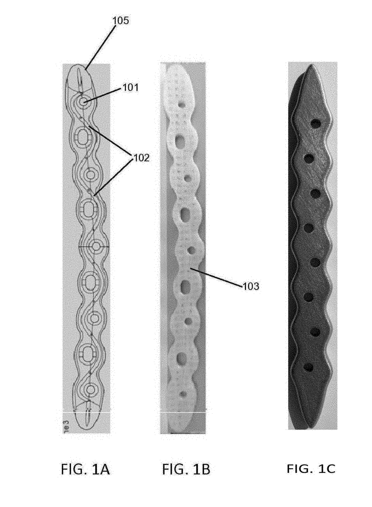
[0104]FIGS. 6A-6D elaborates in at least one such embodiment. In this embodiment, femoral component contain a pair of two reservoirs 602, each reservoir being disposed inside the femoral component proximate to the lateral surface and between the outer edge and inner edge of the surface. Each reservoir form a curve to correspond to the shape of the outer or inner and is connected to two microchannels 601, 606, to facility drug delivery, one of which, 601, is connected to the reservoir at approximate a mid-segment point of the reservoir and extends outward perpendicular to and outside the lateral surface. In this example, the other microchannel 606 is...


PUM

PropertyMeasurementUnit
Fractionaaaaaaaaaa
Fractionaaaaaaaaaa
Diameteraaaaaaaaaa

Abstract

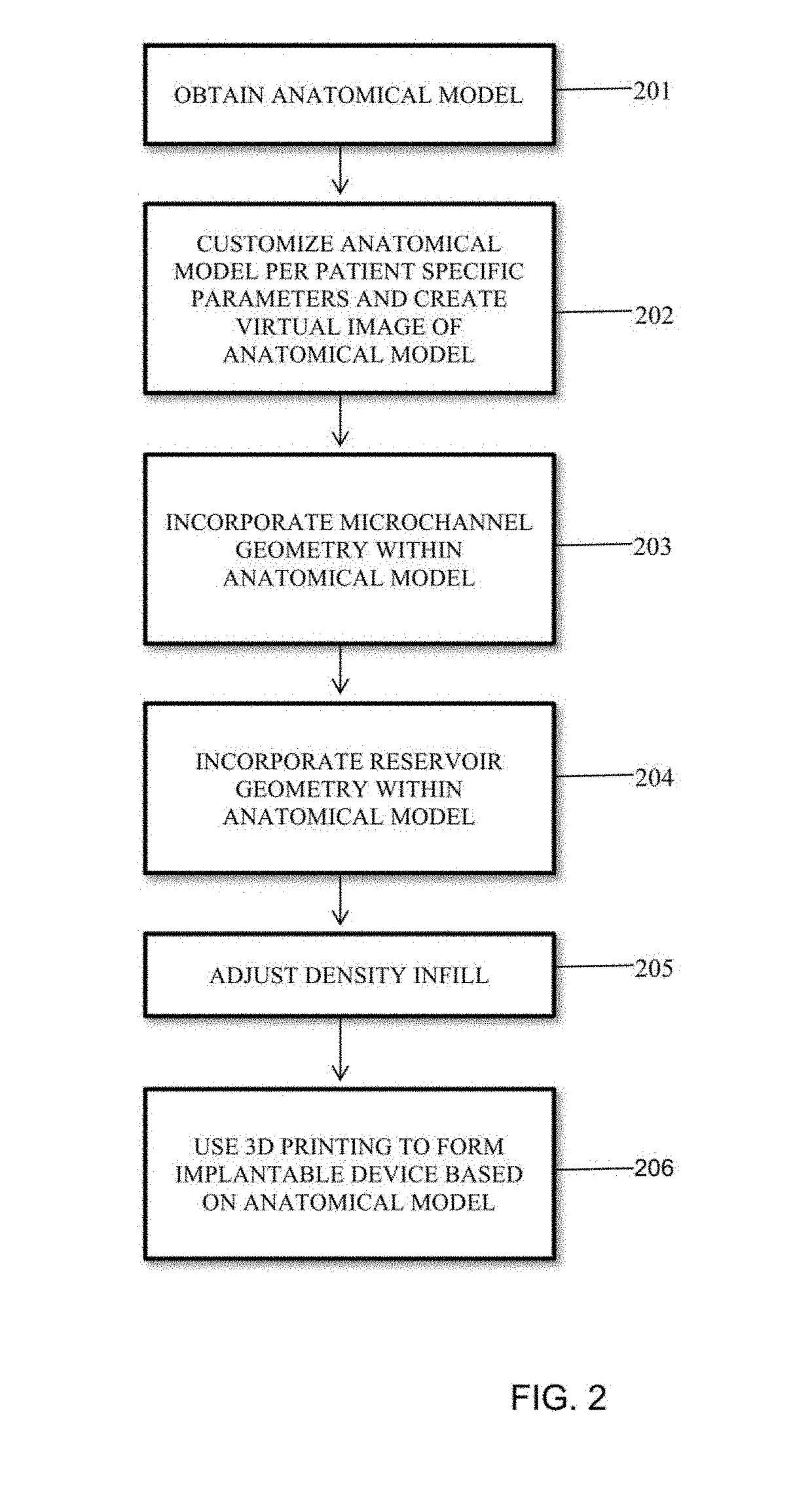
The present invention is directed to a medical implant, methods of use and making of such implants. A method of making an implantable device may include obtaining an anatomical model in a computer aided design (CAD) system, customizing the anatomical model per patient specific parameters and creating a virtual image of the anatomical model, incorporating at least one microchannel geometry within said anatomical model, adjusting the density infill to a measurement ranging per patient specific parameters, and using three-dimensional printing to form the implantable device based on the anatomical model. The implant can be made of suitable metallic or polymeric material, such as PLA.

Description

CROSS-REFERENCE TO RELATED APPLICATION[0001]This application is a national phase of International Application No. PCT / US2016 / 037614, filed Jun. 15, 2016, which claims the benefit of priority of U.S. Provisional Patent Application No. 62 / 175,572, filed Jun. 15, 2015, the entire disclosure of both of which are hereby incorporated herein by reference.FIELD OF THE INVENTION[0002]The present invention relates to medical implants, apparatuses, systems and methods of using and making such implants and apparatus by employing three-dimensional printing methodologies.BACKGROUND OF THE INVENTION[0003]Three-dimensional (3D) printing is a process for printing or building parts of objects in layers to produce a three-dimensional object. Various systems have been developed for three-dimensional printing. In such systems, a predetermined configuration may be designed by the way of Computer Aided Design (CAD) system connected to the printing systems and the configuration is ultimately constructed by...

Claims


Application Information

Patent Timeline
IPC IPC(8): A61F2/28A61B17/80A61M31/00A61B5/00A61L31/06A61L31/02A61L31/14A61L31/16A61K31/7036A61K31/4178
CPCA61L2300/406A61L2430/24A61L2430/02A61F2/28A61B17/8052A61M31/002A61B5/0064A61L31/06A61L31/022A61L31/148A61L31/16A61L31/146A61K31/7036A61K31/4178A61F2002/2817A61F2002/30062A61F2002/3068A61F2002/30784A61F2002/30952A61F2002/30985A61F2002/30113A61F2002/30125A61F2310/00017A61F2310/00023A61F2310/00029A61F2310/00059A61B17/80A61F2/3859A61F2/3886A61F2/30942A61F2/389A61F2002/30677A61F2002/30878A61F2002/30884A61F2002/30962
Inventor RANGANATHAN, SHIVAKUMAR IYERKIM, TAE WON BENJAMINCAMPBELL, DANIEL JOSEPHSMITH, GARRETT CHRISTOPHER
Owner ROWAN UNIVERSITY
Learn More