Systems & Methods for Computational Pathology using Points-of-interest

a computational pathology and system & method technology, applied in the field of systems & methods for computational pathology using points-of-interest, can solve the problems of long examination time, inconvenient manual examination of tissue samples under microscope, and inconvenient us

Active Publication Date: 2018-08-16
SETHI AMIT +1
View PDF38 Cites 86 Cited by
  • Summary
  • Abstract
  • Description
  • Claims
  • Application Information

AI Technical Summary

Benefits of technology

[0006]In further embodiments, the two or more disease classes comprise a disease outcome with or without treatment such as cancer recurrence, effectiveness of treatment options, effectiveness of drugs, follow-up procedures, metastasis status, mortality status, etc. In further embodiments, the disease state comprises a disease condition or an outcome. In further embodiments, the disease state comprises a stage or grade of a disease. In further embodiments, the disease state includes pathological, molecular or genomic sub-type; tumor grade, stage or spatial extent; treatment or survival outcome. In further embodiments, the aggregation algorithm is applied to disease class probabilities at all the points-of-interest located in step (iv). In further embodiments, one or more probability or probability-like metric of disease classifications is the weighted probability or entropy. In further embodiments, the metric of one or more disease classifications is the precision, recall, specificity, F1-score, receiver-operating-characteristic (ROC) or accuracy score. In further embodiments, the imaging apparatus comprises a whole-slide tissue scanning and imaging equipment and the patient tissue is fixed on one or more slides. In further embodiments, the imaging apparatus comprises a microscope with optical zoom and a camera for capturing the one or more images of the patient tissue. In even further embodiments, the at least one processor includes a graphics processing unit (GPU).
[0007]In further embodiments, the magnifying component magnifies with a 1× to 100× objective. In some further embodiments, the magnifying component comprises a microscope and the image capturing component comprises a digital camera or scanner. In further embodiments, the imaging apparatus includes a mobile device with a camera and a microscopic attachment. In a further embodiment, the imaging apparatus includes an infrared (IR), hyperspectral or multispectral scanner. In further embodiments, the instructions executable by the at least one processor includes software for image capturing or storage. In further embodiments, the software for image capturing and storage is capable of image enhancements such as contrast adjustment, image sharpening, and histogram equalization of one or more images of the patient tissue. In further embodiments, one or more images of the patient tissue comprises (0.01 μm)2 to (10 μm)2 per pixel.
[0008]In further embodiments, the patient tissue is fixed on a slide and the one or more images of the patient tissue are digital images of the slide. In further embodiments, the one or more images of the patient tissue comprises an RGB image stored as an m-by-n-by-3 data array that defines red, green, and blue color components for each individual pixel. In further embodiments, the one or more images of the patient tissue include color, fluorescent, hyperspectral, multispectral or confocal images. In further embodiments, the one or more images of the patient tissue comprise images of different sections of one slide with the patient tissue. In further embodiments, the pre-processing module includes stain separation of the one or more images of the patient tissue. In further embodiments, the pre-processing module includes normalization such as color, hue, saturation, image size, etc. In further embodiments, the pre-processing module includes image registration of two or more images of the patient tissue. In certain embodiments, the pre-processing module includes image processing operations such as noise removal, baseline correction, contrast enhancement, super-resolution, etc. In further embodiments, user input includes selection of images of patient tissue, pre-processing steps and parameters, image types and characteristics, tissue staining, disease classes, disease conditions or outcomes, etc. In further embodiments, the magnifying component magnifies slides with a 20× objective for a total magnification of 200×. In further embodiments, the magnifying component projects to an image sensor at (0.25 μm)2 per pixel.
[0009]In further embodiments, the two or more disease classes include cancer types, grades, or cancer stages as determined from an assessment by human experts. In further embodiments, the two or more disease classes comprise cancer types, grades or cancer stages as determined by genetic sequencing of cells. In further embodiments, the two or more disease classes include a disease outcome such as cancer recurrence, effectiveness of treatment options, effectiveness of drugs, follow-up procedures, etc. In further embodiments, the disease classifier is one of several types of neural networks such as a feed forward, deep, convolutional or recurrent neural network. In further embodiments, the aggregation algorithm includes summation, averaging, moving averaging, etc. of the disease classifier estimated probability or probability-like metric of disease classification. In further embodiments, the aggregation algorithm is applied to a sub-set of the points-of-interest located in step (iv) based on the aggregation algorithm. In further embodiments, point-of-interest detector is applied patch-wise on the pre-processed image of the tissue. In further embodiments, the disease classifier is applied to the patches sampled around the points-of-interest located by the point-of-interest detector. In some embodiments, the metric of one or more disease classifications is the probability of a molecular sub-type of cancer as determined by genomic sequencing of cells. In further embodiments, the metric of one or more disease classifications is the probability of cancer recurrence or effectiveness of treatment options. In further embodiments, the metric of one or more disease classifications is the probability of cancer grade or pathological sub-type. In further embodiments, the instructions executable by at least one processor includes a reporting module for generating a report for the disease class scores for the patient tissue. In further embodiments, the report includes one or confidence values for each disease class.
[0010]In another aspect, a method for determining one or more disease class scores of a disease state on a patient tissue is disclosed. The method includes: (a) inputting one or more images of the patient tissue, wherein the tissue has been treated with a stain comprising one or more stain components; (b) generating a pre-processed image from the images of the patient tissue; (c) locating one or more points of interest in the pre-processed image using a first classifier; (d) generating a disease spatial map with the probability of a disease state at the points-of-interest in the tissue image by using a second classifier; and (e) aggregating the probability of the disease condition at each point-of-interest located in step (c) to obtain the disease class scores of the disease state for the patient tissue. In further embodiments, the first classifier is a nucleus detector comprising one or more pre-trained neural networks, convolutional neural networks, or recurrent neural networks etc. In further embodiments, the probability of the disease state is aggregated from two or more images of the patient tissue from the same patient. In further embodiments, the method undergoes periodic or continuous validation.
[0011]In another aspect, a non-transitory computer readable medium is disclosed. The medium having a series of instructions that, when executed by a processor, cause the processor to: (a) input one or more images of the patient tissue, wherein the tissue has been treated with a stain comprising one or more stain components; (b) generate disease spatial maps with the probability of a disease state at one or more points-of-interest by using a combined classifier into one out of two or more disease classes; and (c) aggregate the probability of the disease condition at each point-of-interest located in step (b) to obtain one or more disease class scores of the disease state for the patient tissue. In further embodiments, the one more images of the patient tissue undergo stain separation before step (a). In further embodiments, the points-of-interest are nuclei detected at or near their center points and the combined classifier determines the probability of a disease state at the detected nuclei. In further embodiments, the combined classifier is a pre-trained convolutional neural network. In even further embodiments, the combined classifier locates nuclei patch-wise on the pre-processed image and classifies the detected nuclei into two or more disease classes.

Problems solved by technology

Manual examination of tissue samples under a microscope is fraught with problems such as imprecision, inaccuracies, inter- and intra-pathologist discordance, lengthy examination times, and human inability to pick up subtle visual cues of differences in disease classes.
Some communities in different parts of the globe have limited access to pathologists.
These new disease classes may be too subtly different from each other for human pathologists to reliably recognize compared to an automated quantitative method.

Method used


Image

  • Systems & Methods for Computational Pathology using Points-of-interest
  • Systems & Methods for Computational Pathology using Points-of-interest
  • Systems & Methods for Computational Pathology using Points-of-interest

Examples


example 1

Recommendation

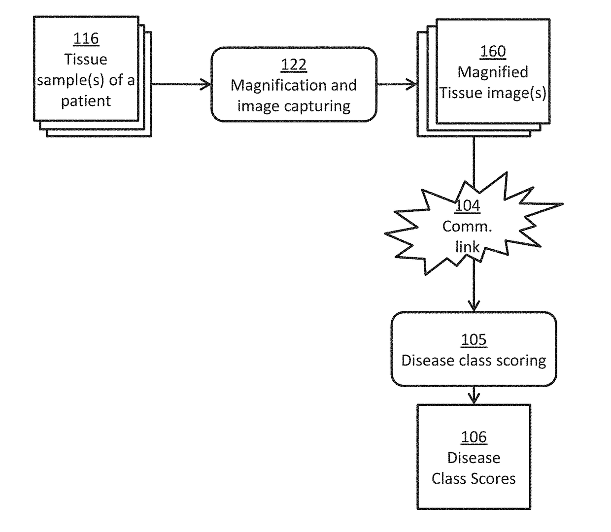
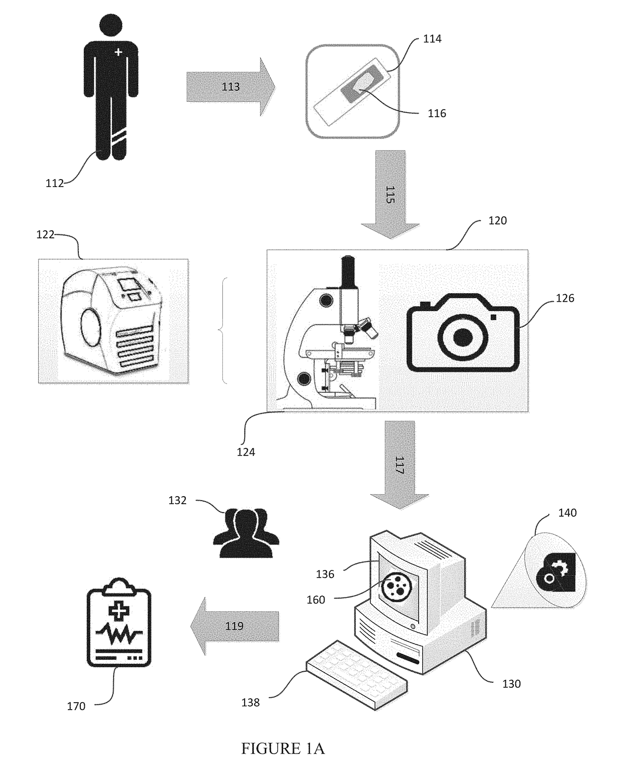
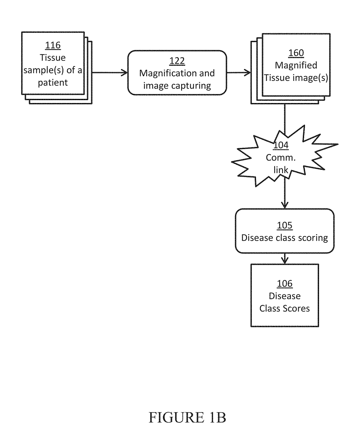
[0086]In this example, a team of pathologist and oncologist use the disclosed system to make treatment recommendations for a prostate cancer patient to improve his chance of remission. Three slides of H&E stained biopsy tissue of prostate of a patient with suspected prostate cancer are prepared and scanned using a whole slide scanner to give the three tissue images for that patient. In a computer, each tissue image is color normalized to give a pre-processed image using H&E color prototypes obtained from a different standard image. In a computer, all possible sub-image windows of size 51×51 from each pre-processed image are extracted one by one, and given as an input to a pre-trained deep convolutional neural network to determine if these have an epithelial nucleus centroid at their centers. All pixel locations designated as centroids of epithelial nuclei are points-of-interest (POIs). In another computer, a second pre-trained deep convolutional neural network is used ...

example 2

& Telemedicine

[0087]In this example, we describe the possibility of setting up mobile cancer screening and telemedicine camps made possible through a potential triage embodiment of the our system for populations without access to good healthcare. A mobile screening camp for identifying people at risk for head-and-neck or cervical cancer is organized in a remote village without access to a good hospital or a pathologist. People go to a lab in a van, where their mouth or cervical swabs (PAP smears) are taken by a nurse. A technician stains the tissues with hematoxylin and eosin in another part of the van, and puts it under a microscope attached to a smartphone, wherein the smartphone is an embodiment of both the image capturing component and the computer component described in this disclosure. One of the modules running on the smartphone is used to enter patient details and capture images of the view from the microscope. These images of a patient are passed to the pre-processing modul...

example 3

Planning

[0088]In this example, we describe how extra information provided to a pathologist or an oncologist using the disclosed system and methods may be used to develop individualized treatment plans for two patients. Current clinical diagnosis practices would have likely led to the same treatment plan for both. A patient with suspected breast lump checks in to a clinic for a mammogram. The mammogram reveals a potential tumor whose biopsy is taken. The tissue extracted from the biopsy is cut into four adjacent sections and stained with hematoxylin+eosin (H&E), HER2neu, ER, and PR stains respectively. Conventionally, the presence of cancer is ascertained through the microscopic examination of H&E, while the clinical decision making starting from determination of sub-type of cancer to treatment planning is done through the examination of ER, PR, and HER2neu. If the patient is assessed to be a case of 3+ on a scale of 0, 1, 2+, and 3+ in HER2 positivity determined by examination of HE...


PUM


Abstract

Systems, methods and devices for determination of disease class scores for patient tissue are disclosed. The disease class scores may be based on the probability or probability-like metric for a disease condition or outcome. The system includes an imaging apparatus and a computing system with instructions executable by a processor. The computer system may locate one or more points-of-interest on the pre-processed images of the patient tissue using a point-of-interest detector and generate one or more disease spatial maps including one or more probability or probability-like metric of disease classifications on the detected points-of-interest. The information in the disease spatial maps is aggregated to produce disease class scores.

Description

CROSS-REFERENCE[0001]This application claims the benefit of Indian patent application No. 201711005034 filed on Feb. 13, 2017 under 35 USC § 365.BACKGROUND[0002]Manual examination of tissue samples under a microscope is fraught with problems such as imprecision, inaccuracies, inter- and intra-pathologist discordance, lengthy examination times, and human inability to pick up subtle visual cues of differences in disease classes. Some communities in different parts of the globe have limited access to pathologists. On the other hand, advances in computer vision, machine learning algorithms, computer hardware technology, and computer networking technology, along with high throughput whole slide scanning technology will enable the emergence of computational pathology systems that can alleviate the aforementioned problems. With the emergence of deep learning and convolutional neural network algorithms and methods and the development of graphical processing unit (GPU) technologies, automate...

Claims


Application Information

Patent Timeline
Patent Type & Authority Applications(United States)
IPC IPC(8): G06T7/00G06N3/08G06N3/04G16H50/20G06K9/46G06K9/62G06V10/764
CPCG06T7/0014G06N3/08G06N3/0454G16H50/20G06K9/4671G06K9/6277G06K9/628G06K9/6219G06K2209/05G06T2207/10056G06T2207/20084G06T2207/30024G06T2207/30096G06T2207/10024G06T2207/20076G06T2207/20081G16H30/40G16H20/10G06N20/10G06N20/20G06V2201/03G06V10/82G06V10/764G06N5/01G06N3/044G06N3/045G06V2201/031G06V2201/032G06F18/24G06F18/2415G06F18/2431
Inventor SETHI, AMITKUMAR, NEERAJ
Owner SETHI AMIT
Why Eureka
Try Eureka