Method of diagnosing and/or monitoring therapy of atherosclerosis

Active Publication Date: 2018-12-27
UNM RAINFOREST INNOVATIONS
View PDF1 Cites 1 Cited by
  • Summary
  • Abstract
  • Description
  • Claims
  • Application Information

AI Technical Summary

Benefits of technology

[0019]It is noted that compounds according to the present invention (and in particular, compound IN or GA, above) exhibit a favorable bioavailability to tissues which have are to be diagnosed for disease states and/conditions, thus providing a ready means by which the infection may be diagnosed and/or monitored for therapeutic success or failure. It is noteworthy that

Problems solved by technology

Early stage diagnosis is challenging, however; symptoms often become clinically evident only at late stages where complete prevention is no longer viable[4].
As vascular disease progresses, the inflammatory and remodeling process also evolves, and the plaque can become unstable or v

Method used


Image

  • Method of diagnosing and/or monitoring therapy of atherosclerosis
  • Method of diagnosing and/or monitoring therapy of atherosclerosis
  • Method of diagnosing and/or monitoring therapy of atherosclerosis

Examples


Example

Example 1

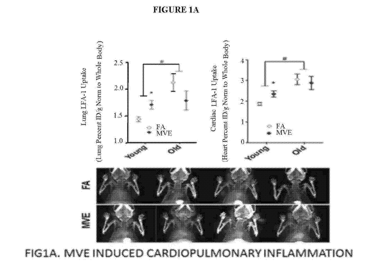
[0150]This first example is presented to show evidence of the use of the present invention in diagnosing the inflammatory process associated with atherosclerosis. In particular, in this example, 111In-DANBIRT is used as an in vivo SPECT / CT imaging tool for the Expression of LFA-1 in the inflammatory process of atheroma development.

[0151]The objective of this experiment was to assess inflammatory leukocyte presence and accumulation in vascular atherosclerotic plaque using 111In-DANBIRT as a non-invasive diagnostic imaging tool.

[0152]Methods: 6 week ApoE KG mice were fed either normal or high fat chow (n=8 per group) for 8 weeks to induce vascular atherosclerotic lesions. SPECT / CT imaging was performed 3-hrs post injection of ˜700 uCi of 111In-DANBIRT at baseline, 4 weeks and 8 weeks. Whole body Autoradiography was performed 24-hrs post injection after 8-week time point. Image processing and analysis was performed by region of interest (ROI) determination in relationship to v...

Example

Example 2

[0155]The aim of this example is to validate 111In-DANBIRT as a non-invasive diagnostic tool using SPECT / CT imaging for inflammation assessment in vulnerable atherosclerotic plaque in a murine model.

Method for Example 2

[0156]SPECT / CT images were obtained from 2 different cohorts over 3 identical imaging time points (0 weeks (baseline), 4 weeks and 8 weeks),[0157]6 week old ApoE KO C57 black male mice[0158]We focused on 3 specific areas in our SPECT / CT imaging analysis: Heart, aortic arch and descending aorta; normalized to muscle.

1st Cohort n = 40HighHigh FatNor-HighFatHigh FatDiet +malFatDiet +Diet +Statin +DietDietStatinGW501516GW501516TotalSPECT / CT4444420and biodistributionSerum4444420analysisTotal8888840[0159]Animals were imaged for ˜75 minutes 3 hrs post tail vein injection of ˜700 uCi (˜25.9 Mbq) of 111In-DANBIRT[0160]Atorvastatin was administered in the drinking water and PPAR-Delta agonist (GW1516) was incorporated into food pellets by diet manufacturing company.[01...

Example

Example 3

Materials and Methods

Animals

[0179]Male Apo-E− / 31 mice on a C57BL / 6 background (Taconic Laboratories, Indianapolis, Ind.) aged 6 weeks were housed two per cage and allowed to acclimate over the course of one week after delivery. Mice were fed either a normal chow diet or HFD (Harlan-Teklad, TD.88137: 1.5 g / kg of cholesterol and 42% kcal from fat) for 8 weeks; food and water were provided ad libitum. Food was changed every 3 days and stored at −20° C. and thawed before administration to the mice. For isolated blood distribution studies, male Sprague Dawley rats (Taconic Laboratories, Indianapolis, Ind.) aged 6-8 weeks were allowed 1 week of acclimation following delivery. Rats were housed two per cage and given food and water ad libitum. Both rats and mice were maintained on a 12 h light:dark schedule in AAALAC-approved facilities, and euthanized via cardiac exsanguination while under deep anesthesia with Isofluorane (Piramidal Healthcare, NDC 66794-093-25). The UNM Institut...


PUM

PropertyMeasurementUnit
Lengthaaaaaaaaaa
Volumeaaaaaaaaaa
Currentaaaaaaaaaa

Abstract

The present invention is directed to novel non-invasive diagnostic took to dispose numerous disease states and/or conditions. The presets invention, represents a clear advance in the art which presently relies on tissue biopsy for diagnoses of these disease states. The novel imaging probe is capable of detecting infected cells, as well tissue. The methods described herein are able to diagnose, treat and/or monitor the therapy of numerous diseases and conditions including atherosclerosis, atherothrombosis, cerebral vascular disease, cerebral ischemia, cerebral infarct and meningitis as well as pneumonitis, pericarditis, multiple sclerosis, lupus erythematosus and pancreatitis, among others.

Description

RELATED APPLICATIONS AND GRANT SUPPORT[0001]The present application claims the benefit of priority of provisional application U.S. 62 / 271,573 of identical title, filed Dec. 28,2015, the entire contents of which is incorporated by reference herein.[0002]This invention was made with government support under grant nos. P30CA118100-06 and ES0014639 awarded by the National Institute of Health (NIH). The government has certain rights in the invention.FIELD OF THE INVENTION[0003]The present invention is directed to diagnosing and imaging tissue suspected of or known to affected by atherosclerosis, atherothrombosis and related disease states and conditions. The present invention can be used to diagnose the presence and extent of atherosclerotic tissue, atherosclerotic plaque in tissue (including vascular atherosclerotic lesions) in blood vessels and / or cardiovascular tissue (especially cardiopulmonary tissue) of a patient suspected of having atherosclerosis and / or atherthrombosis The presen...

Claims


Application Information

Patent Timeline
IPC IPC(8): A61K51/04
CPCA61K51/0482A61K51/0453A61K47/22A61K49/106
Inventor NORENBERG, JEFFREY P.
Owner UNM RAINFOREST INNOVATIONS
Why Eureka
Try Eureka