
Automated segmentation of three dimensional bony structure images

a three-dimensional image and automatic segmentation technology, applied in the field of automatic segmentation of three-dimensional images of a bony structure, can solve the problems of reducing the accuracy and efficacy of navigated tools and implants, difficulty in adequately addressing, and difficulty in using low-quality image datasets in machine learning applications

Active Publication Date: 2019-04-11
HOLO SURGICAL INC
View PDF0 Cites 34 Cited by
  • Summary
  • Abstract
  • Description
  • Claims
  • Application Information

AI Technical Summary

Benefits of technology

The patent describes a method for training a machine learning system to segment parts of a three-dimensional bony structure using a deep learning network. The method involves receiving data of labeled anatomical image sets, which include information about the parts of the bone structure being segmented. The data is then used to train a segmentation neural network model, which can accurately identify and segment the different parts of the bone structure. This method can be used in a variety of applications, such as in the field of medical imaging. The technical effects of this patent include improved accuracy in identifying and segmenting parts of a bone structure, which can aid in the diagnosis and treatment of bone-related diseases.

Problems solved by technology

In the field of image guided surgery, low quality images may make it difficult to adequately identify key anatomic landmarks, which may in turn lead to decreased accuracy and efficacy of the navigated tools and implants.
Furthermore, low quality image datasets may be difficult to use in machine learning applications.
The downside is increased radiation exposure to the patient.
These survivors, who are estimated to have experienced doses slightly larger than those encountered in CT, have demonstrated a small but increased radiation-related excess relative risk for cancer mortality.
However, low dose settings may have an impact on the quality of the final image available for the surgeon.
This in turn can limit the value of the scan in diagnosis and treatment.
As these coils are rapidly switched on and off they create the characteristic repetitive noise of an MRI scan.
For example, a 0.3 T magnet strength will result in lower quality imaging then a 1.5 T. Low quality images may pose a diagnostic challenge as it may be difficult to identify key anatomical structures or a pathologic process.
Low quality images also make it difficult to use the data during computer assisted surgery.

Method used


Image

  • Automated segmentation of three dimensional bony structure images
  • Automated segmentation of three dimensional bony structure images
  • Automated segmentation of three dimensional bony structure images

Examples


Embodiment Construction

[0054]The following detailed description is of the best currently contemplated modes of carrying out the invention. The description is not to be taken in a limiting sense, but is made merely for the purpose of illustrating the general principles of the invention.

[0055]The invention relates to processing images of a bony structure, such as a spine, skull, pelvis, long bones, shoulder joint, hip joint, knee joint etc. The foregoing description will present examples related mostly to a spine, but a skilled person will realize how to adapt the embodiments to be applicable to the other bony structures as well.

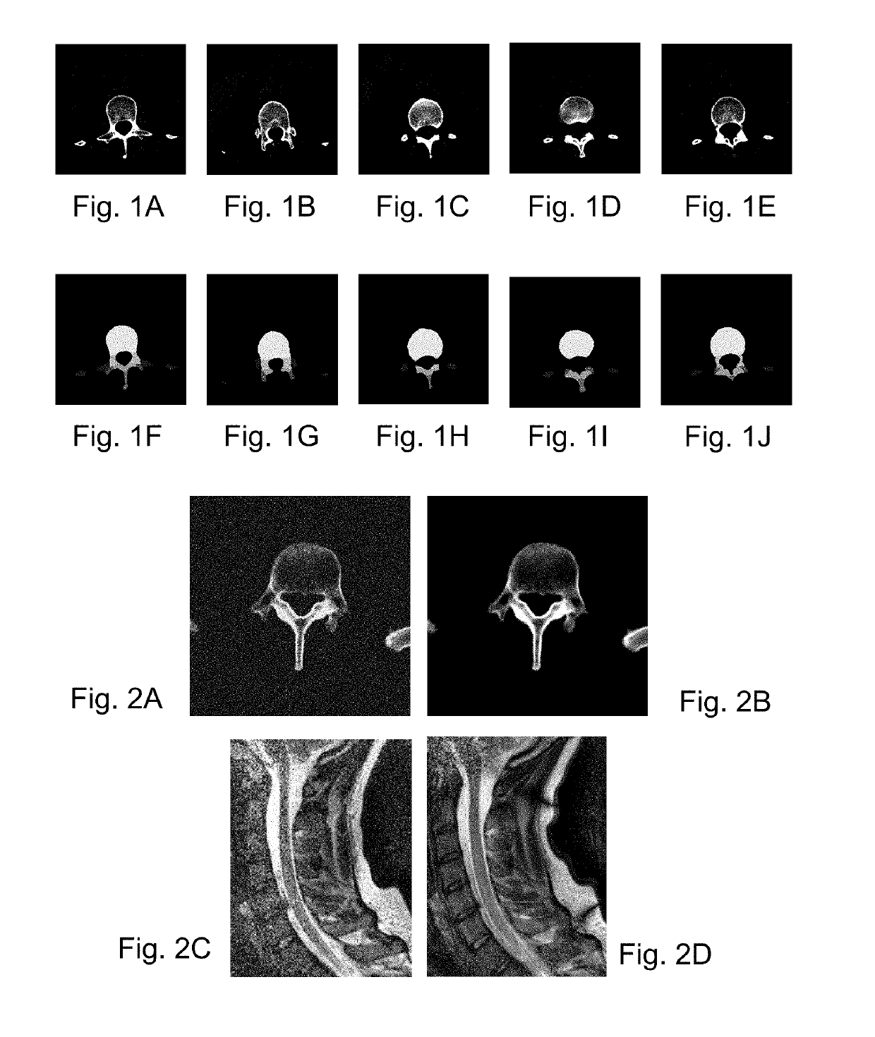
[0056]Moreover, the invention may include, before segmentation, pre-processing of lower quality images to improve their quality. For example, the lower quality images may be low dose computer tomography (LDCT) images or magnetic resonance images captured with a relatively low power scanner. The foregoing description will present examples related to computer tomography (CT) images, b...


PUM


Abstract

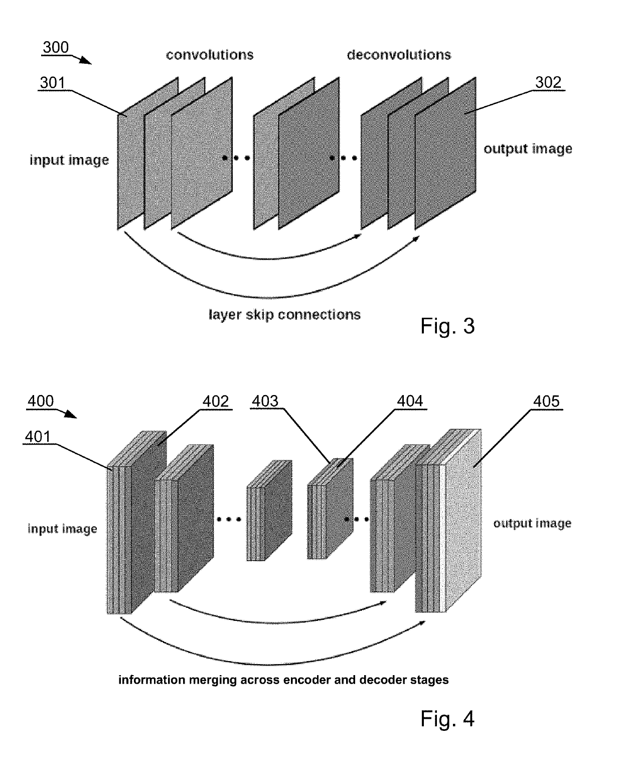
A computer-implemented system: at least one processor communicably coupled to at least one nontransitory processor-readable storage medium storing processor-executable instructions or data receives segmentation learning data comprising a plurality of batches of labeled anatomical image sets, each image set comprising image data representative of a series of slices of a three-dimensional bony structure, and each image set including at least one label which identifies the region of a particular part of the bony structure depicted in each image of the image set, wherein the label indicates one of a plurality of classes indicating parts of the bone anatomy; trains a segmentation CNN, that is a fully convolutional neural network model with layer skip connections, to segment semantically at least one part of the bony structure utilizing the received segmentation learning data; and stores the trained segmentation CNN in at least one nontransitory processor-readable storage medium of the machine learning system.

Description

TECHNICAL FIELD[0001]The present disclosure generally relates to automated segmentation of three dimensional images of a bony structure, useful in particular for the field of computer assisted surgery, diagnostics, and surgical planning.BACKGROUND[0002]Image guided or computer assisted surgery is a surgical procedure where the surgeon uses tracked surgical instruments in conjunction with preoperative or intraoperative images in order to indirectly guide the procedure. Image guided surgery can utilize images acquired intraoperatively, provided for example from computer tomography (CT) scanners.[0003]Specialized computer systems can be used to process the CT images to develop three-dimensional models of the anatomy fragment subject to the surgery procedure.[0004]For this purpose, various machine learning technologies are developed, such as a convolutional neural network (CNN) that is a class of deep, feed-forward artificial neural networks. CNNs use a variation of multilayer perceptro...

Claims


Application Information

Patent Timeline
IPC IPC(8): A61B6/00G06T3/40A61B6/03
CPCA61B6/5229G06T3/4046A61B6/032A61B6/542A61B6/5258G06T5/002G06T2207/10081G06T5/50G06T11/008G06K9/66G06T2207/30012G06T2207/20084G06T7/11G06T5/70
Inventor SIEMIONOW, KRIS B.LUCIANO, CRISTIAN J.KRAFT, MAREK
Owner HOLO SURGICAL INC
Learn More