Pd-1 car-t cell, preparation method therefor, and application thereof

a t cell and pd-1 technology, applied in the field of cellular drug for treating tumors, can solve the problems of poor prognosis, poor tumor prognosis, and death of t cells, and achieve the effect of more efficient tumor killing activity

Inactive Publication Date: 2019-04-25
ASCLEPIUS SUZHOU TECH CO GRP CO LTD
View PDF0 Cites 1 Cited by
  • Summary
  • Abstract
  • Description
  • Claims
  • Application Information

AI Technical Summary

Benefits of technology

[0019]The beneficial effects of the present invention are: the PD-1 CAR-T cells of the present invention and preparation methods and applications thereof, including modifying and altering T cells with chimeric antigen receptors and expressing a PD-1-CD8™-4-1BB-CD3ζ molecule in the T cells, so that the modified T cells can specifically recognize and kill tumors, and the obtained cells have more efficient tumor killing activity.

Problems solved by technology

In addition to expression in malignant melanoma, PD-L1 also was expressed in other different tumors, including glioblastoma, pancreatic cancer, ovarian cancer, breast cancer, renal cell carcinoma, head and neck and esophageal squamous cell carcinoma, and non-small cell lung cancer, and high expression of PD-L1 on tumor cells is associated with poor prognosis.
However, tumors realize immune escape mainly from two aspects: (1) The antigen presentation mechanism of tumor cell is down-regulated or even lost (HLA-negative), which causes T cells to fail to recognize tumor cells; (2) Many tumor cells are abnormally highly expressed PD-L1 molecules, which activates PD-1 molecules on the surface of T cells, and leads to the depletion of T cells function and even death of T cells.

Method used


Image

  • Pd-1 car-t cell, preparation method therefor, and application thereof
  • Pd-1 car-t cell, preparation method therefor, and application thereof
  • Pd-1 car-t cell, preparation method therefor, and application thereof

Examples


embodiment 1

Preparation of a Lentiviral Expression Vector

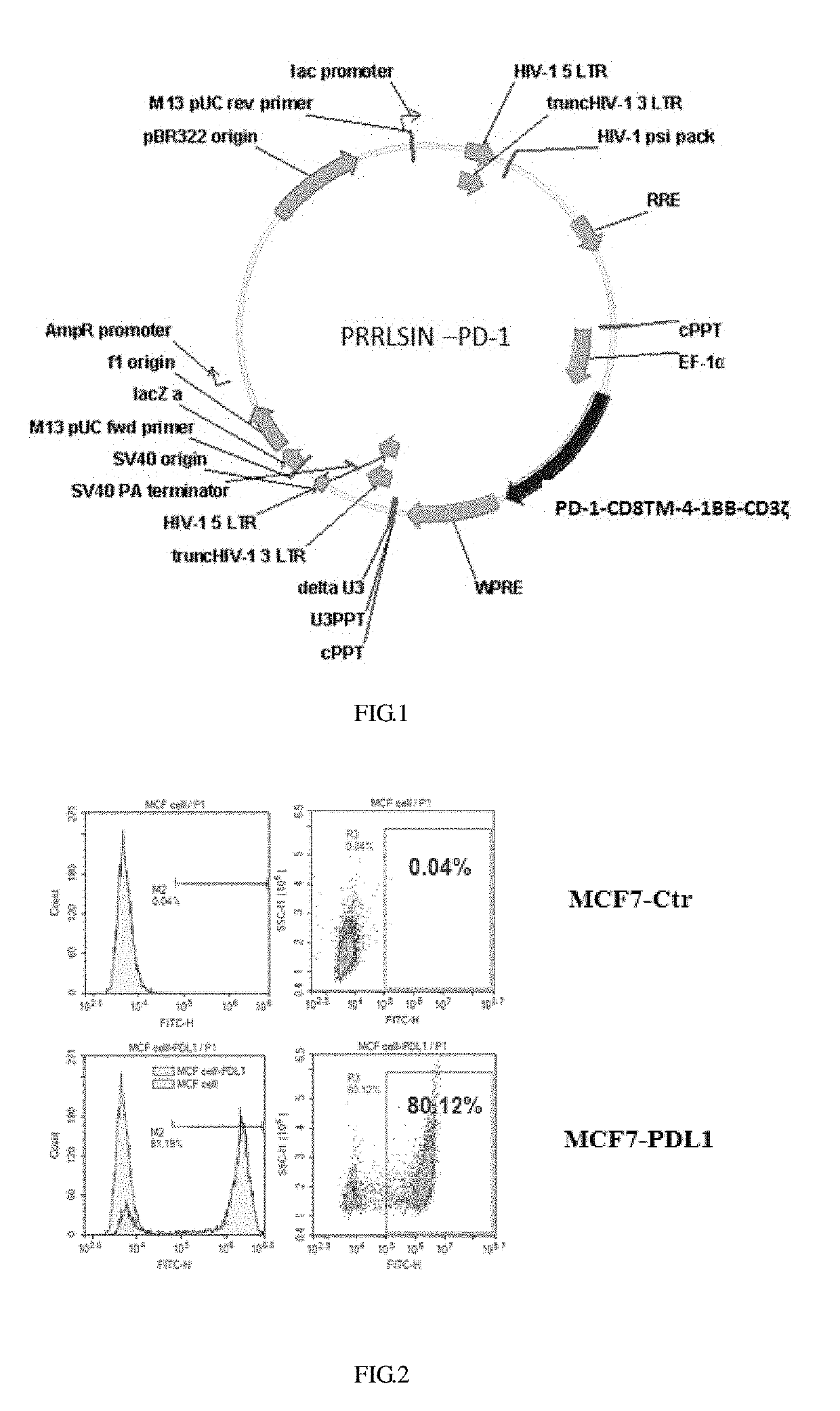
[0029]The gene encoding the PD1-CD8™-4-1BB-CD3ζ was synthesized, then the gene was ligated into the PRRSLIN vector by enzyme restriction and transformation, and the upstream of the gene is EP-1α promoter. The vector was transformed into Stbl3 Escherichia coli strain, and screened by ampicillin to obtain positive clones, then the plasmids were extracted and identified by restriction enzyme digestion, and PRRLSIN-PD-1 lentiviral transfection vector was obtained, the structure of which is as shown in FIG. 1.

embodiment 2

Preparation of Lentivirus

[0030](1) Twenty-four hours before transfection, seeding 293T cells into 15 cm culture dishes at a cell density of approximately 8×10⁶ cell per dish, which could ensure that the cells are at about 80% of confluence and distributed uniformly in the culture dish during transfection.

(2) Prepare solution A and solution B

Solution A: 6.25 ml of 2×HEPES buffer (using 5 large dishes that are packed together could achieve the best effects).

Solution B: adding the following plasmids, respectively, and mixing: 112.5 μg of pRRLSIN-EF-ROBO1 (target plasmid); 39.5 μg of pMD2.G (VSV-G envelop); 73 μg of pCMVR8.74 (gag, pol, tat, rev); 625 μl of 2M calcium ion solution. Total volume of solution A: 6.25 ml.

[0031]The solution B was mixed completely, and the solution A was added dropwise while the solution A was gently rocked, then let the solution sit for 5-15 minutes. The above mixed solution of A and B was gently rocked and added to the dish containing 293T cells dropwise, t...

embodiment 3

Preparation of PD-1 CAR-T Cells

[0032]0.5 ml of blood was taken and tested for pathogenic microorganisms rapidly to exclude microbial infections such as HBV, HCV, HDV and HEV, HIV-1 / 2, treponema pallidum and parasites; 50 ml of blood was collected with heparin bottle (heparin anticoagulation) under sterile conditions, and immediately (4° C., within 24 hours) sent to the cell preparation laboratory to ensure that this process was free of pathogenic microbial contamination. After the patient's blood was obtained, the surface of the heparin bottle was wiped with an alcohol cotton ball for disinfection in the GMP preparation room, then the heparin bottle was placed in a biological safety cabinet. Two 50 ml centrifuge tubes were opened in advance, then the blood was transferred into the two 50 ml centrifuge tubes and tightened up. The above 50 ml centrifuge tubes filled with blood were placed in a centrifuge and centrifuged at 400 g (2000 rpm) for 10 min at room temperature, then the supe...


PUM

PropertyMeasurementUnit
concentrationaaaaaaaaaa
densityaaaaaaaaaa
densityaaaaaaaaaa

Abstract

Provided are a PD-1 CAR-T cell, a preparation method thereof, and an application thereof. By means of chimeric antigen receptor-modified T cell transformation, PD-1-CD8™-4-1BB-CD3ζ molecules are expressed in a T cell. The CAR-T cell prepared by the method can specifically recognize and bind with tumor cells with high PDL-1 protein expression, and is applicable to preparation of a drug for preventing and treating tumor diseases.

Description

FIELD OF THE INVENTION[0001]The invention relates to the field of cellular drug for treating tumor, in particular to a PD-1 CAR-T cell and a preparation method and application thereof.BACKGROUND OF THE INVENTION[0002]With the gradual progress of tumor immunotherapy research, programmed death growth factor-1 (PD-1 / CD 279) and its ligand PD-L1 / 2 (B7-H1 / CD274) won the favor of many researchers as important members in the tumor microenvironment. On Sep. 4, 2014, the U.S. Food and Drug Administration (FDA) approved Keytruda (pembrolizum ab) for the treatment of terminal or unrespectable melanoma patients who do not respond to other drugs, and Keytruda became the first drug approved by FDA to block the PD-1 cell pathway. PD-1 was first discovered in 1992, and mainly expressed in T cells, regulatory T cells, “depleted” T cells, B cells, activated mononuclear cells, dendritic cells, natural killer cells, natural killer T cells and so on. PD-1 is generally expressed in activated T cells, whi...

Claims


Application Information

Patent Timeline
Patent Type & Authority Applications(United States)
IPC IPC(8): A61K35/17A61P35/00C12N15/85C12N15/62
CPCA61K35/17A61P35/00C12N15/85C12N15/62
Inventor LI, HUASHUN
Owner ASCLEPIUS SUZHOU TECH CO GRP CO LTD
Why Eureka
Try Eureka