
Enzyme-specific intracellularly-retained red fluorescent probe

a red fluorescent and enzyme-specific technology, applied in the field of new fluorescent probes, can solve the problems of difficult co-staining with gfp (green fluorescent protein), which is often used in live imaging, and living cells and the like are difficult to clearly image at a single-cell level, and achieve significant industrial utility value and economic effect, and the effect of sufficient cell permeability

Active Publication Date: 2019-08-22
THE UNIV OF TOKYO
View PDF0 Cites 2 Cited by
  • Summary
  • Abstract
  • Description
  • Claims
  • Application Information

AI Technical Summary

Benefits of technology

The present invention provides an intracellularly-retainable red fluorescent probe with the advantage of having high cell permeability and retention. This probe allows selective fluorescence imaging of target cells at a single-cell level in living cells and biological tissues. It enables tracking changes in individual cells over time, resulting in significant industrial utility value and economic effect.

Problems solved by technology

However, from problem points such as low cell membrane permeability and low intracellular retention of a fluorescent dye generated after an enzyme reaction, in the conventional β-galactosidase fluorescent probes, living cells and the like are difficult to clearly image at a single-cell level.
The probe can detect a target cell at a single-cell level, but luminescence in fluorescence detection is in a green fluorescence region, which makes it difficult to perform co-staining together with GFP (green fluorescent protein) which is frequently used in live imaging.

Method used


Image

  • Enzyme-specific intracellularly-retained red fluorescent probe
  • Enzyme-specific intracellularly-retained red fluorescent probe
  • Enzyme-specific intracellularly-retained red fluorescent probe

Examples


example 1

1. Synthesis of Probe Molecule

[0089]4-CH2F-SPiDER-RED-βGal and 2-CH2F-SPiDER-RED-βGal used as an intracellularly-retainable red fluorescent probe of the present invention were synthesized as shown below.

(1) Synthesis of 4-CH2F-SPiDER-RED-βGal

[0090]According to the following scheme, 4-CH2F-SPiDER-RED-βGal which was the intracellularly-retainable red fluorescent probe of the present invention was synthesized.

[Synthesis of Compound 1]

[0091]

[0092]A compound 1 was synthesized in accordance with the previously reported literature (Chemical Communications 47, 4162-4164 (2011)). Specifically, 3-bromoaniline (4.0 mL, 37 mmol) and allyl bromide (11 mL, 131 mmol) were dissolved in MeCN (40 mL) to obtain a dissolved product, to which potassium carbonate (11 g, 80 mmol) was then added. The reaction solution was stirred overnight at 80° C. under an argon atmosphere. After the reaction solution was cooled to room temperature, an insoluble matter was removed by Celite filtration to obtain a filtrat...

example 2

[0147]Changes in Absorption and Fluorescence Spectra by Enzyme Reaction

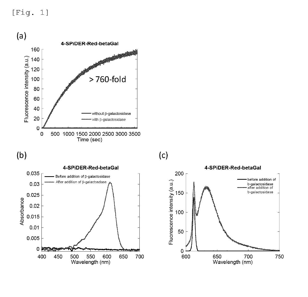
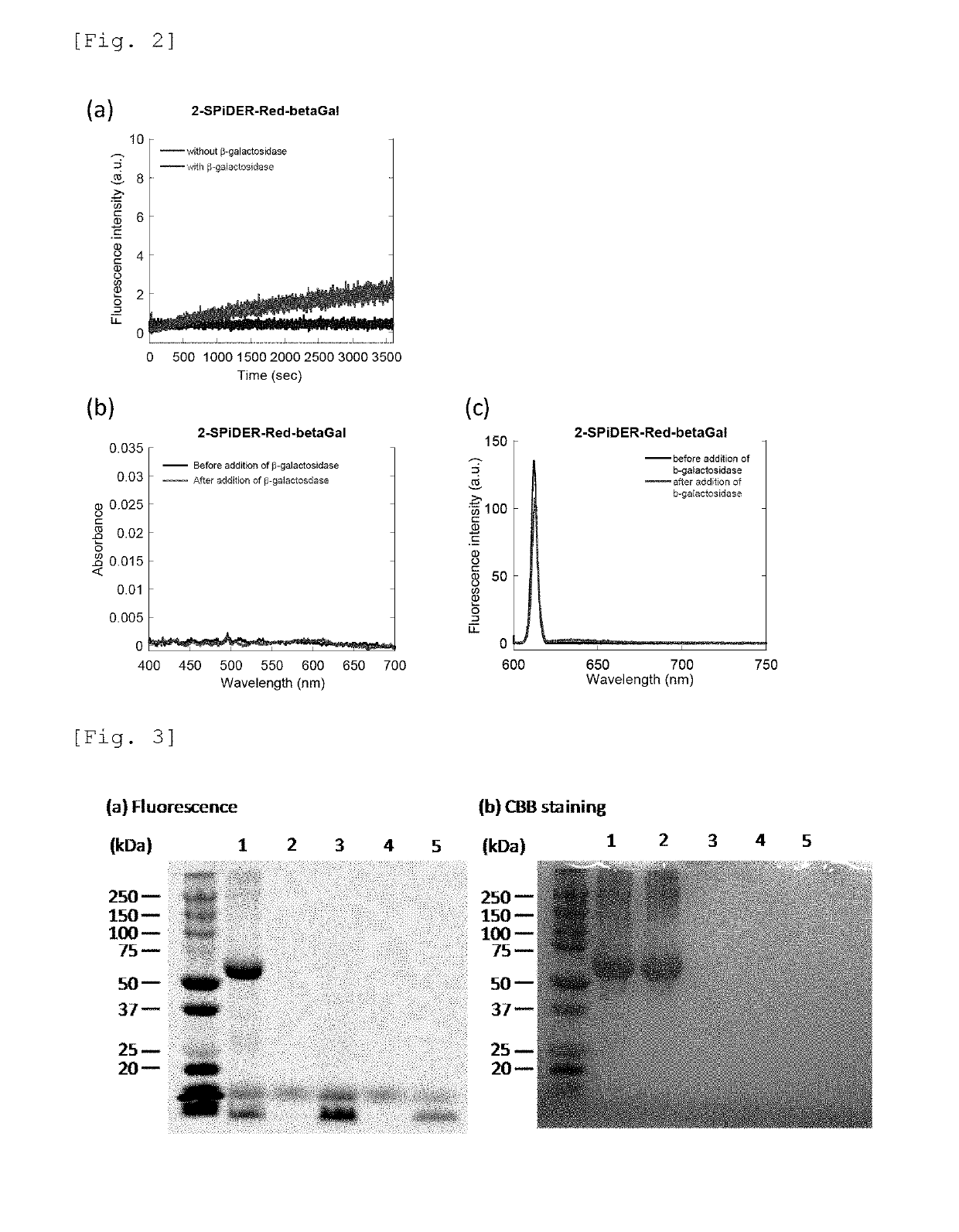
[0148]For the probe compounds of the present invention obtained in Example 1 (4-CH2F-SPiDER-RED-βGal and 2-CH2F-SPiDER-RED-βGal), changes in absorption and fluorescence spectra according to the addition of β-galactosidase were measured. The measurement results of 4-CH2F-SPiDER-RED-βGal are shown in FIG. 1, and the measurement results of 2-CH2F-SPiDER-RED-βGal are shown in FIG. 2.

[0149]FIG. 1(a) shows the fluorescence intensity of 4-CH2F-SPiDER-RED-βGal by an enzyme reaction upon lapse of time. Measurements were carried out by adding 5U (3-galactosidase to 4-CH2F-SPiDER-RED-βGal prepared at 1 μM in a PBS buffer solution. The measurement conditions are as follows: an excitation wavelength of 610 nm, a fluorescence wavelength of 638 nm, a measurement time of 3600 seconds, a slit (excitation / fluorescence) of 2.5 nm / 2.5 nm, and a photomultiplier voltage of 700 V. FIG. 1(b) shows an absorption spectrum of a 1 μM 4-CH2F...

example 3

[0152]Labeling of Fluorescent Dye to BSA Protein by (3-Galactosidase Enzyme Reaction in Presence of BSA Protein

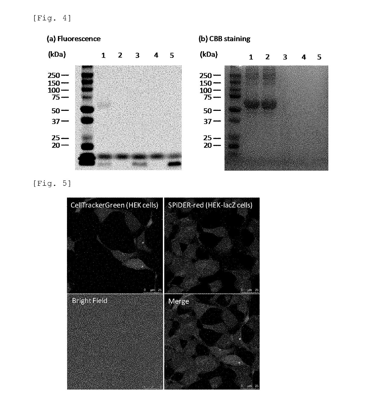
[0153]Using 4-CH2F-SPiDER-RED-βGal and 2-CH2F-SPiDER-RED-βGal which are the probe compounds of the present invention obtained in Example 1, we have demonstrated that these compounds cleaved by β-galactosidase fluorescently label bovine serum albumin (BSA) protein coexisting therewith in a solution. The measurement results of 4-CH2F-SPiDER-RED-βGal are shown in FIG. 3, and the measurement results of 2-CH2F-SPiDER-RED-βGal are shown in FIG. 4.

[0154]Using 1) 10 μL of a PBS buffer solution containing 10 μM 4-CH2F-SPiDER-RED-1Gal, 1 mg / mL BSA, and 5U β-galactosidase, 2) 10 μL of a PBS buffer solution containing 10 μM 4-CH2F-SPiDER-RED-βGal and 1 mg / mL BSA, 3) 10 μL of a PBS buffer solution containing 10 μM 4-CH2F-SPiDER-RED-βGal and 5U β-galactosidase, 4) 10 μL of a PBS buffer solution containing only 10 μM 4-CH2F-SPiDER-RED-βGal, and 5) 10 μL of a PBS buffer solution containing...


PUM

PropertyMeasurementUnit
Fluorescenceaaaaaaaaaa

Abstract

It is an object of the present invention to provide a fluorescence imaging probe capable of selectively visualizing target cells such as cells expressing β-galactosidase (lacZ expressing cells) at a single-cell level in a red fluorescence region, and of performing co-staining together with GFP.An intracellularly-retainable red fluorescent probe comprising a compound represented by the following formula (I) or a salt thereof:wherein: A represents a monovalent group cleaved by an enzyme; R1 representsa hydrogen atom, or one to four of the same or different substituents bonded to a benzene ring; R3, R4, R5, and R6 each independently represent —CFR10R11, —CF2R12, a hydrogen atom, a hydroxyl group, an alkyl group, or a halogen atom, wherein at least one of R3, R4, R5, and R6 is —CFR10R11 or —CF2R12;R2 and R7 each independently represent a hydrogen atom, a hydroxyl group, an alkyl group, or a halogen atom; R8 and R9 each independently represent a hydrogen atom or an alkyl group; R10, R11, and R12 each independently represent a hydrogen atom, an alkyl group, or an alkenyl group; X represents Si(Ra) (Rb), wherein Ra and Rb each independently represent a hydrogen atom or an alkyl group; and Y is —C(═O)— or —RcC(═O)—, wherein Rc is an alkylene group having 1-3 carbon atoms.

Description

TECHNICAL FIELD[0001]The present invention relates to a novel red fluorescent probe which can be retained in a target cell and can act specifically in the cell, a method for specifically imaging a target cell expressing a specific enzyme using the fluorescent probe, and a detection kit including the probe.BACKGROUND ART[0002]Reporter proteins have made an immeasurable contribution to the development of life sciences. The most commonly used reporter protein is β-galactosidase (lacZ). A relationship between aging and expression of β-galactosidase in cells has recently been suggested (see Non-Patent Literature 1), and imaging probes which are enzyme-specific to β-galactosidase are important molecular tools for elucidating mechanisms of cell aging. Furthermore, β-galactosidase activity has been shown to be elevated in certain types of cancer cells (see Non-Patent Literatures 2 and 3), and an imaging probe which is enzyme-specific to β-galactosidase is thought to be usable also as a canc...

Claims


Application Information

Patent Timeline
IPC IPC(8): C07H17/04C12Q1/34G01N33/58
CPCC07H17/04C12Q1/34G01N33/582C12Q2563/107C07H17/00C09K11/06C12Q1/54C12Q1/42C12Q1/28C12Q1/66G01N33/52G01N33/58G01N33/574
Inventor URANO, YASUTERUKAMIYA, MAKOITO, HIROKIKAWAMATA, YU
Owner THE UNIV OF TOKYO
Learn More