Single-impulse panoramic photoacoustic computed tomography (sip-pact)

a computed tomography and single-impulse technology, applied in the field of single-impulse panoramic photoacoustic computed tomography, can solve the problems of large limitations, long data acquisition time, and high cost of high-strength magnetic fields and other problems, to achieve the effect of high spatiotemporal resolution and high spatial resolution

Active Publication Date: 2019-10-10
WASHINGTON UNIV IN SAINT LOUIS
View PDF0 Cites 15 Cited by
  • Summary
  • Abstract
  • Description
  • Claims
  • Application Information

AI Technical Summary

Benefits of technology

[0007]The ability to directly visualize dynamics of internal structures with high spatiotemporal resolution in various small-animal models at the whole-body scale may provide insights into biolog...

Problems solved by technology

Although these techniques provide deep penetration, they suffer from significant limitations.
For example, adapting MRI to achieve microscopic resolution requires costly high-strength magnetic fields and long data acquisition time, ranging from seconds to minutes; this data acquisition time is too slow for imaging dynamics.
X-ray CT can achieve microscopic resolution, but lacks functional contrast.
UST does not provide cannot image blood oxygenation or extravascular molecular contrasts.
However, the application of conventional optical imaging technologies to small-animal whole-body imaging is impeded by the strong optical scattering of tissue, which prevents high-resolution imaging beyond the optical diffusion limit of about one mm in depth.
Although existing diffusive ...

Method used


Image

  • Single-impulse panoramic photoacoustic computed tomography (sip-pact)
  • Single-impulse panoramic photoacoustic computed tomography (sip-pact)
  • Single-impulse panoramic photoacoustic computed tomography (sip-pact)

Examples


example 1

Body PACT Using SIP-PACT System

[0245]To demonstrate 3D whole body photoacoustic computed tomographic (PACT) imaging using the SIP-PACT system and methods described herein, the following experiments were conducted.

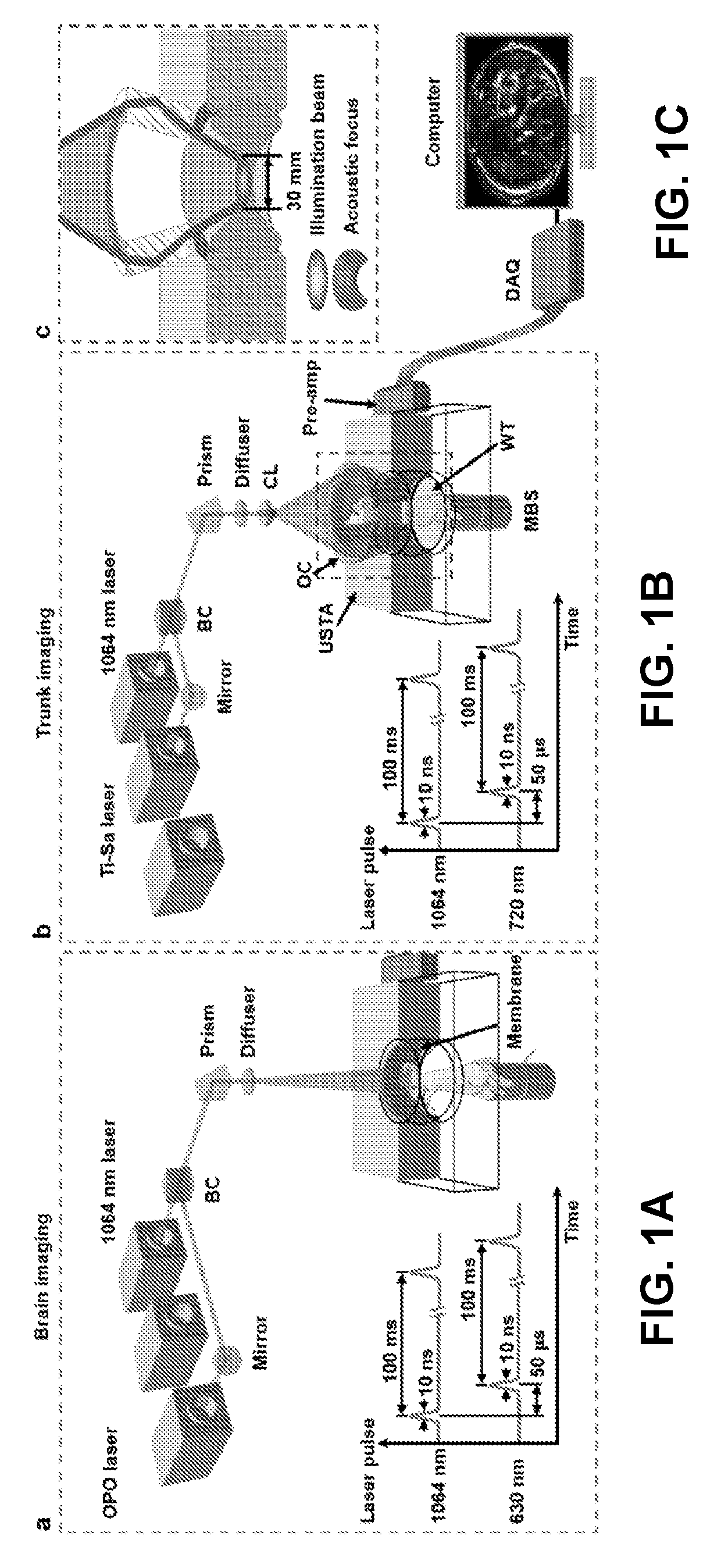
[0246]Adult, 8-10-week old nude mice (Hsd:Athymic Nude-FoxlNU, Harlan Co.; 20-30 g body weight) were used for whole body imaging in vivo experiments. Throughout the experiment, each mouse was maintained under anesthesia with 1.5% vaporized isoflurane. For brain imaging, the mouse was secured to a lab-made imaging platform (see FIG. 25), and the cortical surface was positioned in alignment with the ring transducer array's focal plane. During the whole body imaging experiments, the mouse's fore and hind legs were respectively taped to the top and bottom parts of the lab-made holder that held the animal upright during imaging. The top of the holder included an aluminum tube affixed to the animal's nose and mouth, and the bottom of the holder included an aluminum cylinder attac...

example 2

esolved Imaging of Cardiac and Respiratory Cycles Using SIP-PACT System

[0250]To demonstrate 2D whole body time-resolved imaging of cardiac and respiratory cycles using the SIP-PACT system and methods described herein, the following experiments were conducted.

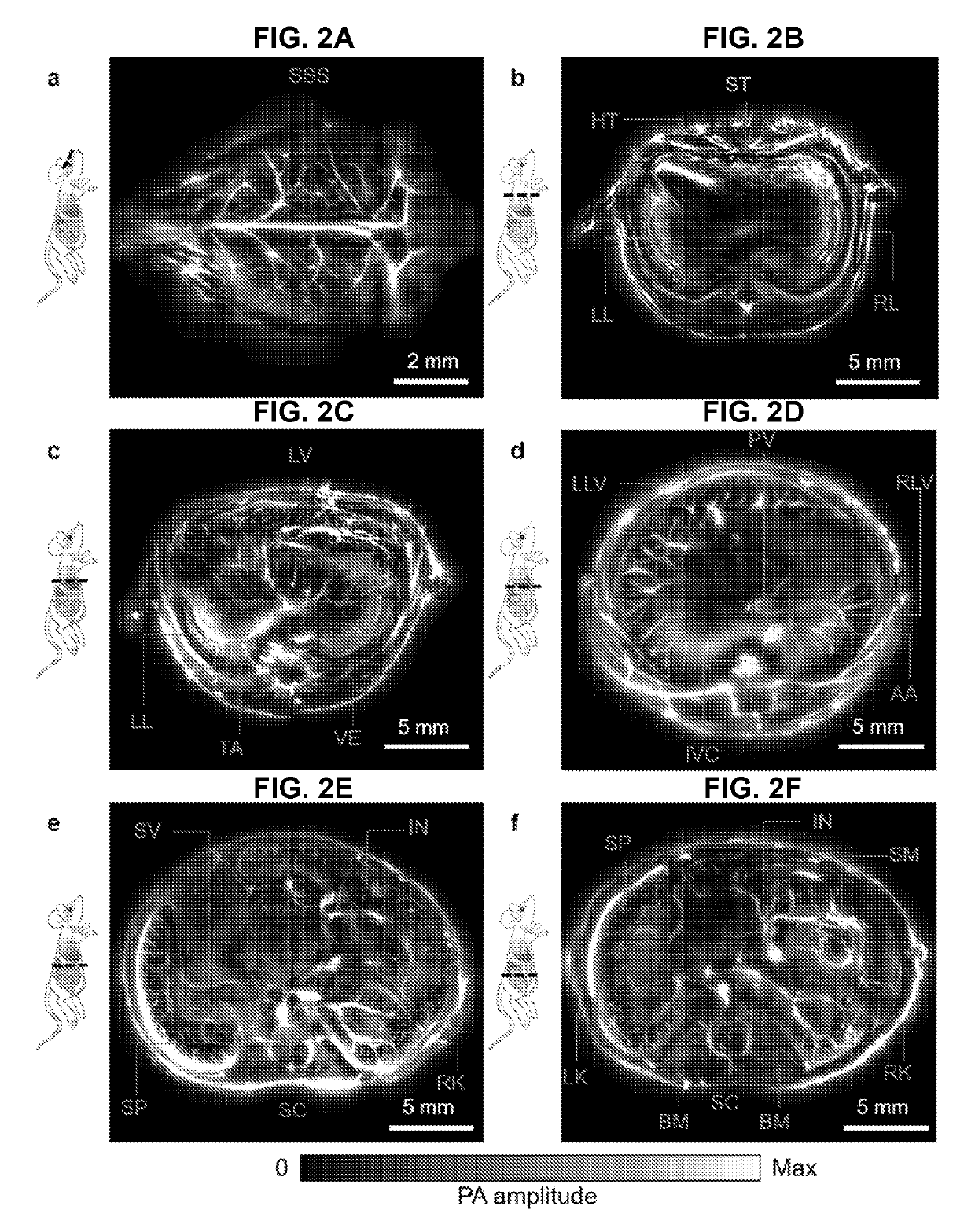
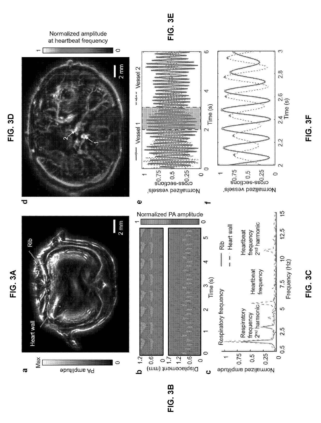
[0251]Nude mice were prepared and mounted in the SIP-PACT system illustrated in FIG. 1B using a protocol similar to the protocol described in Ex. 1. The mouse was positioned in the SIP-PACT system so that the focal plane of the ring transducer array passed through the heart of the patient, resulting in a PA image similar to the PA image shown in FIG. 2B. The SIP-PACT system was operated in a manner similar to Ex. 1 using full-ring side illumination and side detection with 1064 nm laser pulses delivered at a 50 Hz repetition rate to the same transverse plane of the mouse's thoracic cavity to obtain a time-series of images.

[0252]FIG. 3A is a representative image of the transverse slice through the thoracic cavity of the mouse. At ...

example 3

l Imaging Using SIP-PACT System

[0257]To demonstrate functional imaging using the SIP-PACT system and methods described herein, the following experiments were conducted.

[0258]Adult, 3-4-month-old Swiss Webster mice (Hsd: ND4, Swiss Webster, Harlan Co.; 20-30 g body weight) were used for in vivo functional brain and CTC imaging. Prior to brain functional imaging using the SIP-PACT system, the hair of each mouse was removed by clippers and depilatory cream. Each mouse was then secured to a lab-made imaging platform, and the brain's cortical surface was positioned aligned with the transducer array's focal plane as described in Ex. 1. Each mouse breathed an inhalation gas containing varying concentrations of oxygen to systemically modulate the oxygen saturation of hemoglobin (sO2) within the mice during functional SIP-PACT imaging, as described in detail below.

[0259]A SIP-PACT system similar to the system described in Ex. 1 (see FIG. 1A) was used to obtain time series PA images of the mo...


PUM


Abstract

A single-impulse panoramic photoacoustic computed tomography (SIP-PACT) system for small-animal whole-body imaging is disclosed. In addition, a dual-speed of sound image universal back-projection reconstruction method is disclosed. Further, a PACT system for imaging a breast of a subject is disclosed.

Description

CROSS-REFERENCE TO RELATED APPLICATION[0001]This application claims the benefit of U.S. Provisional Application No. 62 / 427,470 filed Nov. 29, 2016, which is incorporated herein in its entirety.STATEMENT REGARDING FEDERALLY SPONSORED RESEARCH & DEVELOPMENT[0002]This invention was made with government support under grant EBO16963 awarded by National Institutes of Health. The government has certain rights in the invention.BACKGROUND OF THE INVENTION[0003]The field of this disclosure relates generally to a photoacoustic tomography imaging system used to capture whole-body images of small animals with high frame rate and high resolution.[0004]Small animals, in particular rodents, are essential models for preclinical studies, and they play an important role in modeling human physiology and development, in guiding the study of human diseases and in advancing the quest for effective treatments. The ability to directly visualize dynamics with high spatiotemporal resolution in these small-ani...

Claims


Application Information

Patent Timeline
IPC IPC(8): A61B5/00G01N29/24G06T11/00
CPCG06T2211/421A61B5/0095G06T11/006G01N29/2418G01N2291/02475A61B5/0091G01N29/06G01N29/0672G01N21/1702G01N21/4795A61B5/721A61B5/024A61B5/113A61B2562/0247A61B5/4312A61B5/318
Inventor WANG, LIHONGLI, LEIZHU, LIRENMA, CHENG
Owner WASHINGTON UNIV IN SAINT LOUIS
Why Eureka
Try Eureka