
Compounds as fluorescent probes, synthesis and applications thereof

a fluorescent probe and compound technology, applied in the field of chemical sciences and imaging technology, can solve the problems of uneven staining, unsuitable existing dyes based on rhodamine scaffolds, and inoptimal staining of sensitive cells like primary cells and stem cells

Active Publication Date: 2019-12-12
INST FOR STEM CELL BIOLOGY & REGENERATIVE MEDICINE INSTEM
View PDF0 Cites 11 Cited by
  • Summary
  • Abstract
  • Description
  • Claims
  • Application Information

AI Technical Summary

Benefits of technology

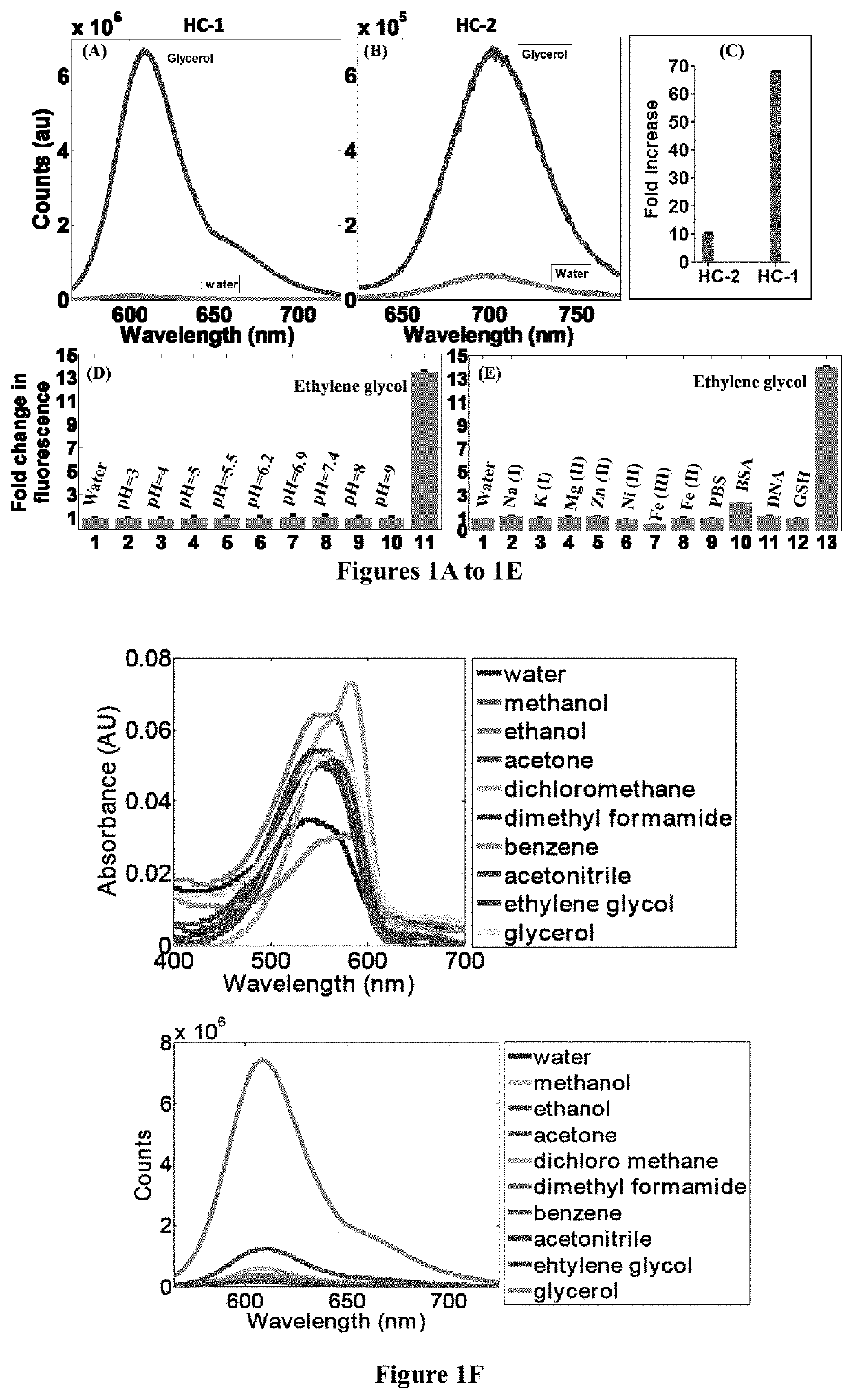
The present patent text describes a new compound that can be used as a fluorescent probe to stain and image mitochondria in eukaryotic cells, tissues, or animals. The compound has a specific structure and contains different chemical moieties that can attach to the outer membrane of mitochondria and make them visible under a microscope or in image analysis. This new compound can help researchers better understand the function, order, and dynamics of mitochondria, which are important in cellular metabolism and survival.

Problems solved by technology

Though the existing dyes are widely used for staining mitochondria or for measuring mitochondrial potential, they have several disadvantages including the following:(i) the existing dyes are not optimal for staining sensitive cells like primary cells and stem cells, including embryonic stem cells and induced pluripotent stem cells due to their toxicity and photo-induced damage that they cause.
Further, it is known in the art that existing dyes based on Rhodamine scaffold are unsuitable for uniform staining of embryonic stem cell and induced pluripotent stem cells since they are exported out of the cell.
This results in uneven staining and under sampling of the mitochondria,(ii) the existing dyes are unsuitable for imaging mitochondria in live cells where functionality is a major concern since these dyes perturb the mitochondrial function,(iii) further, the existing dyes are unsuitable for fast tracking of the mitochondria, due to mitochondrial damage caused by repeated light exposure when these dyes are used.
This segregation and movement is linked to the cell state, energetics and function but currently there is no method to obtain this information using cell permeable dyes.
There is now growing evidence that understanding mitochondrial dynamics is linked to disease pathologies,(iv) furthermore, tracking of mitochondria in stem cells with existing dyes causes differentiation of stem cells through reactive oxygen species (ROS) generation, making them unsuitable for repeated and rapid imaging of stem cells.
While mitochondrial potential is important, it does not fully describe the mitochondrial state and function.
These mitochondrial parameters are critical in multiple disease pathologies but existing dyes are inadequate to describe these features.
In several cases, mitochondrial potential is insufficient to distinguish different mitochondrial states.
Long wavelength probes reduce imaging noise and allow deep tissue imaging, but often require complex synthesis.

Method used


Image

  • Compounds as fluorescent probes, synthesis and applications thereof
  • Compounds as fluorescent probes, synthesis and applications thereof
  • Compounds as fluorescent probes, synthesis and applications thereof

Examples


example 1

General Synthetic Procedure for Preparing Compound of Formula-I:

[0144]A mixture of compound of Formula-III (5.00 mmol), and substituted / unsubstituted aldehyde derivative (5.00 mmol) was added in presence of 10 ml anhydrous ethanol. 0.5 ml of piperidine was added to this mixture and the resultant reaction mixture was refluxed for about 2 to 4 hours followed by cooling to obtain the precipitate. The precipitated solid was filtered and then purified by column chromatography using DCM / Methanol and the eluent yielded compound of Formula I.

wherein[0145]‘R4’, ‘R5’ and ‘R6’ are individually selected from a group consisting of hydrogen, alkoxy and alkyl; and[0146]‘X’ is selected from a group consisting of I, Cl, Br and F.

example 2

Synthesis of HC-1

(E)-2-(4-(Dimethylamino)styryl)-1,1,3-trimethyl-1H-benzo [e]indol-3-ium iodide (HC-1)

[0147]

[0148]A mixture of 1,1,2-trimethyl-1H-benzo[e]indolium iodide (5.00 mmol), 4-(dimethylamino)benzaldehyde (5.00 mmol) was added with 10 ml anhydride ethanol, and to this mixture 0.5 ml of piperidine was added and was refluxed for about 3 hours. After cooling to room temperature, the red solid was precipitated. The precipitated red solid was filtered and then purified by column chromatography using DCM / Methanol as the eluent yielded brick red solid HC-1. Mass Spectroscopy (MS), proton and C13 nuclear magnetic resonance (NMR) spectroscopy of HC-1 are provided in FIGS. 16, 18 and 19, respectively.

example 3

Synthesis of HC-2

2-((1E,3E)-4-(4-(Dimethylamino)phenyl)buta-1,3-dien-1-yl)-1,1,3-trimethyl-1H-benzo[e]indol-3-ium iodide

[0149]

[0150]A mixture of 1,1,2-trimethyl-1H-benzo[e]indolium iodide (5.00 mmol), 4-(dimethylamino)cinnamaldehyde (5.00 mmol) was added with 10 ml anhydride ethanol, and to this mixture 0.5 ml of piperidine was added and was refluxed for 3 hours. After cooling to room temperature, the dark blue solid was precipitated. The precipitated dark blue solid was filtered and then purified by column chromatography using DCM / Methanol as the eluent yielded dark blue solid HC-2. Mass Spectroscopy (MS), proton and C13 nuclear magnetic resonance (NMR) spectroscopy of HC-2 are provided in FIGS. 17, 20 and 21, respectively.


PUM


Abstract

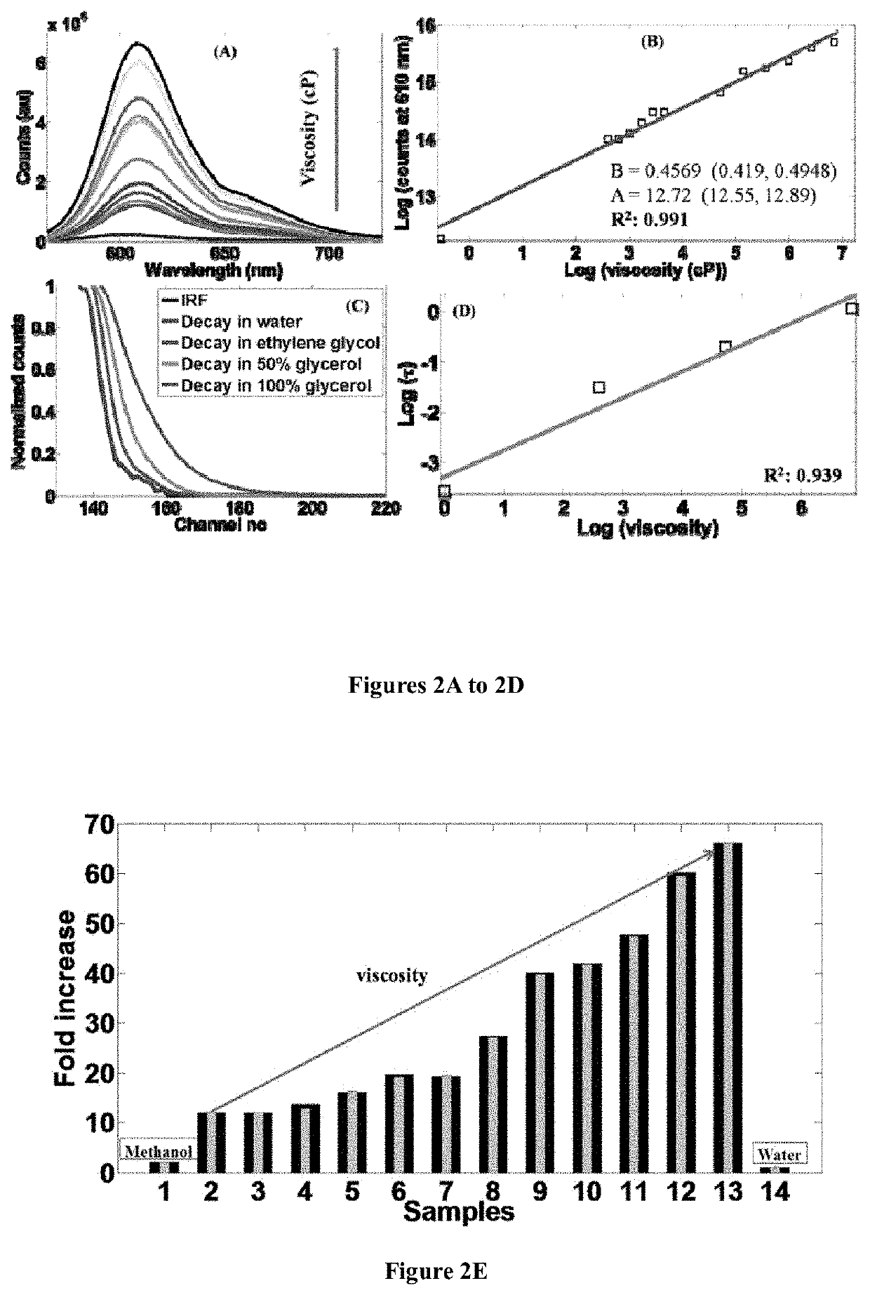
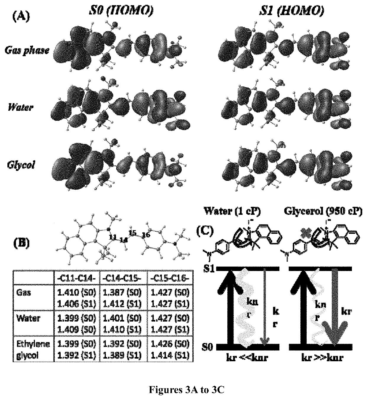
The present disclosure relates to chemical dyes useful for staining and imaging of cells. In particular, the disclosure relates to compound of Formula I, method of preparation thereof, and it's use as a fluorescent probe for staining and / or imaging mitochondria in cells, tissues or animals, resulting in a range of applications including, but not limiting, to sensing local ordering or viscosity of mitochondria, tracking mitochondrial mobility, comparing & evaluating mitochondrial function, local ordering, microviscosity and dynamics. Said dyes have additional advantages including, but not limiting, to low toxicity, longer shelf-life, generate little or no reactive species upon long term light irradiation and do not perturb the functionality of the mitochondria in cells compared to prior art dyes.

Description

CROSS REFERENCE TO RELATED APPLICATION[0001]This application claims priority to and benefit of Indian Complete Application No. 201841021840, filed Jun. 11, 2018, the disclosure of which is hereby incorporated herein by reference in their entirety.FIELD OF THE INVENTION[0002]The present disclosure is in the field of chemical sciences and imaging technology. The present disclosure generally relates to chemical dyes useful for staining and imaging of cells. In particular, the disclosure relates to Formula I compounds, method of preparing thereof, and their application as fluorescent probes for staining and / or imaging mitochondria in cells, tissues or animals. Said fluorescent probes also have applications in sensing local ordering or viscosity of mitochondria, tracking mitochondrial mobility, comparing & evaluating mitochondrial function, local ordering and dynamics. The dyes have low toxicity, longer shelf-life, generate little or no reactive species upon long term and repeated light ...

Claims


Application Information

Patent Timeline
IPC IPC(8): C09B23/02C09B23/01G01N33/58
CPCC09B23/0066G01N33/582C09B23/02C09B23/105C09B23/107G01N33/5076
Inventor GULYANI, AKASHRAJA, SUFI OASIMSIVARAMAN, GANDHI
Owner INST FOR STEM CELL BIOLOGY & REGENERATIVE MEDICINE INSTEM
Learn More