
Biomarkers for photoreceptor cells

a technology of photoreceptor cells and biomarkers, which is applied in the field of identification of photoreceptors or cone photoreceptors in cell populations, can solve the problems of slowing down disease progression, not cure the condition or reverse the effect, and affecting the use of the condition, etc., and achieves the effects of good manufacturing practice compatibility, wide applicability and ease of us

Pending Publication Date: 2020-02-27
UCL BUSINESS PLC
View PDF0 Cites 2 Cited by
  • Summary
  • Abstract
  • Description
  • Claims
  • Application Information

AI Technical Summary

Benefits of technology

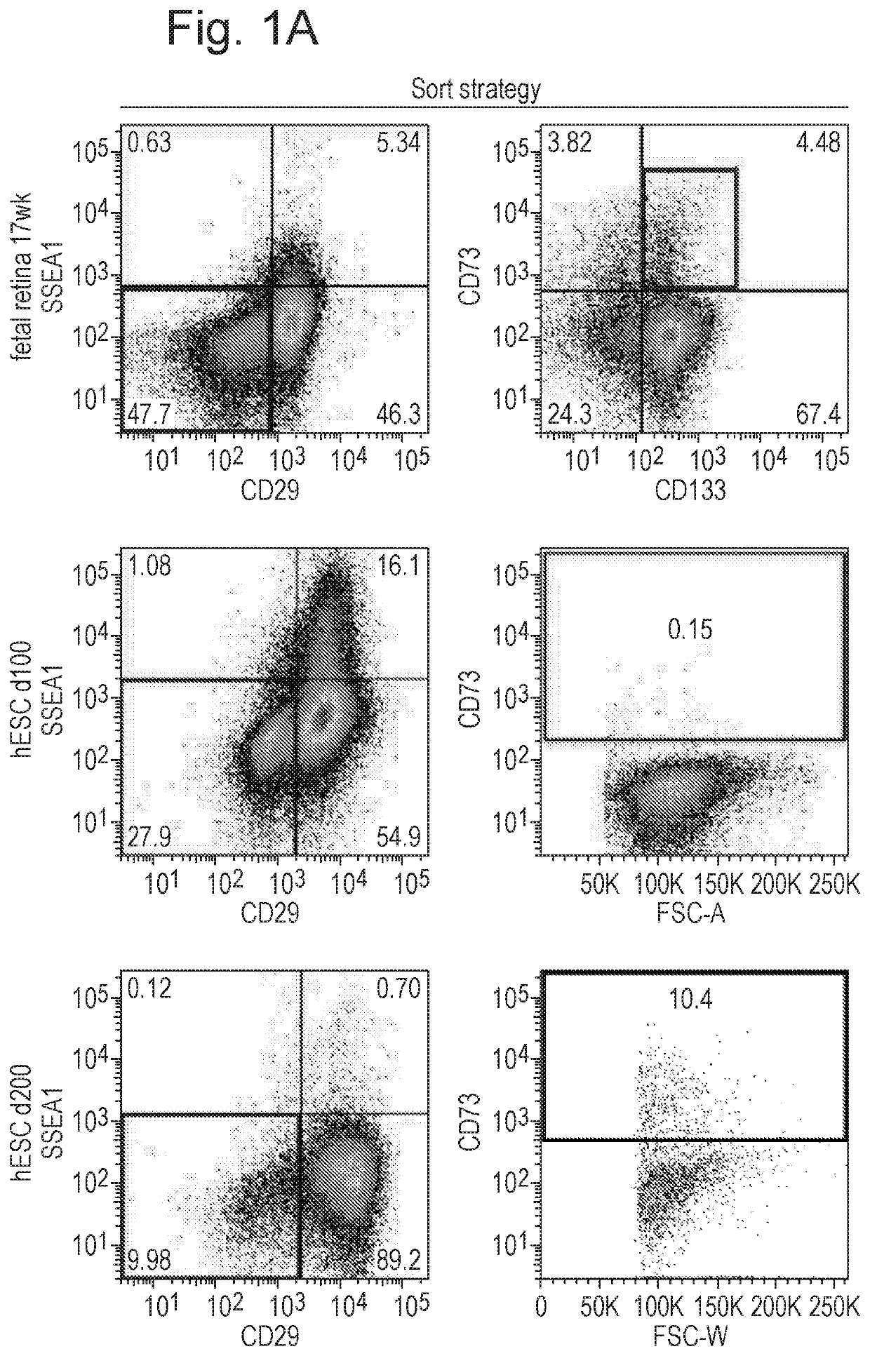
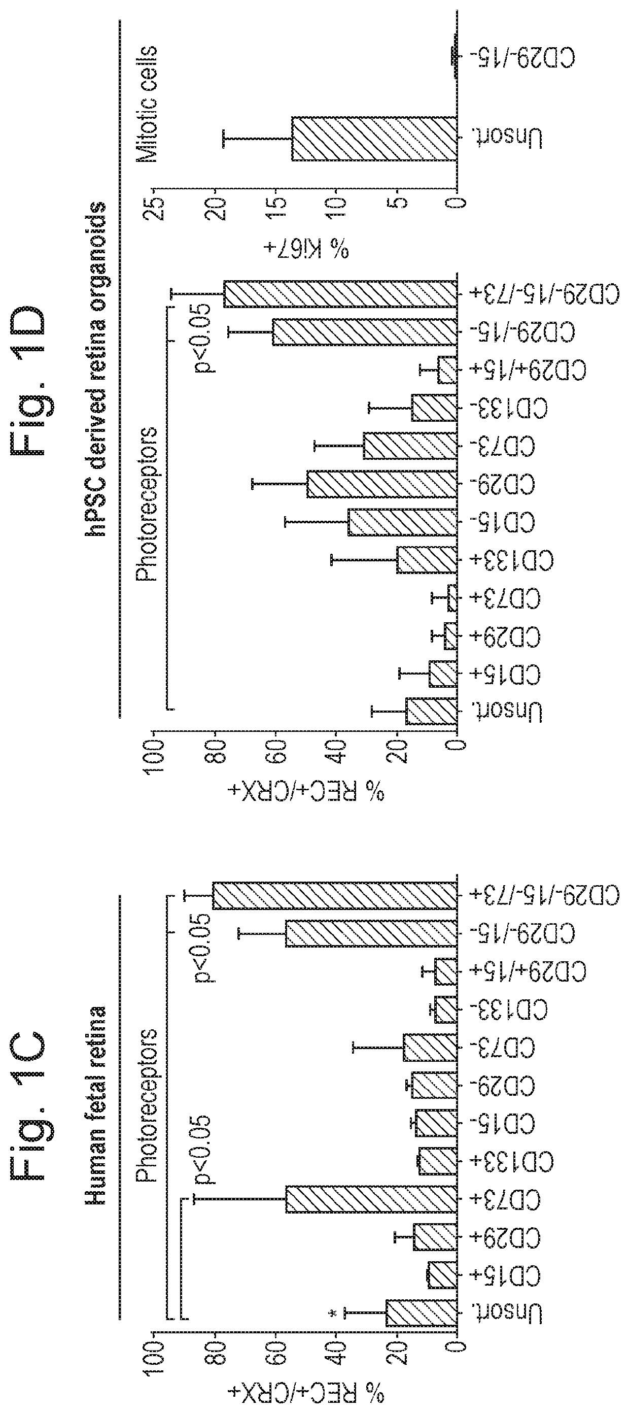
The surface biomarker purification approaches described in this patent are useful across different cell production platforms, and are compatible with industry standards for good manufacturing practice. These methods rely on naturally present biomarkers on the surface of cells, which means that they do not require any genetic modification of the cells.

Problems solved by technology

As the mammalian nervous system has limited regenerative capacity, any disease or injury to retinal photoreceptors leads inevitably to visual impairment or even blindness.
However, current therapies can at best slow down the disease progression but not cure the condition or reverse its effects.
This is mainly due to the fact that the details of retinal histogenesis, maintenance and pathology are not sufficiently well understood, for the development of effective therapies.
However, many critical challenges exist that prevent the development of cell replacement therapies to replace photoreceptors or cone photoreceptors, including challenges in developing the strategies required for the identification and purification of photoreceptors or cone photoreceptors to allow for cell therapy or transplantation into the eye.
Furthermore, as donor cells are always produced alongside other cell populations, some with potentially detrimental properties, the successful translation of this approach to the clinic is dependent on the development of stringent cell selection and purification methods.
For example, while inclusion in donor cell preparations of non-photoreceptors may prevent or hinder establishment of connectivity to host retinal circuitry, presence of mitotically active cell types poses the risk of tumor formation in the host retina after transplantation.
There are currently no photoreceptor or cone photoreceptor purification methods available that would be suitable for human use, as the current methods require genetic manipulation in order to isolate the required cells.
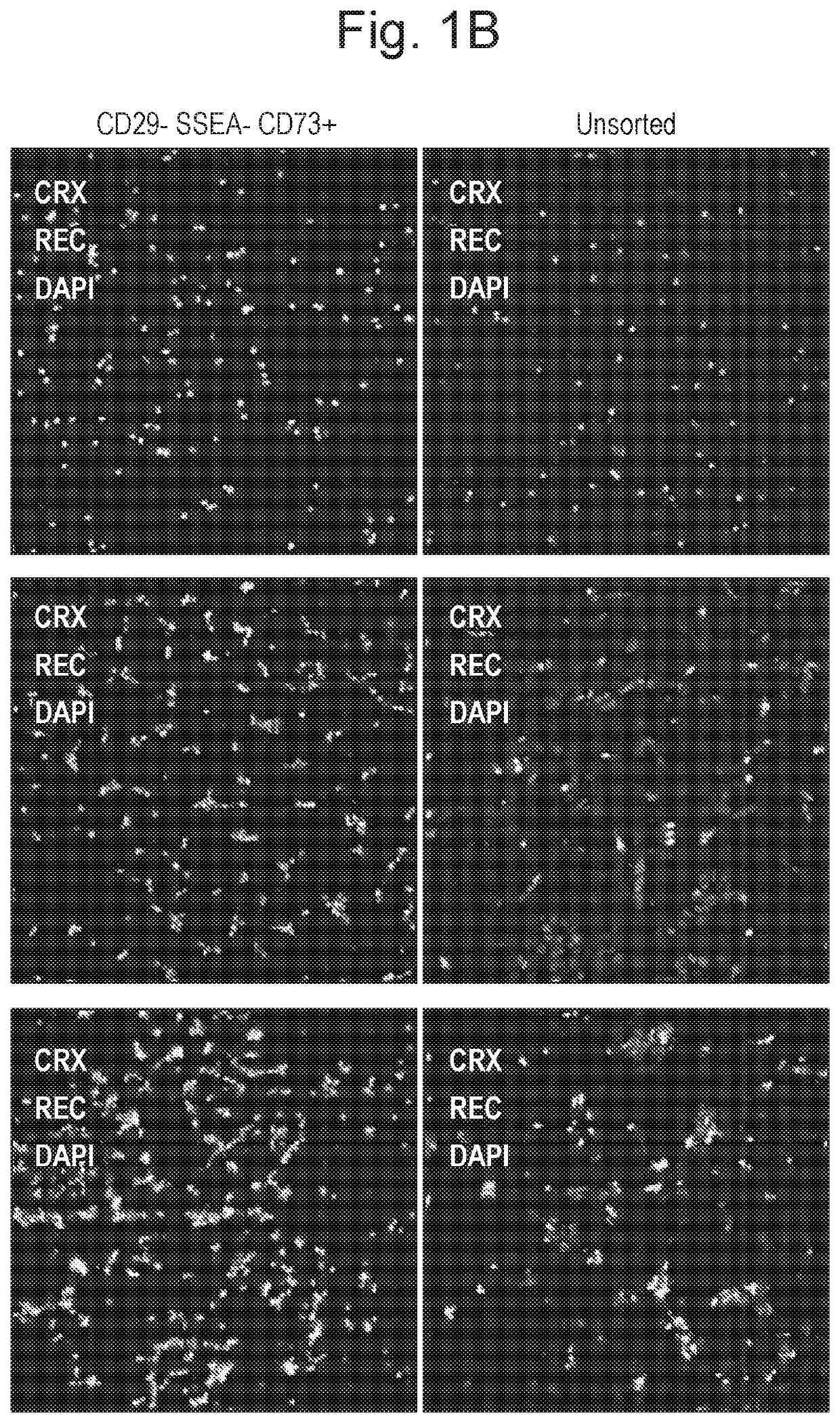
However, subsequent experiments with human retinal tissue showed that the combination of markers developed in the mouse model was not directly translatable into the human system, due on the one hand to differences in expression of cell surface antigens between human and mouse retina, and on the other hand to the more ubiquitous expression of biomarkers such as CD73 in the heterogeneous human stem cell differentiation cultures, compared with isolated human foetal retina.

Method used


Image

  • Biomarkers for photoreceptor cells
  • Biomarkers for photoreceptor cells
  • Biomarkers for photoreceptor cells

Examples


example 1

[0067]Photoreceptor Identification

[0068]Methods

[0069]Animals

[0070]Experimental mice were kept in University College London animal facilities and all experiments were conducted in agreement with the Animals (Scientific Procedures) Act 1986 and the Association for Research in Vision and Ophthalmology Statement for the Use of Animals in Ophthalmic and Vision Research. C57Bl / 6J, and C3H / HeNCrl (RD1; Pde6brdl / rdl) recipient mice at 3 weeks of age at the time of transplantation were obtained from Charles Rivers laboratory.

[0071]Human Pluripotent Stem Cell Culture

[0072]All pluripotent stem cells were maintained as previously described for hESC cultures. Human embryonic stem cells and induced pluripotent cells were either cultured on a feeder layer of irradiated mouse embryonic fibroblasts (MEFs) in embryonic stem cell medium (DMEM / F12 (1:1), 20% knockout serum replacement, 0.1 mM mercaptoethanol, 1 mM L-glutamine, MEM nonessential amino acids, and 4 ng / mL FGF2) or under feeder-free conditi...

example 2

[0106]Cone Photoreceptor Identification

[0107]Methods

[0108]Human Foetal Tissue Preparation

[0109]Human foetal eyes were obtained from the Wellcome Trust and Medical Research Council Funded Human Developmental Biology Resource (http: / / www.hdbr.org / ). Human adult eyes were obtained from Moorfields BioBank.

[0110]Human Foetal Retinal Explant Cultures

[0111]Human foetal eyes were dissected in sterile conditions and all ocular tissue was removed in order to obtain retina. Intact human foetal retina were cultured free floating in 12 well plates with retinal differentiation media (RDM) containing DMEM-F12, Glutamax, 1×N2, 1×B27 neural supplements (Invitrogen), 10% FBS (Invitrogen) and 1.5× penicillin / streptomycin (Invitrogen). Cell culture media was changed every 2 days.

[0112]iPSC Maintenance and Retinal Differentiation

[0113]Human iPSCs were cultured in 6 well plates on gelatin with irradiated mouse embryonic fibroblast layer (IRR MEFs; GlobalStem; 150,000 IRR MEFs per well) and passaged using...


PUM

PropertyMeasurementUnit
temperatureaaaaaaaaaa
thickaaaaaaaaaa
temperatureaaaaaaaaaa

Abstract

The present invention relates to the identification of photoreceptors or cone photoreceptors in populations of cells. In particular the present invention relates to methods of identification of photoreceptors or cone photoreceptors and methods of isolating photoreceptors or cone photoreceptors. Photoreceptors or cone photoreceptors isolated by the methods of the present invention are useful for transplantation and the treatment of retinal dystrophies. Also claimed are human cell populations enriched for photoreceptors or cone photoreceptors and kits for identifying photoreceptors or cone photoreceptors.

Description

FIELD OF THE INVENTION[0001]The present invention relates to the identification of photoreceptors or cone photoreceptors in populations of cells. In particular the present invention relates to methods of identification of photoreceptors or cone photoreceptors and methods of isolating photoreceptors or cone photoreceptors. Photoreceptors or cone photoreceptors isolated by the methods of the present invention are useful for transplantation and the treatment of retinal dystrophies. Also claimed are human cell populations enriched for photoreceptors or cone photoreceptors and kits for identifying photoreceptors or cone photoreceptors.BACKGROUND OF THE INVENTION[0002]Rod and cone photoreceptors, which are located in the outer nuclear layer within the retina, are the primary sensory cells of the mammalian visual system. Stimulation by light results in the initiation of the visual transduction cascade, whose information is relayed to bipolar cells, processed by horizontal and amacrine inte...

Claims


Application Information

Patent Timeline
Patent Type & Authority Applications(United States)
IPC IPC(8): C12N5/0793G01N33/569G01N33/68A61K35/30A61K9/00
CPCC12N5/062G01N33/56966G01N33/68A61K9/0048A61K35/30C12N2506/02C12N2506/45C12N2501/58C12N2501/599
Inventor SOWDEN, JANELAKOWSKI, JORNALI, ROBINWELBY, EMILYCORDERO, ANAI GONZALEZ
Owner UCL BUSINESS PLC
Learn More