

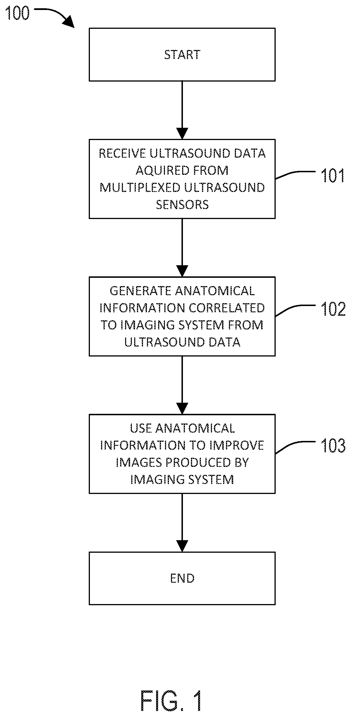
Systems and Methods for Controlling Imaging Artifacts Using an Array of Sensor Data
an array and sensor data technology, applied in the field of systems and methods for controlling imaging artifacts using an array of sensor data, can solve the problems of artifacts in the resulting image, motion-related problems that plague other imaging modalities, hardware systems, etc., and achieve the effects of reducing image artifacts, improving image quality, and high temporal resolution
- Summary
- Abstract
- Description
- Claims
- Application Information
AI Technical Summary
Benefits of technology
Problems solved by technology
Method used
Image

Examples
example 1
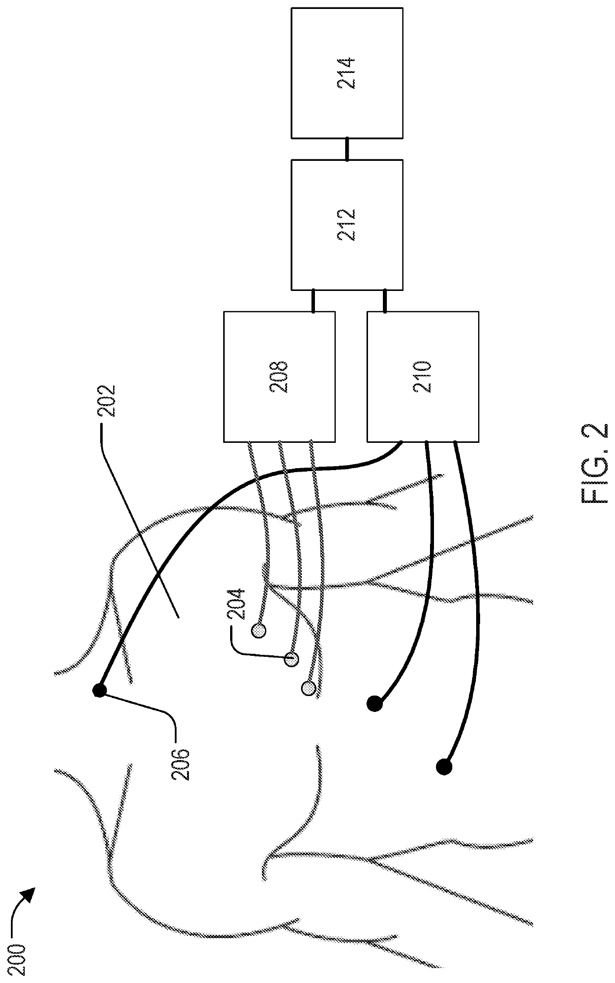
[0055]A study was conducted to investigate the use of a custom 1 MHz single-element ultrasound (US) transducer, referred to here as an organ-configuration motion (OCM) sensor, for echocardiography-based cardiac MR-triggering. The sensor was placed over the heart, and the resulting signal was processed to generate triggers at the end of atrial systole, at the onset of isovolumic contraction. The device is MR compatible and can replace ECG gating when the latter proves impractical, for the purpose of cardiac MRI.
[0056]As shown in FIGS. 6A-E, the sensor is assembled from a 3D printed capsule of our own design, an MR-compatible 1 MHz US transducer, two-way tape, ultrasound gel and a membrane mostly transparent to ultrasound energy. Application to the skin can be done very easily: one peels the protective layer of the tape and applies the sensor onto the skin. Making the OCM sensors as easy to use as possible is important, as it may prove crucial to future clinical adoption. Our OCM sens...
example 2
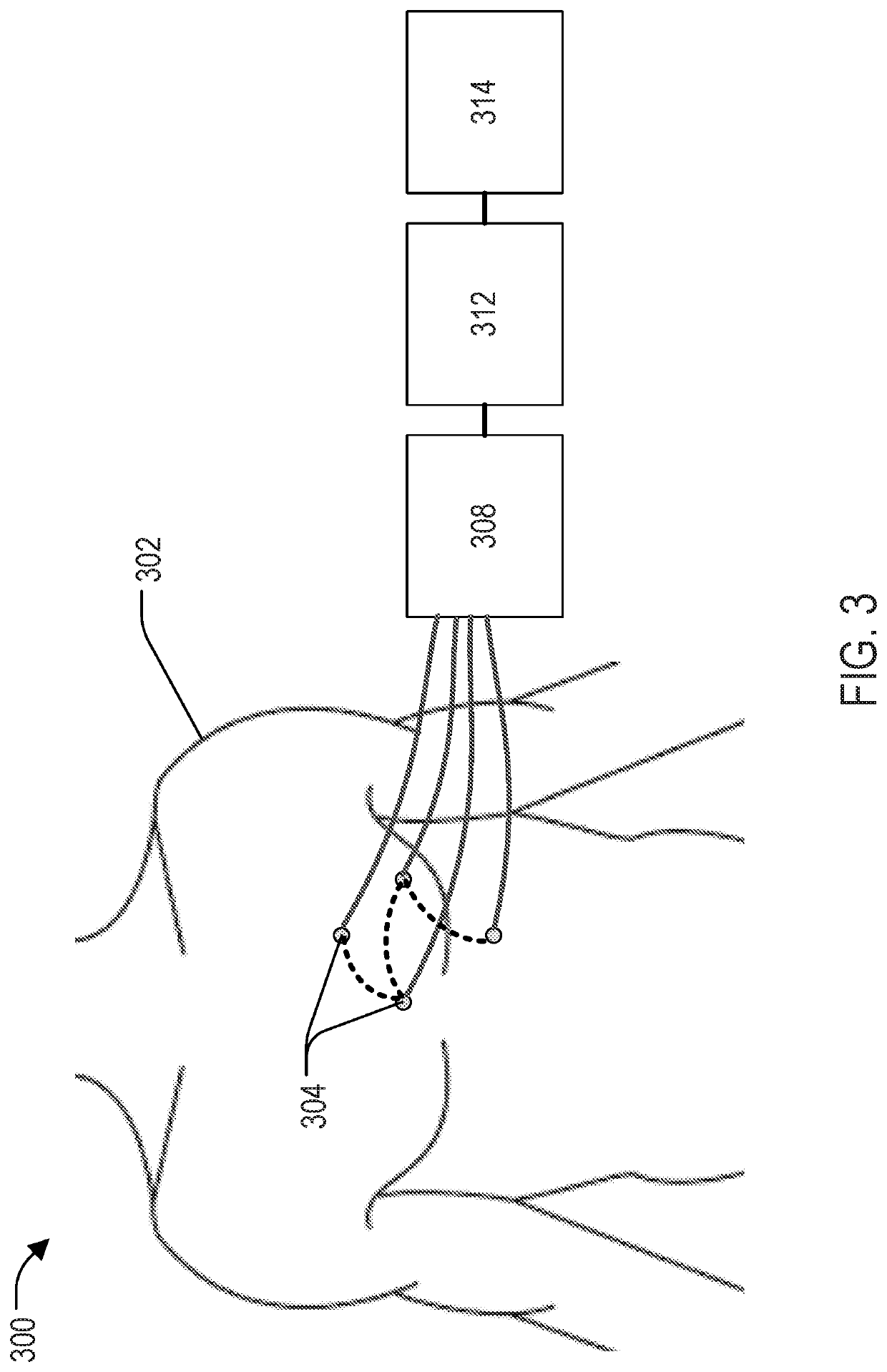
[0060]Simultaneous ultrasound and ECG sensor acquisition was studied. For the ultrasound sensors, a pulser-receiver (5077PR, Olympus) triggered the 1 MHz transducer at a pulse repetition frequency of 100 Hz. The signal was recorded using a standard PC equipped with a digitizer card (5122, National Instruments), acquiring 5000 samples at 20 MS / s, for a maximum depth of 19.25 cm. The ultrasound sensor was attached to the chest, directly over the heart. Three ECG leads were placed in the standard manner. Simultaneous OCM and ECG data were acquired, with the volunteer outside and then inside the 3T scanner, for 2 minutes each. The R-wave detection algorithm of the scanner, as applied to the minimally-distorted ECG traces acquired outside the bore, produced ground truth for validation purposes. A learning-based algorithm detected each heartbeat as follows. 200 OCM samples were selected around a depth of 16.5 cm. The mean absolute difference between subsequent OCM traces was used as a fea...
PUM
 Abstract
Description
Claims
Application Information