
Method for producing cell flaps

a cell flap and cell technology, applied in the field of cell flap production, can solve the problems of inadequate flap release, achieve superior product performance and biological stability, reduce the risk of microbiological contamination, and reduce the risk of transport breakage

Pending Publication Date: 2020-09-17
HOLOSTEM TERAPIE AVANZATE
View PDF0 Cites 0 Cited by
  • Summary
  • Abstract
  • Description
  • Claims
  • Application Information

AI Technical Summary

Benefits of technology

The patent describes a method for growing keratinocytes on a flap of fibrin, which provides support during transport and detachment from the support. This method prevents premature differentiation of keratinocytes and allows for a high level of proliferation and regenerative potential. The use of fibrin as a support is advantageous because it is compact, solid, and biodegradable, providing flexibility and maneuverability during preparation and transport. Compared to flaps grown on plastic, the fibrin flap performs better and is more stable, with reduced risk of contamination and breakage. The method described in the patent allows for efficient and quick growth of cellular flaps of different cell types without the need for expensive culturing plates.

Problems solved by technology

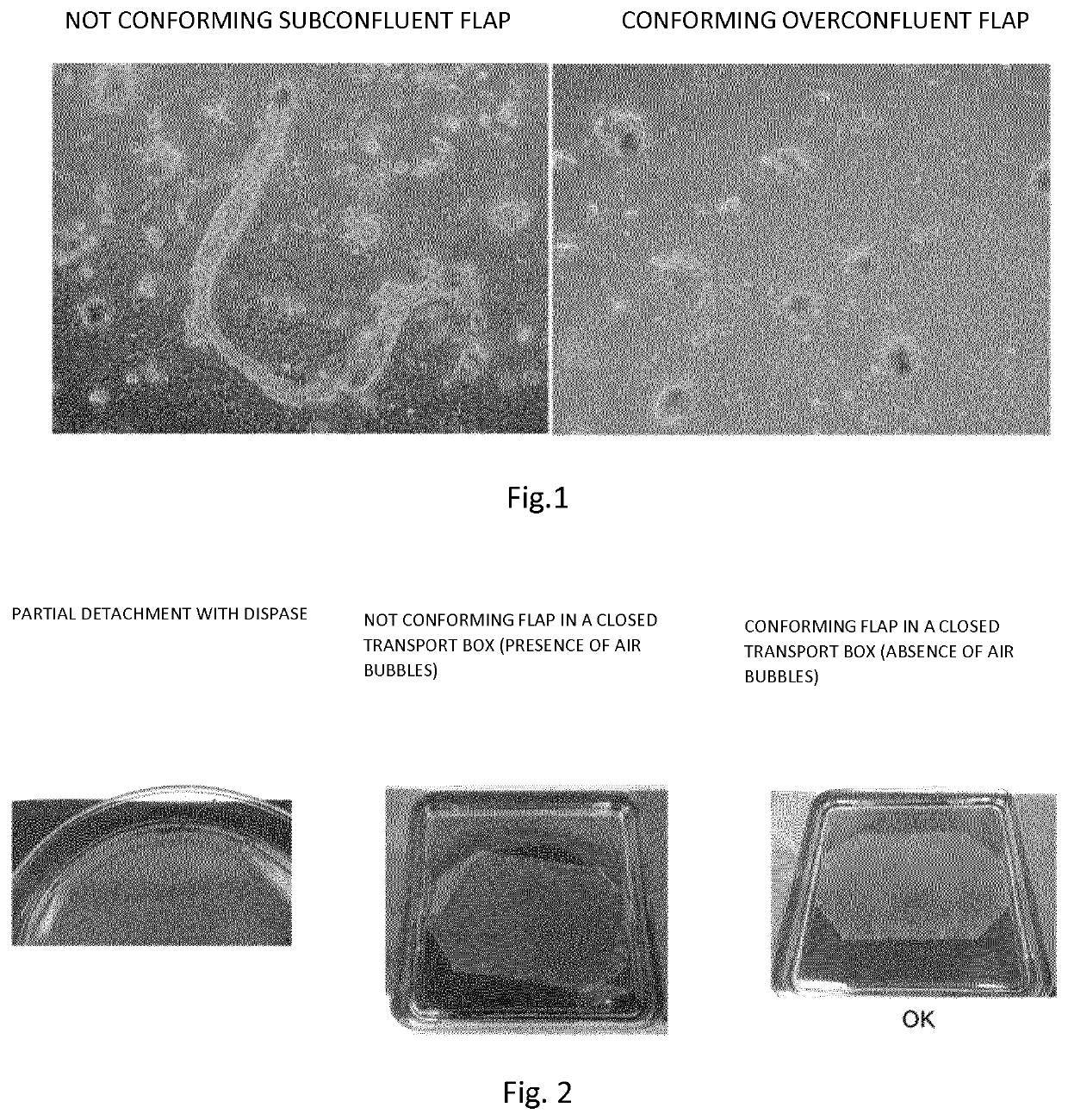
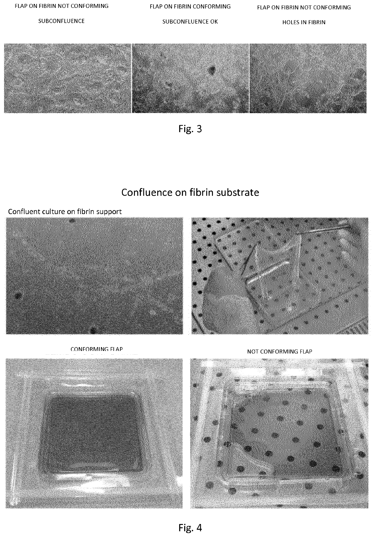
The presence of bubbles would render the flap release not adequate for therapeutic applications, such as transplants (FIG. 4).

Method used


Image

  • Method for producing cell flaps
  • Method for producing cell flaps

Examples


examples

[0122]Materials and Methods

[0123]Isolation of Epidermal Keratinocytes from Biopsy of Human Skin

[0124]Primary human keratinocytes are isolated from 2-9 cm2 skin biopsies after submission and adhesion to informed consent. The biopsy is subjected to enzymatic digestion in Trypsin-EDTA solution at 37° C. To obtain maximum yield and minimize the risk of toxicity from exposure to trypsin-EDTA or prolonged suspension time of keratinocytes during extraction, till 6 sequential trypsinizations of 30′ each are performed. After each trypsinization the cellular material is recovered and the trypsinizations 1-3 and 4-6 are combined and plated in plastic supports according to the cell yield at a density of 1.33×10⁴ cells / cm2 on a feeder layer of lethal irradiated murine cells 3T3-J2 (Rheinwald, J. et al. 1975). The medium used consists of a mixture of Dulbecco's modified Eagle (DMEM) and Ham F12 (2:1) supplemented with 10% fetal bovine serum, 0.5% penicillin-streptomycin, 2% glutamine, insulin (5 ...


PUM

PropertyMeasurementUnit
thickaaaaaaaaaa
temperatureaaaaaaaaaa
timeaaaaaaaaaa

Abstract

The present invention refers to an in vitro method for producing a flap of genetically modified cells on fibrin substrate and to the flap so obtained.

Description

FIELD OF THE INVENTION[0001]The present invention refers to an in vitro or ex vivo method for producing a flap of genetically modified cells on fibrin substrate and the flap so obtained.BACKGROUND ART[0002]To date, the procedure for the preparation of ex vivo genetically modified epidermis flaps involves the culture of cells on plastic supports with the aim of obtaining a genetically modified flap of the epidermis. The procedure described to date, for example in Mavilio et al. 2006(12) and Bauer et al. 2017(10), consists in plating, on plastic supports of 75-175 cm2, keratinocytes genetically corrected on feeder layers and allowing them to grow and reach full confluence (9-14 days). The attainment of the confluence represents a fundamental step to ensure the stability of the flap (FIG. 1). The reason is due to the intrinsic stratification / differentiation process in keratinocytes, which, once they reach the confluence, slow down their proliferation in favor of stratification / differen...

Claims


Application Information

Patent Timeline
Patent Type & Authority Applications(United States)
IPC IPC(8): C12N5/071C12N15/86
CPCC12N2533/56C12N15/86C12N2740/10043C12N5/0629C12N2510/00C12N5/0698
Inventor DE LUCA, MICHELEPELLEGRINI, GRAZIELLAALESSANDRINI, ANDREA
Owner HOLOSTEM TERAPIE AVANZATE
Learn More