Palmitoyl Protein Biomarkers in Purified Extracellular Vesicles for Early Identification of Clinically Significant Prostate Cancer
a prostate cancer and protein biomarker technology, applied in the field of palmitoyl protein biomarkers in purified extracellular vesicles for early identification of clinically significant prostate cancer, can solve the problems of expensive and time-consuming existing imaging techniques, and achieve the effect of reducing the number of cancers and affecting the survival rate of patients
- Summary
- Abstract
- Description
- Claims
- Application Information
AI Technical Summary
Benefits of technology
Problems solved by technology
Method used
Image
Examples
example 1
Methods of the Invention
Cell Culture
[0147]LNCaP, DU145, PC3 and WPMY-1 were from the American Type Culture Collection (ATCC) (Di Vizio D et al., Oncosome formation in prostate cancer: association with a region of frequent chromosomal deletion in metastatic disease, Cancer Res 2009, 69:5601-5609; Adam R M et al., Cholesterol sensitivity of endogenous and myristoylated Akt, Cancer Res 2007, 67:6238-6246; Di Vizio D et al., Caveolin-1 interacts with a lipid raft-associated population of fatty acid synthase, Cell Cycle 2008, 7:2257-2267). LNCaP / LacZ and LNCaP / MyrAkt1 lines were described (Adam R M et al., Cholesterol sensitivity of endogenous and myristoylated Akt, Cancer Res 2007, 67:6238-6246). For the generation of LNCaP / Vo and LNCaP / Cav-1, parental LNCaP were plated at 70-80% confluence and transfected using Lipofectamine™ 2000 Transfection Reagent (Invitrogen). Stable populations were isolated following selection with Gentamicin G418 0.5 mg / ml. LNCaP were cultured in RPMI-1640 medi...
example 2
Filtration-Based Isolation Protocol
[0163]Serum-free conditioned media or human platelet-poor plasma are centrifuged at low speed (2,800 g / 10 min) to eliminate cells and cell-debris. In comparison with the ultra-centrifugation based protocol, in which extracellular vesicles (EV) are recovered from the supernatant after centrifugation at 10,000 g / 30 min or 100,000 g / 60 min, in our new protocol supernatant is filtered at 8,000 g / 30 sec using Vivaspin ultrafiltration spin columns. Large oncosomes are recovered from the top of the filter membrane (upper chamber), whereas small EV, are eluted (lower chamber) (FIGS. 10 and 11).
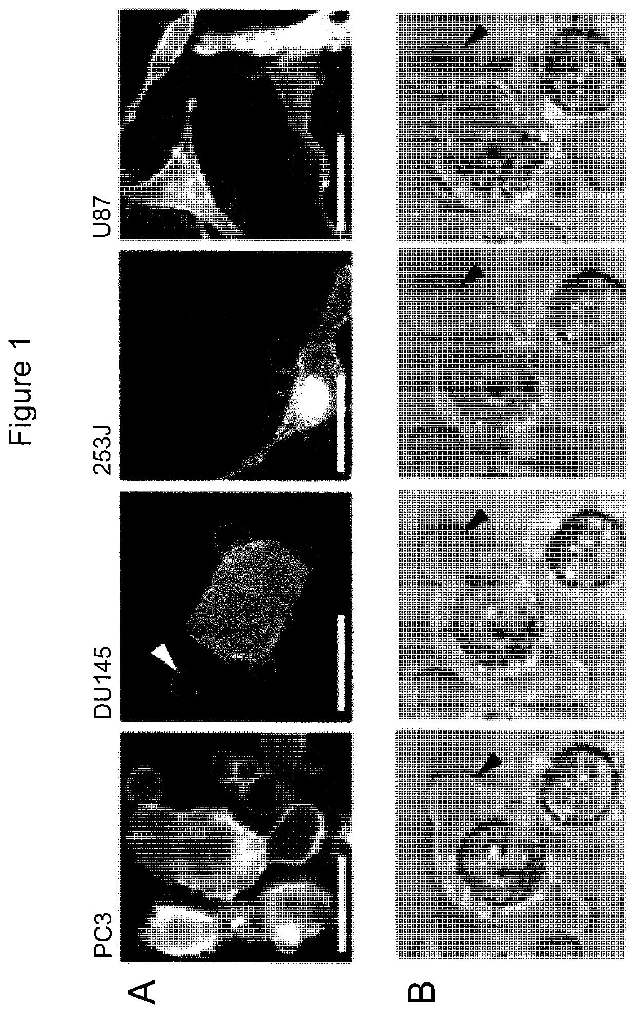
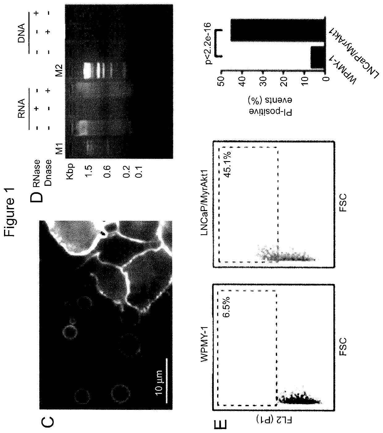
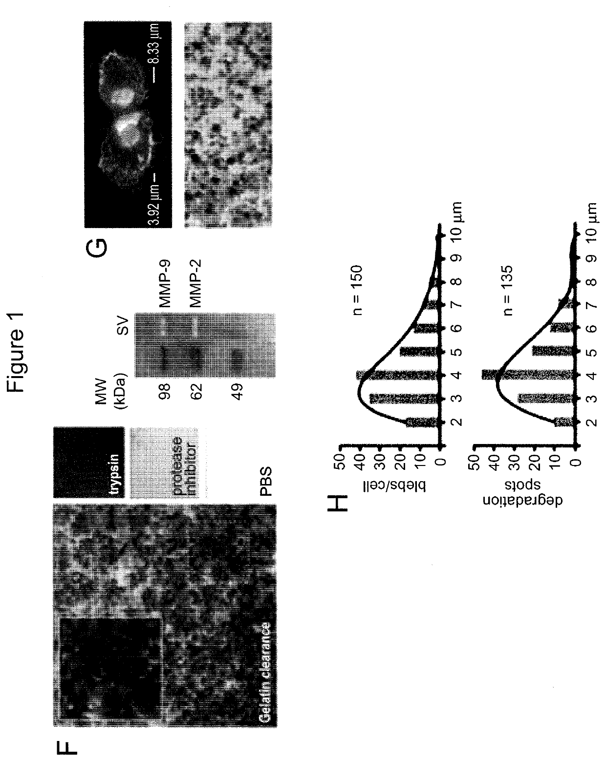
[0164]Tumor cells that exhibit the amoeboid feature of membrane blebbing can release large, plasma membrane-derived vesicles into the extracellular space (Di Vizio D et al., Oncosome formation in prostate cancer: association with a region of frequent chromosomal deletion in metastatic disease, Cancer Res 2009, 69:5601-5609) (FIGS. 1A, 1B, 2A). To determine whether th...
example 3
Large Oncosomes in the Circulation and In Situ
[0168]Microvesicles range from about 20 nm to about 10 μm. Exosomes (a type of microvesicles) range from about 20 nm to about 100 nm. Large oncosomes (another type of microvesicles) range in size from about 1 μm to about 10 μm. To distinguish large oncosomes from smaller TMV unequivocally, ultracentrifugation and immuno-flow cytometry were used in combination with sizing beads to create a calibrated domain of analysis based on size. Following biochemical purification, shed vesicles were stained with DAPI to verify the absence of cellular contamination and subsequently labeled with an HA antibody, for detection of membrane-localized MyrAkt1. MyrAkt1-negative and -positive populations were then distinguished on a cell sorter, with gates set using 1 and 10 μm beads (FIG. 3A). Because aggregates may potentially be a significant component of the vesicle preparation, we excluded them from the analysis. To do this, forward scatter signal (FSS) ...
PUM
 Abstract
Description
Claims
Application Information
 - R&D
- Intellectual Property
- Life Sciences
- Materials
- Tech Scout
- Unparalleled Data Quality
- Higher Quality Content
- 60% Fewer Hallucinations
Browse by: Latest US Patents, China's latest patents, Technical Efficacy Thesaurus, Application Domain, Technology Topic, Popular Technical Reports.
© 2025 PatSnap. All rights reserved.Legal|Privacy policy|Modern Slavery Act Transparency Statement|Sitemap|About US| Contact US: help@patsnap.com


