Systems and methods for simultaneous near-infrared light and visible light imaging

a near-infrared light and visible light imaging technology, applied in the field of systems and methods for simultaneous near-infrared light and visible light imaging, can solve the problems of inefficient and non-optimal infrared images, and the image generated can be less than ideal in at least some instances

Pending Publication Date: 2021-01-21
BLAZE BIOSCI
View PDF2 Cites 16 Cited by
  • Summary
  • Abstract
  • Description
  • Claims
  • Application Information

AI Technical Summary

Benefits of technology

[0004]The present disclosure describes systems and methods for fluorescence and visible light imaging which solve at least some of the problems in prior systems. The systems and methods disclosed herein are capable of generating and combining visible and fluorescent images with imperceptible delays, and providing high fluorescence sensitivity, decreasing disruption to the surgical workflow, and improving ease of use with an operating microscope. The systems and methods can either be used as a stand-alone imaging device or in combination with a surgical instrument, such as an operating microscope, exoscope, or a surgical robot. In some embodiments, excitation light is directed to the sample coaxially with fluorescence light received from the sample, which can decrease shadows and can help to ensure that tissue tagged with a fluorescent marker can be properly identified. In some embodiments, the viewing axis of the visible light imaging optics can be coaxial with the excitation light and fluorescent light axes in order to improve registration of the fluorescence image and the visible image over a range of distances extending between the optics and the imaged tissue. The systems and methods can comprise a beam splitter to transmit visible light toward eye pieces and reflect fluorescent light toward a detector, in which a portion of the visible light is reflected toward a detector to generate a visible image with the reflected light. The amount of reflected visible light can be much less than the transmitted light, in order for the user such as a surgeon to readily view the tissue through the eyepieces while the visible light image is being generated with the detector for combination with the fluorescence image. In some embodiments, the excitation light and the fluorescent light comprise light having wavelengths longer than about 650 nm in order to provide an increased penetration depth into the tissue as compared with light used to generate the visible image.

Problems solved by technology

Although such prior instrumentation can be able to produce images from such infrared light sources, sensitivity can be less than ideal due to inefficient halogen lighting as well as lower energy light sources surrounding excitation wavelengths, leading to inefficient and non-optimal infrared images.
Although lasers have been used to achieve higher absorption and as a result increase fluorescence of the infrared or near infrared dyes, the images generated can be less than ideal in at least some instances.

Method used


Image

  • Systems and methods for simultaneous near-infrared light and visible light imaging
  • Systems and methods for simultaneous near-infrared light and visible light imaging
  • Systems and methods for simultaneous near-infrared light and visible light imaging

Examples


example 1

stem During Pediatric Brain Tumor Resection

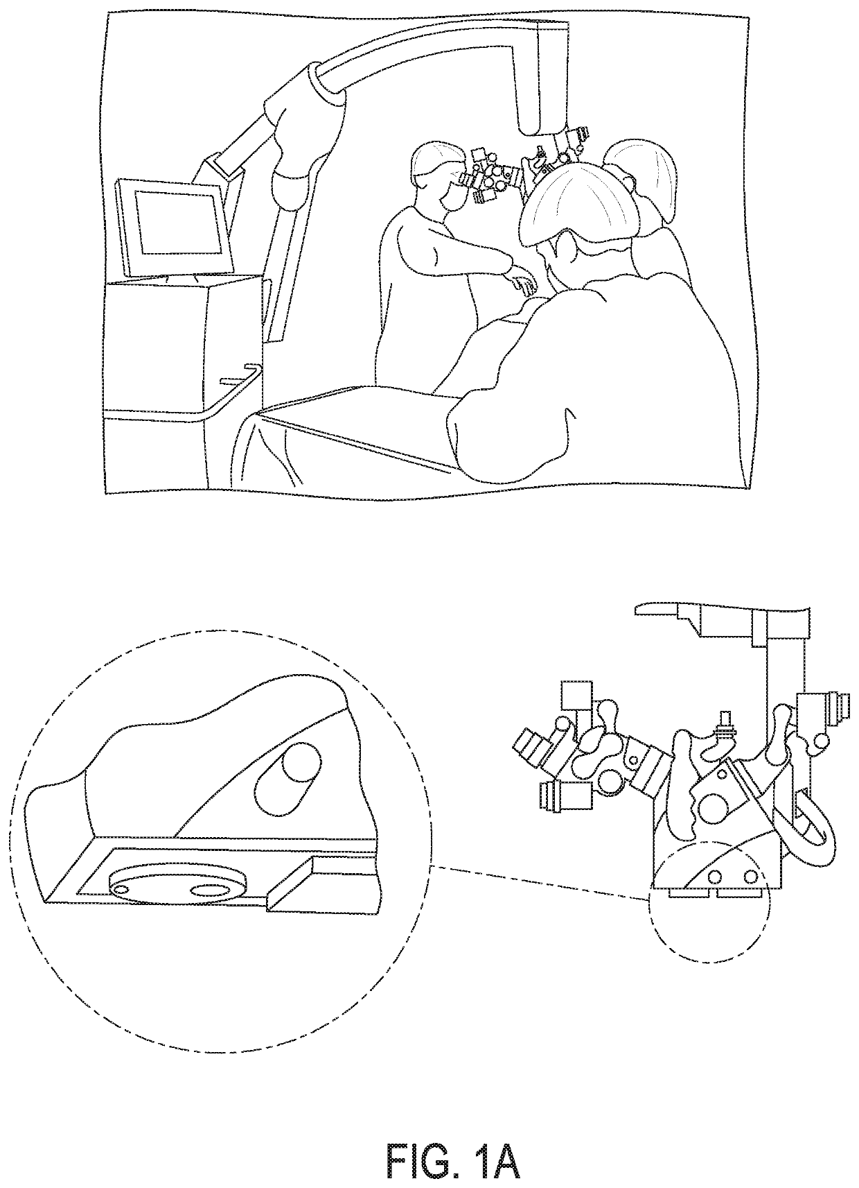
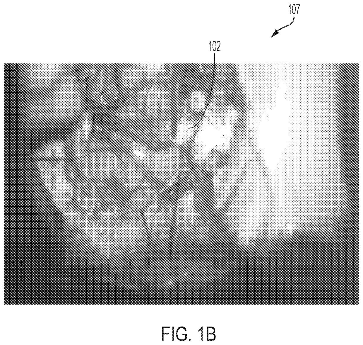
[0259]This example describes use of the imaging system and / or method disclosed herein for coaxial illumination and visualization of tozuleristide fluorescence during surgical resection of a pediatric brain tumor. The imaging system of the present invention was used to image brain tissue to detect a cancer using fluorescence imaging. Surgery was performed to remove cancer from the subject.

[0260]Subject T613 was diagnosed with a Grade 4 Atypical Teratoid Rhabdoid Tumor (ATRT) in the posterior fossa / brain stem. Tozuleristide which is a peptide-fluorophore detectable agent (15 mg / m2 dose), was given by intravenous (IV) bolus injection about 13.5 hours prior to the start of surgery. The imaging head was attached to the Zeiss Pentero surgical microscope along with two eyepieces prior the start of surgery.

[0261]After the tumor was exposed, the imaging system was initialized and used continuously. The imaging system enabled the surgeon to view fluo...


PUM

PropertyMeasurementUnit
wavelengthaaaaaaaaaa

Abstract

Disclosed herein are imaging systems and methods for simultaneous near-infrared light and visible light imaging of a sample comprising: a detector to form a fluorescence image of the sample and a visible image of the sample; a light source configured to emit infrared light to induce fluorescence from the sample; and a plurality of optics arranged to direct the infrared light toward the sample and form the fluorescence image of the sample and the visible light image of the sample on the detector, wherein the infrared light is directed to the sample substantially coaxially with fluorescence light received from the sample in order to decrease shadows.

Description

CROSS-REFERENCE[0001]This application claims the benefit of U.S. Provisional Application No. 62 / 650,974, filed Mar. 30, 2018 and U.S. Provisional Application No. 62 / 679,671 filed Jun. 1, 2018, which are hereby incorporated by reference in their entirety herein.BACKGROUND[0002]Fluorescence, including the use of fluorescent molecules tagged to other structures such as cells, nanoparticles, small molecules and peptides can be useful for organ, organ substructure, tissue and potentially cellular identification in medical imaging. For example, fluorescent dyes can emit in visible (e.g., blue, green, yellow, red) and / or infrared, ultraviolet, or near infrared wavelengths. Although visible light fluorescence can be generally detected by naked eye, detection of infrared (IR) light and near infrared (NIR) light typically requires additional instrumentation for viewing. Infrared and near infrared can be a beneficial wavelength range for medical imaging. The benefits of infrared, near infrared...

Claims


Application Information

Patent Timeline
Patent Type & Authority Applications(United States)
IPC IPC(8): A61B1/04G02B21/00A61B1/06
CPCA61B1/043G02B21/0076G02B23/2407A61B1/0669G02B21/0012A61B1/051A61B1/07A61B1/00165A61B1/042A61B1/063A61B1/0638A61B1/0684A61B1/0646A61B1/00186G02B21/16G02B5/208G02B21/082A61B1/046A61B90/361A61B90/30
Inventor BUTTE, PRAMODKITTLE, DAVIDPERRY, JEFFREY
Owner BLAZE BIOSCI
Why Eureka
Try Eureka