

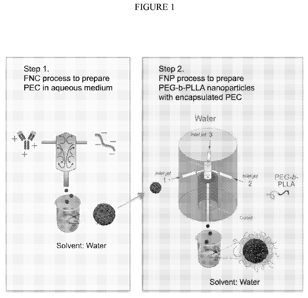
Polymeric nanoparticle compositions for encapsulation and sustained release of protein therapeutics
a technology of protein therapeutics and nanoparticles, which is applied in the direction of capsule delivery, microcapsules, peptide/protein ingredients, etc., can solve the problems of difficult encapsulation of protein therapeutics in sustained release devices, high manufacturing cost, and inability to readily obtain spatiotemporal controlled delivery vehicles of proteins, etc., to achieve sustained release of protein from nanoparticles, efficient mixing, and efficient loading of pec
- Summary
- Abstract
- Description
- Claims
- Application Information
AI Technical Summary
Benefits of technology
Problems solved by technology
Method used
Image

Examples
example 1
on of Lysozyme / DS:PEG-b-PLLA Nanoparticles
Methods
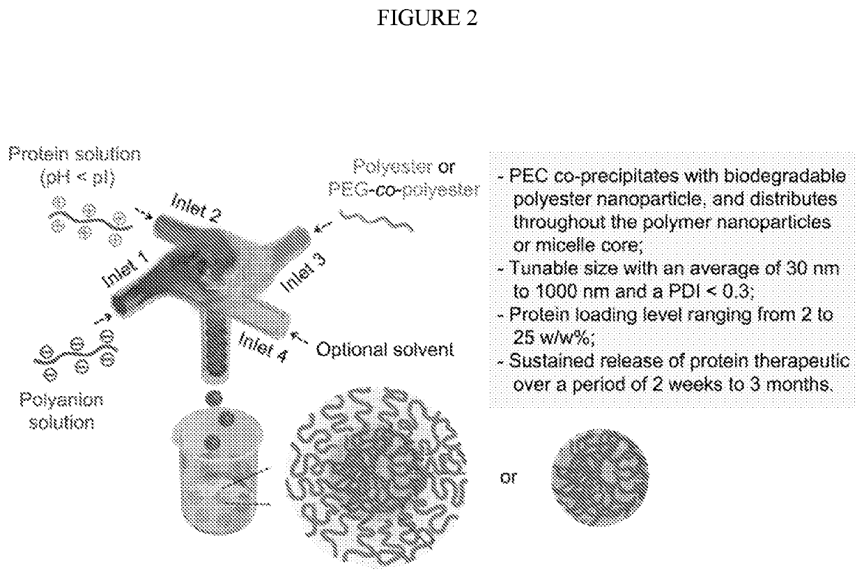
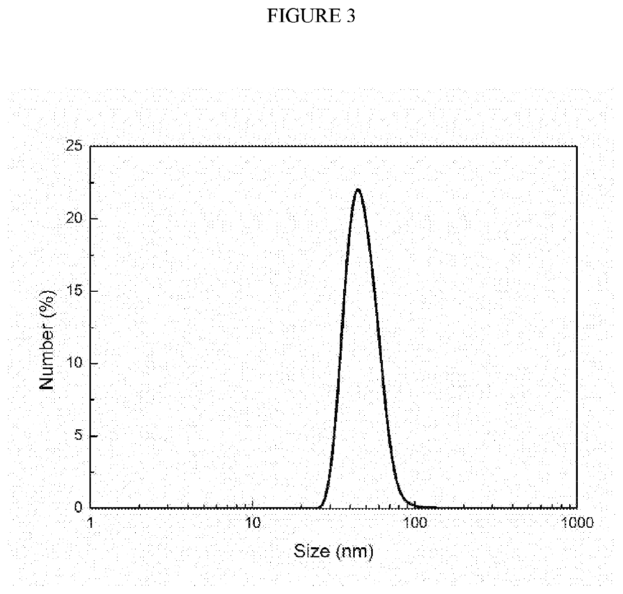
[0044]Preparation and characterization of PEC nanoparticle core: Lysozyme was dissolved in deionized (DI) water at a concentration of 5 mg / mL, followed by adjusting pH to 4.0 by adding 0.1 M HCl solution. One milliliter of the lysozyme solution was then rapidly mixed with an equal volume of DS solution (20 mg / mL, pH was adjusted to 4.0) through a CIJ mixer with two inlets at a flow rate of 5 mL / min. The size of the obtained lysozyme / DS PEC nanoparticles was characterized using a dynamic light scattering (DLS) Zetasizer Nano (Malvern Instruments, Worcestershire, UK). Each sample was measured for three runs and the data was reported as the mean±standard deviation of three readings.
[0045]Preparation and characterization of PEG-b-PLLA micellar nanoparticles: One milliliter of the lysozyme / DS PEC nanoparticle suspension obtained in the previous step (2.5 mg / mL) was rapidly mixed with an equal volume of a solution of PEG5k-b-PLLA20k dissolv...
example 2
on and Characterization of IgG-Encapsulated PEG-b-PLLA Nanoparticles
Methods
[0048]Preparation and characterization of IgG / DS PEC nanoparticles: IgG was dissolved in DI water at a concentration of 5 mg / mL, followed by adjusting pH to 4.0 by adding 0.1 M HCl solution. One milliliter of the IgG solution was then rapidly mixed with an equal volume of DS solution (20 mg / mL, pH was adjusted to 4.0) through a CIJ mixer with two inlets at a flow rate of 5 mL / min. The obtained PEC nanoparticle suspension was used immediately for PEG-b-PLLA nanoparticle preparation.
[0049]Preparation and characterization of IgG / DS:PEG-b-PLLA nanoparticles: One mL of the IgG / DS PEC nanoparticle suspension obtained in the previous step (2.5 mg / mL) was rapidly mixed with an equal volume of PEG5k-b-PLLA20k DMSO solution at two different concentrations (12.5 and 25 mg / mL) through a two-inlet CIJ mixer at a flow rate of 5 or 10 mL / min to obtained four different formulations of core-shell nanoparticles (NP4 to NP7, Ta...
example 3
on of IgG / DS:PEG-b-PCL Nanoparticles
Methods
[0053]Preparation and characterization of IgG / DS:PEG-b-PCL nanoparticles: The IgG / DS:PEG-b-PCL nanoparticles were prepared and characterized using the same procedures as described in the method section in Example 2, except that PEG5k-b-PLLA20k was replaced with PEG5k-b-PCL20k. Four nanoparticles NP8-NP11 were prepared with two different IgG to polymer ratios (1:5 and 1:10) and two different flow rates (5 and 10 mL / min) (FIG. 5B).
[0054]In vitro release of IgG: One mL of IgG / DS:PEG-b-PCL nanoparticle solution containing 200 micrograms of IgG was added into a 1-mL dialysis tube (SpectrumLab, MWCO 300 kDa), which was then submerged in a vial containing 5 mL of PBS (pH 7.4). The vial was put into an incubator with an agitation rate of 100 rpm at 37° C. The PBS filtrate was collected and refreshed daily. The collected medium was concentrated by lyophilization and further reconstituted using 400 microliters of DI water. Micro BCA assay was employe...
PUM
| Property | Measurement | Unit |
|---|---|---|
| size | aaaaa | aaaaa |
| size | aaaaa | aaaaa |
| particle size | aaaaa | aaaaa |
Abstract
Description
Claims
Application Information