
Next-generation biomarkers to detect sun damage and predict skin cancer risk

a biomarker and sun damage technology, applied in the direction of microbiological testing/measurement, biochemistry apparatus and processes, etc., can solve the problems of difficult to produce consistent and reliable information on uvr, and achieve the effect of being reliable uvr biomarkers, facilitating characterization, and improving reliability and accuracy

Active Publication Date: 2021-02-25
THE TRUSTEES OF COLUMBIA UNIV IN THE CITY OF NEW YORK
View PDF0 Cites 3 Cited by
  • Summary
  • Abstract
  • Description
  • Claims
  • Application Information

AI Technical Summary

Benefits of technology

The patent text describes a study that has attempted to identify biomarkers to detect skin damage and cancer risk caused by UVR exposure. However, individual markers have limitations in providing consistent and reliable information due to the complex effects of UVR on multiple biological pathways. At present, a panel of UVR-responsive genes has been identified through comprehensive transcriptomic profiling studies, which has been subjected to rigorous validation and persists long after UVR. This panel of genes can serve as an industrial standard for testing UVR-protective effects of sunscreen products and can also be used in clinical diagnosis. A gene array system is being designed to allow simultaneous detection of the expression of the UVR biomarker genes from multiple samples. Overall, the UVR biomarker panel and the high capacity screening assay system can revolutionize the way we assess sun damage and predict skin cancer risk, ultimately achieving effective prevention and reduction of skin cancer-related illness, death, and health care costs.

Problems solved by technology

Such individual markers are simple and easy to characterize, but it is difficult for them to produce consistent and reliable information on UVR damage and skin cancer risk due to the complex effects of UVR on multiple biological pathways leading to skin neoplastic growth in addition to the variations in skin type-dependent UVR sensitivity.

Method used


Image

  • Next-generation biomarkers to detect sun damage and predict skin cancer risk
  • Next-generation biomarkers to detect sun damage and predict skin cancer risk
  • Next-generation biomarkers to detect sun damage and predict skin cancer risk

Examples


example 1

Materials and Methods

Human Keratinocyte Cultures, Human SCC and Normal Skin Tissues

[0070]Primary human keratinocytes were established from neonatal foreskins through the Columbia University Skin Disease Research Center tissue culture core facility. The protocol was exempt by our Institutional Review Board. Keratinocytes were isolated from separate individual neonatal foreskins (N0, N1, N2, and N6), and cells from each individual were maintained and analyzed separately for assessing individual variations. Keratinocytes were cultured in 154CF medium supplemented with human keratinocyte growth supplement (Life Technologies, Grand Island, N.Y.). Human SCC tumor tissues and matched normal skin tissues from two patients were obtained from the Molecular Pathology Shared Resource / Tissue Bank of the Herbert Irving Comprehensive Cancer Center of Columbia University under CUMC IRB protocol AAAB2667.

UVB Radiation

[0071]Keratinocytes were rinsed once with PBS and irradiated with UVB supplied by f...

example 2

Transcriptomic Responses to Different UVR Conditions

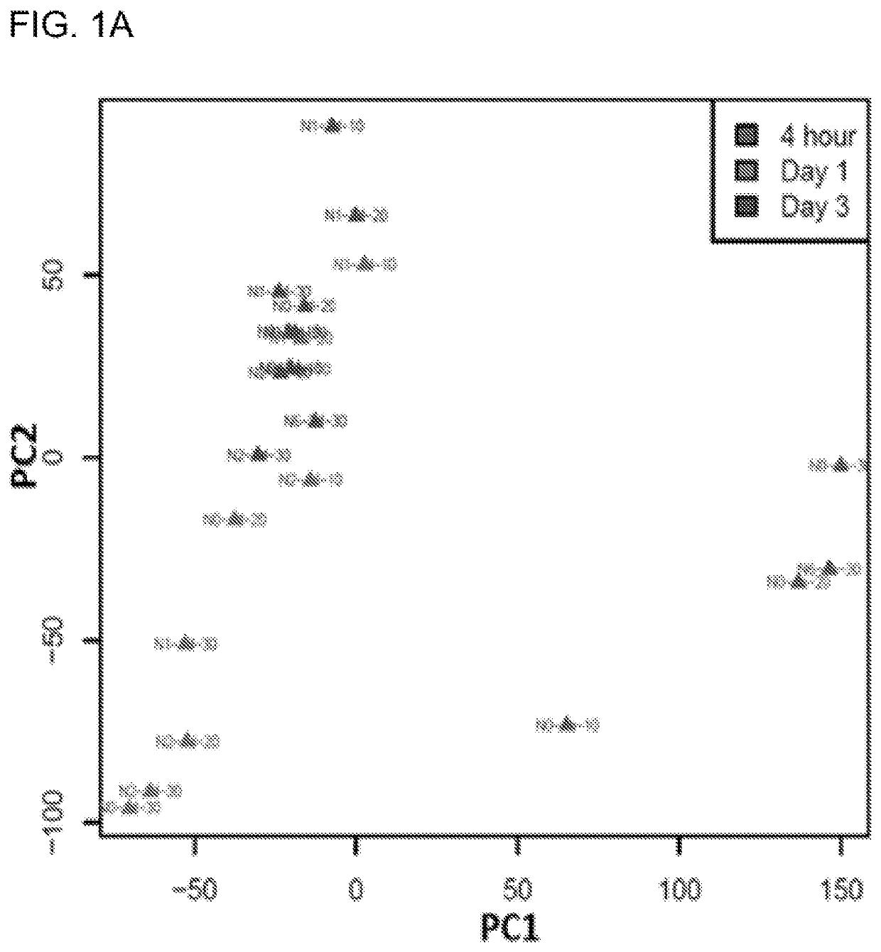
[0074]In addition to its mutagenic effect, UVR has been shown to cause transcriptomic instability affecting thousands of genes. To fully characterize UVR-induced transcriptomic changes, we took advantage of the recent advances in RNA-Seq to profile UVR-induced kinetic changes in human primary keratinocytes exposed to different UVR conditions (Table 1). We used keratinocytes isolated from four individual neonatal foreskins to generate UVR-induced differentially expressed gene (DEG) lists in response to each of the UVR conditions (Table 1). Together with four DEG lists representing transcriptomic profiles at four hours after exposure, we performed principle component analysis (PCA) to differentiate the DEG profiles under various UVR conditions. As shown in the PCA plot (FIG. 1A), DEG profiles from Day 1 and 3 groups, but not the 4 hour group, demonstrated great similarities with each other in the first principle component (PC1). Alon...

example 3

Time-Dependent Transcriptomic Changes in Response to UVR

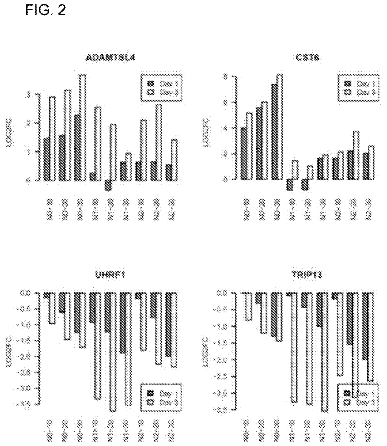
[0076]Our PCA analysis in FIG. 1A revealed time-dependent variations in UVR-responsiveness. To identify genes exhibiting time-dependent cumulative UVR responsiveness, we performed paired t-tests to compare the gene expression signatures of Day 3 versus those of Day 1 for each keratinocyte cell line (N0, N1 and N2) under the same UVR dose. 164 out of the 531 up-regulated genes showed higher expressions at Day 3 than at Day 1 (FDR-corrected p-value <0.05); while 239 out of the 610 down-regulated genes were more repressed at Day 3 than at Day 1 at the same p-value threshold (Table 4). Two examples of time-dependent up-regulation include ADAMTSL4, encoding a disintegrin and metalloproteinase; and CST6, encoding a cystatin superfamily protein. Examples of time-dependent down-regulation include UHRF1, encoding a member of a subfamily of RING-finger type E3 ubiquitin ligases; and TRIP13, which encodes a protein that interacts with thy...


PUM

PropertyMeasurementUnit
Fractionaaaaaaaaaa
Fractionaaaaaaaaaa
Fractionaaaaaaaaaa

Abstract

The present invention provides methods of detecting ultraviolet radiation (UVR)-induced skin damage in a subject. The method includes the steps of: a) obtaining a skin sample from the subject; b) analyzing expression levels in the skin sample of UVR-induced differentially expressed genes (DEGs) listed in Table 8 or a subset thereof; and c) comparing the expression levels of the UVR-induced DEGs to a control skin sample; wherein, when the expression levels of the UVR-induced DEGs in the skin sample is above or below the level of each of the UVR-induced DEGs in the control sample, the subject is identified as likely being afflicted with UVR-induced skin damage. Also provided are methods for measuring the effectiveness of a test agent in reducing ultraviolet radiation (UVR)-induced damage.

Description

CROSS-REFERENCE TO RELATED APPLICATIONS[0001]The present application is the National Stage of International Application No. PCT / US17 / 22848 filed Mar. 16, 2017, which claims benefit to U.S. Provisional Application No. 62 / 458,535 filed Feb. 13, 2017, and U.S. Provisional Application No. 62 / 313,425 filed Mar. 25, 2016. The entire contents of the aforementioned applications are incorporated by reference as if recited in full herein.GOVERNMENT FUNDING[0002]This invention was made with government support under ES009089 and AR064315 awarded by National Institutes of Health. The government has certain rights in the invention.BACKGROUND OF THE INVENTION[0003]Skin cancer is the most prevalent cancer worldwide. (Guy 2015; Rogers 2015) Every year in the United States, nearly 5 million people are treated for skin cancer, at an estimated cost of $8.1 billion. (The surgeon General 2014) Solar ultraviolet radiation (UVR), especially the UVB spectrum of sunlight, is widely recognized as the major ca...

Claims


Application Information

Patent Timeline
IPC IPC(8): C12Q1/6886
CPCC12Q1/6886C12Q2600/158
Inventor LIU, LIANGSHEN, YAO
Owner THE TRUSTEES OF COLUMBIA UNIV IN THE CITY OF NEW YORK
Learn More