Check patentability & draft patents in minutes with Patsnap Eureka AI!

Systems and methods for planning peripheral endovascular procedures with magnetic resonance imaging

a technology of magnetic resonance imaging and peripheral endovascular surgery, which is applied in the field of systems and methods for planning peripheral endovascular surgery with magnetic resonance imaging, can solve the problems of high re-intervention rate (20%, high immediate failure rate, etc.) and is less invasive than pvi, and achieves the effect of less invasiveness, high re-intervention rate (20%) and high immediate failure ra

Pending Publication Date: 2021-04-08
SUNNYBROOK RES INST
View PDF0 Cites 1 Cited by
  • Summary
  • Abstract
  • Description
  • Claims
  • Application Information

AI Technical Summary

Benefits of technology

This patent describes a method for using magnetic resonance imaging (MRI) to characterize a lesion in a patient. The method involves first obtaining a set of ultrashort echo time images and then a set of longer echo time images of the patient's volume of interest. These images are then combined using a computer system to produce a combined image. This combined image is used to identify and characterize the lesion based on the comparison of magnetic resonance signal behaviors between the images. Additionally, this patent also describes a method for generating an endovascular procedure plan using MRI. This involves obtaining magnetic resonance angiography (MRA) images and combining them with the ultrashort echo time images to create a three-dimensional angiogram. This angiogram is then used to identify and characterize a lesion. The combined image data and the MRA images are then used to generate a report indicating the endovascular procedure plan for the patient. The technical effects of this patent include improved accuracy in identifying and characterizing lesions and improved guidance in endovascular procedures.

Problems solved by technology

PVI is less invasive but has high immediate technical failure rates (20%) and high re-intervention rates (20%).
The most common mode of immediate failure is the inability to enter / cross the lesion.
With current imaging (x-ray angiography, CTA, duplex ultrasound) it is difficult to predict which lesions will be soft enough to cross with a wire to make PVI possible.
This requires more procedures per index limb at significant cost to healthcare systems and delays definitive revascularization.
Additionally, there is evidence that surgical bypass after failed PVI results in worse outcomes, including higher amputation rates within 1 year.
However, the length and degree of stenosis / occlusion of a lesion is not equivalent to its burden, mechanical properties or morphology, all of which influence PVI success.
Intravascular imaging devices, however, require invasive arterial access which makes the “percutaneous-first” strategy and associated complications impossible to avoid.
The added procedure time and cost of intravascular imaging devices also limit their widespread clinical use, which provides motivation to improve non-invasive lesion characterization imaging.
Existing plaque analysis techniques with CT or MRI are tailored to characterize atherosclerotic plaque, but are not tailored to characterize peripheral arterial lesions, specifically.
100081 Though the mechanical properties of atherosclerotic plaques have been described, the prognostic value of mechanical properties for planning endovascular treatment has not been comprehensively investigated.
However, ultrasound elastography is limited due to issues with severely calcified vessel acoustic shadowing, repeatability and user dependence, penetration depth, and inability to perform 3D lesion analysis.
It is challenging to accurately evaluate heavily calcified small-caliber vessels using non-invasive techniques, including CTA and duplex ultrasound.

Method used


Image

  • Systems and methods for planning peripheral endovascular procedures with magnetic resonance imaging
  • Systems and methods for planning peripheral endovascular procedures with magnetic resonance imaging
  • Systems and methods for planning peripheral endovascular procedures with magnetic resonance imaging

Examples


Embodiment Construction

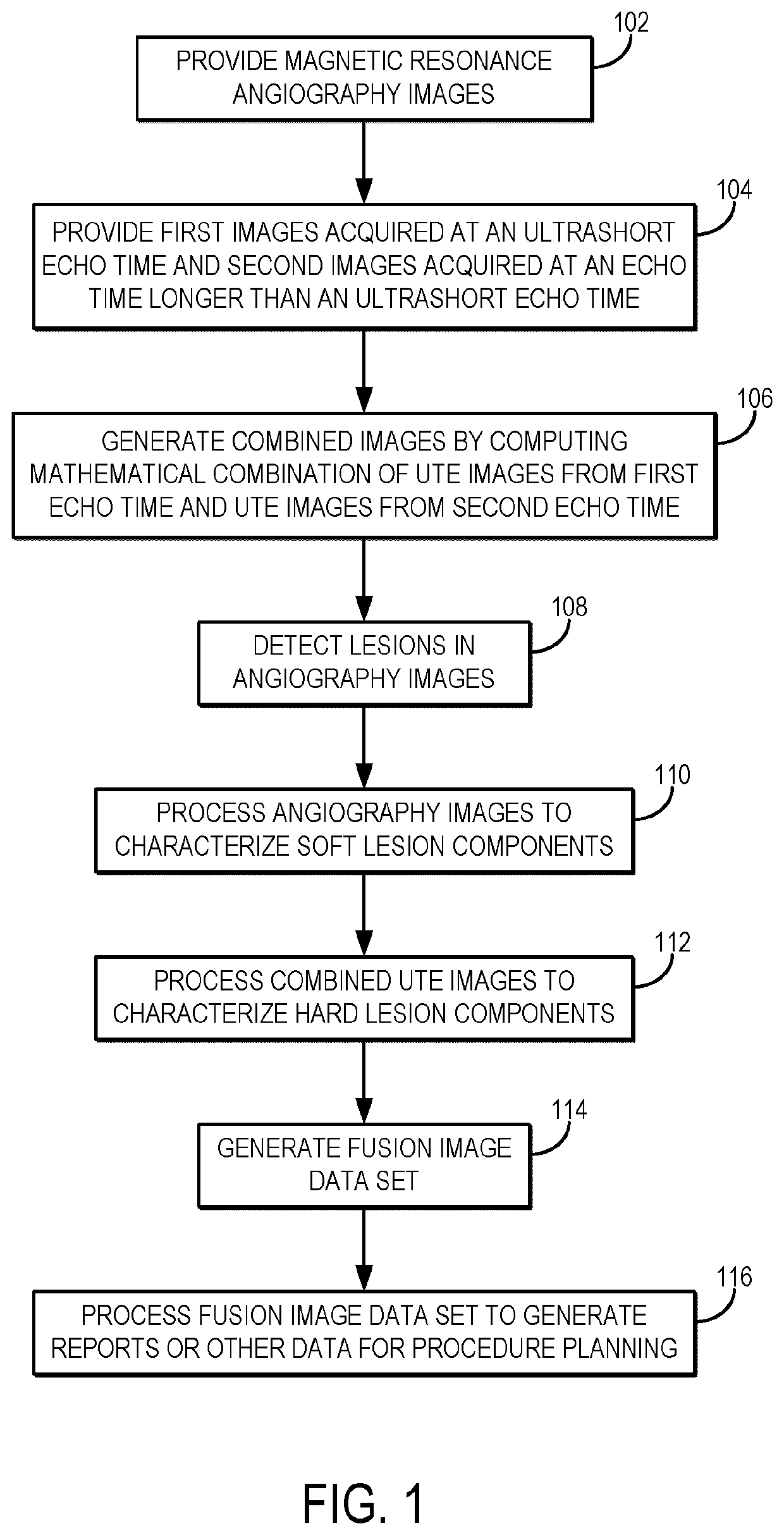
[0023]Described here are methods for characterizing the mechanical properties lesions or other regions of tissue, as well as assessing patency, using magnetic resonance imaging (“MRI”). Such methods can be implemented for planning peripheral endovascular, or other vascular, procedures. The methods described in the present disclosure include acquiring magnetic resonance images using different contrast weightings and analyzing those images together in a single analytical framework to characterize properties of the subject's vasculature. The properties that can be characterized include patency (e.g., the degree of stenosis, occlusion, or both), mechanical properties (e.g., stiffness, which can be used to differentiate hard plaque components from soft plaque components), tissue content (e.g., calcification content, collagen content), and morphology (e.g., eccentricity, stump morphology). The methods described in the present disclosure also provide improved visualization of the vascular ...


PUM


Abstract

Systems and methods for planning peripheral endovascular, and other, procedures based on magnetic resonance imaging (“MRI”] are provided. Mechanical properties of lesions, morphology, and vessel patency are characterized based on non-contrast angiography and ultrashort echo time (“UTE”] images. The methods described in the present disclosure also provide improved visualization of the vascular tree and microchannels.

Description

CROSS-REFERENCE TO RELATED APPLICATIONS[0001]This application claims the benefit of U.S. Provisional Patent Application Ser. No. 62 / 481,899, filed on Apr. 5, 2017, and entitled “SYSTEMS AND METHODS FOR PLANNING PERIPHERAL END OVASCULAR PROCEDURES WITH MAGNETIC RESONANCE IMAGING,” which is herein incorporated by reference in its entirety.BACKGROUND[0002]There are two treatment options for revascularizing patients with critical limb ischemia: bypass surgery and percutaneous vascular intervention (“PVI”). PVI is less invasive but has high immediate technical failure rates (20%) and high re-intervention rates (20%). The most common mode of immediate failure is the inability to enter / cross the lesion.[0003]With current imaging (x-ray angiography, CTA, duplex ultrasound) it is difficult to predict which lesions will be soft enough to cross with a wire to make PVI possible. Physicians have responded with a “percutaneous-first” strategy where they attempt PVI in all patients and perform sur...

Claims


Application Information

Patent Timeline
IPC IPC(8): A61B34/10G01R33/56G01R33/561G01R33/563G01R33/483G01R33/54A61B5/00A61B5/107
CPCA61B34/10G01R33/5608G01R33/5614G01R33/5635A61B2034/105G01R33/546A61B5/004A61B5/1075A61B5/1079G01R33/483A61B5/055G01R33/4818A61B5/02007G01R33/56G06T7/254
Inventor ROY, TRISHADUECK, ANDREWWRIGHT, GRAHAM A.LIU, GARRY
Owner SUNNYBROOK RES INST
Features
  • R&D
  • Intellectual Property
  • Life Sciences
  • Materials
  • Tech Scout
  • Unparalleled Data Quality
  • Higher Quality Content
  • 60% Fewer Hallucinations
Learn More