Methods for Enhanced Endothelialization of Implanted Material

a technology of endothelialization and implanted materials, which is applied in the field of cardiac and vascular surgery, can solve the problems of large luminal area of prosthetic vascular grafts remaining without endothelium, significant increase in the risk of thrombosis, and the greatest risk of thrombosis, and achieve the effect of increasing endothelialization

Pending Publication Date: 2021-08-26
RUSH UNIV MEDICAL CENT
View PDF0 Cites 0 Cited by
  • Summary
  • Abstract
  • Description
  • Claims
  • Application Information

AI Technical Summary

Benefits of technology

[0005]Methods of increasing endothelialization on a surface of a medical device are provided. The methods include delivering the medical device to an intravascular site. The delivered medical device has an α-gal epitope attached to the surface of the medical device.

Problems solved by technology

Complete endothelialization of synthetic vascular or cardiac grafts is not achieved in many patients even decades after grafting, resulting in large luminal areas of prosthetic vascular grafts remaining without an endothelium (Marois et al., ASAIO J.45: 272.1999; Zilla et al., Biomaterials, 28: 5009, 2007).
In the absence of endothelial surfacing of the graft, there is a significant increase in the risk of thrombosis.
In general, the risk of thrombosis is greatest early after device / material deployment, and decreases over time due to ingrowth of an endothelial layer that separates and protects the device and synthetic grafts from the thrombogenic elements in the bloodstream.
All of these medications carry a risk of bleeding, and not all patients are good candidates for these medications due to prior bleeding events or bleeding risk.

Method used


Image

  • Methods for Enhanced Endothelialization of Implanted Material
  • Methods for Enhanced Endothelialization of Implanted Material

Examples


example 1

[0050]Exemplary α-gal epitopes are generated from extracts of rabbit red blood cell (RBC) membranes. These membranes are used since they contain glycolipids carrying from one to more than seven α-gal epitopes per molecule as disclosed in Eto et al., Biochem. (Tokyo) 64, 205, (1968); Stellner et al., Arch. Biochem. Biophys. 133, 464 (1973); Dabrowski et al., J. Biol. Chem. 259, 7648 (1984) and Hanfland et al., Carbohydr. Res. 178, 1 (1988), all of which are hereby incorporated by reference. However, α-gal epitopes may be produced from any natural or synthetic source of α-gal epitopes and may include the addition of phospholipids in the presence or absence of cholesterol, after processing as described herein. As a non-limiting example, rabbit RBC are used at a volume of 0.25 liter packed cells. The RBC are lysed by repeated washes with distilled water. The rabbit RBC membranes are then mixed with a solution of 600 ml chloroform and 900 ml methanol for 20 h with constant stirring to di...

example 2

[0051]In some embodiments, human or rabbit or bovine RBC may be used as a source of α-gal epitopes. Isolation of α-gal epitopes from rabbit RBC are used by way of non-limiting example herein. Batches of 1 liter rabbit RBC were lysed in water and washed repeatedly to remove hemoglobin. For the extraction process, rabbit RBC membranes (RBC ghosts) were mixed with 1000 ml chloroform and 1000 ml methanol (1:1 chloroform:methanol) for 2 h, then 1000 ml methanol was added for overnight incubation with constant stirring (1:2 chloroform:methanol). The extract was filtered under vacuum through Whatman filter paper for removing residual RBC membranes and precipitated proteins. For use for attaching the α-gal epitopes to a medical device, the isolated α-gal epitopes will be resuspended in a solution suitable for attaching to the medical device. The α-gal epitope suspension will be sterile because all proteins were denatured and removed in the chloroform:methanol extraction process.

example 3

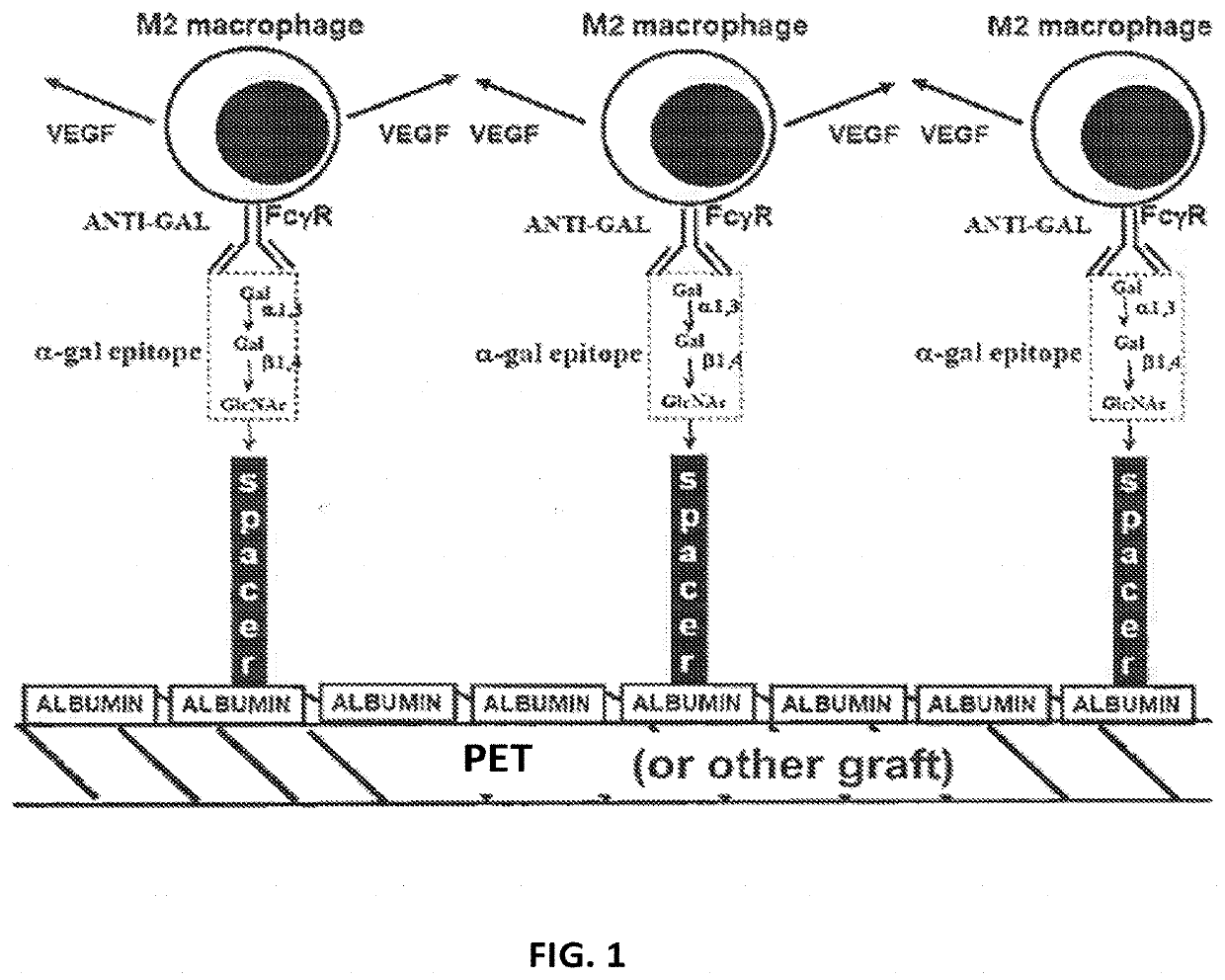
[0052]In some embodiments, α-gal glycolipids may be used, for example from human, bovine or rabbit sources. By way of non-limiting example a rabbit source of α-gal glycolipids is described herein. A rich source for α-gal glycolipids is rabbit red blood cells. The two major glycosphingolipids (GSL), i.e. glycolipids with ceramide tail, in rabbit red blood cell membranes are ceramide (Cer) trihexoside (CTH, with 3 sugars [hexoses], present also in human red blood cells) with the structure Galα1-4Galβ1-4Glc-Cer and ceramide pentahexoside (CPH, with 5 sugars [hexoses]) with α-gal epitopes as terminal part of the CPH structure of Galα1-3Galβ1-4GlcNAcβ1-3Galβ1-4Glc-Cer (Eto T, Iichikawa Y, Nishimura K, Ando S, Yamakawa T J. Biochem. (Tokyo) 1968; 64: 205-13, and Stellner K, Saito H, Hakomori S. Arch. Biochem. Biophys. 1973; 133: 464-72) (See FIG. 1 and FIG. 6A, lane 1). Immunostaining of rabbit α-gal glycolipids on thin layer chromatography (TLC) plates with human natural anti-Gal resulte...


PUM

PropertyMeasurementUnit
sizeaaaaaaaaaa
diameteraaaaaaaaaa
diameteraaaaaaaaaa

Abstract

Methods and devices for increasing endothelialization on a surface of a medical device are provided. The methods include delivering the medical device to an intravascular site and the medical device has an α-gal epitope attached to the surface of the medical device.

Description

RELATED APPLICATIONS[0001]The present patent application claims the benefit of the filing date of U.S. Provisional Patent Application No. 62 / 684,042, filed Jun. 12, 2018, the contents of which is hereby incorporated by reference.TECHNICAL FIELD[0002]The present disclosure relates to the fields of cardiology and vascular surgery, and in particular to methods and devices for enhanced endothelialization of the devices.BACKGROUND[0003]Prosthetic synthetic grafts such as poly(ethylene terephthalate) (PET also called DACRON) and poly(tetrafluoroethylene) (ePTFE also called GORE-TEX) are widely used in cardiac and vascular surgery since the mid-1970s. When implanting any vascular prosthetic grafts, one important goal to ensure long-term patency is achieving complete endothelialization of the luminal surface. Complete endothelialization of synthetic vascular or cardiac grafts is not achieved in many patients even decades after grafting, resulting in large luminal areas of prosthetic vascula...

Claims


Application Information

Patent Timeline
Patent Type & Authority Applications(United States)
IPC IPC(8): A61L27/50A61L27/36A61F2/00A61L27/18
CPCA61L27/507A61L27/3687A61F2/0077A61F2002/0086A61L2430/20A61L2430/22A61L2400/18A61L27/18A61K9/0024
Inventor SOBLE, JEFFREY SCOTTGALILI, URISCHAER, GARY
Owner RUSH UNIV MEDICAL CENT
Why Eureka
Try Eureka