

Artificial flourescent image systems and methods
a technology of flourescent images and flourescent light, applied in the field of artificial flourescent light image systems and methods, can solve the problems of artificially inflating the amount of observed cell death, consuming a lot of time, and affecting the quality of the imag
- Summary
- Abstract
- Description
- Claims
- Application Information
AI Technical Summary
Benefits of technology
Problems solved by technology
Method used
Image

Examples
example 1
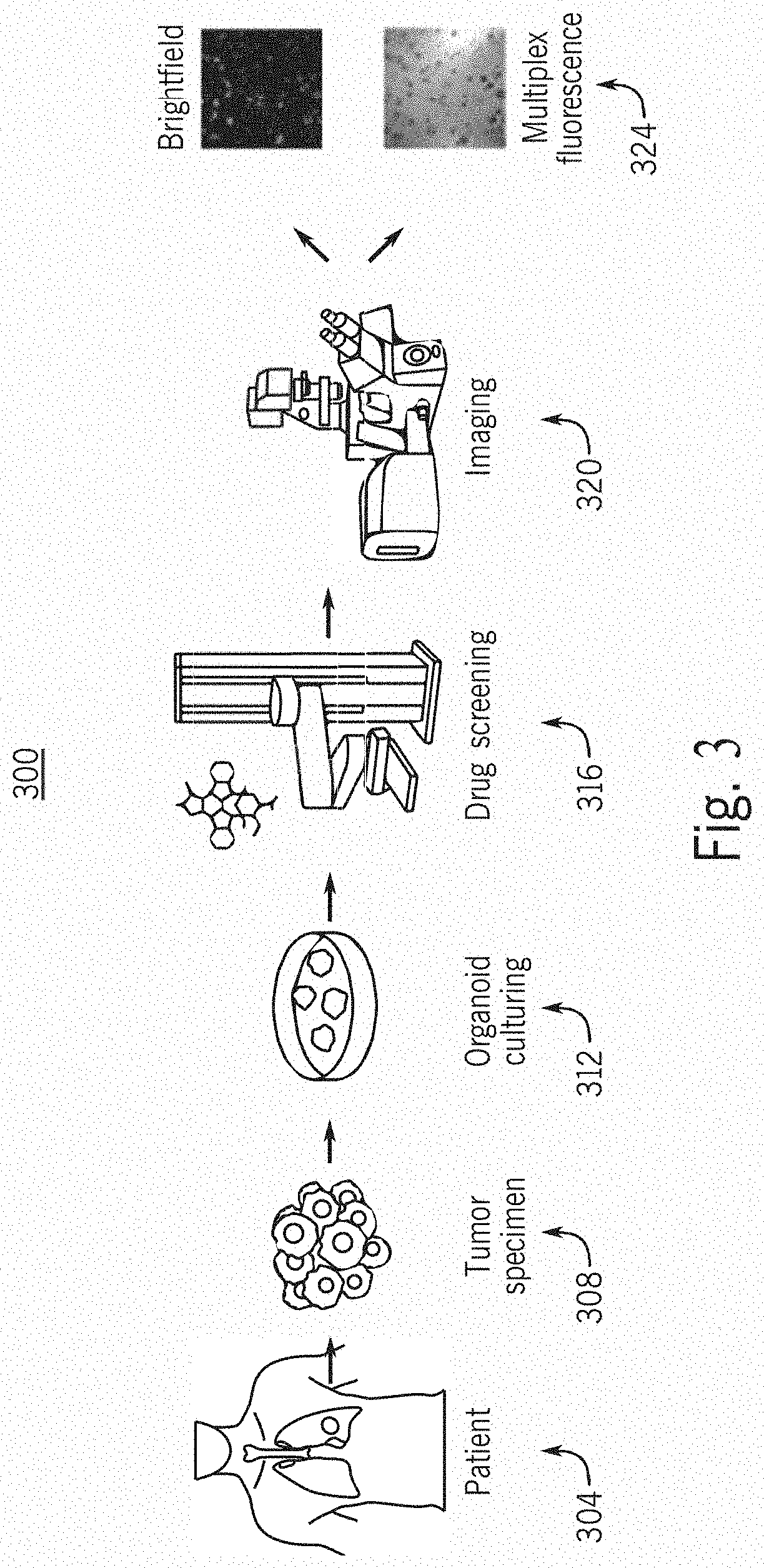
[0153]In this example, Tumor Organoids in each well were stained using three fluorophores for high content fluorescent confocal imaging analysis. In order to obtain the fluorescent readouts a high content imager (ImageXpress Confocal, Molecular Devices) was utilized for data acquisition. Images were acquired at 10× magnification with a 50 micron slit spinning disk aperture. Four channels were acquired using incandescent brightfield, and LED light sources using manufacturer's default settings for 4′,6-diamidino-2-phenylindole (DAPI), Fluorescein isothiocyanate (FITC), and Cyanine 5 (CY5) to acquire data from Hoechst 33342 (Thermo), Caspase-3 / 7 reagent (Essen Bioscience), or TO-PRO-3 (Thermo) respectively.
[0154]In this example, the experimental setup used a 384 well plate, with 330 usable wells within each plate. Since each well has two sites that get imaged, each plate has a total of 660 paired brightfield and fluorescence images. At a magnification of 10×, two images are taken per w...
example 2
[0163]Experiments were performed to try and improve model performance. Candidate models included a GANLoss+SSIM model, a GANLoss+SSIM+L1 model trained using a GANLoss+0.17L1+0.83 SSIM model, a GANLoss+MS-SSIM model, and a GANLoss+0.83MS-SSIM+0.17L1 model.
[0164]Initially, three separate Pix2Pix models were employed to train the individual fluorescent channels. The Avg SSIM and RMSE results over the same 743 blind test images as described in Example 1 are shown below. Tables 5-8 show results of the candidate models implemented in three-model fashion. Table 5 shows results of the GANLoss+SSIM model. Table 6 shows results of the GANLoss+0.83SSIM+0.17L1 model. Table 7 shows results of the GANLoss+MS-SSIM model. Table 8 shows results of the GANLoss+0.83 MS-SSIM+0.17 L1 model.
TABLE 5ExperimentAvg RMSEAvg SSIMDead Cells1.651330.91736Dead / Dying Cells 1.128270.91784All cells1.736300.91097
TABLE 6ExperimentAvg RMSEAvg SSIMDead cells1.567810.92950Dead / Dying Cells 1.107350.92782All cells1.706040....
example 3
[0166]In Example 2, the process of training 3 separate pix2pix models for multiple different objective functions proved to require several GPU's (3 per model) and extra effort in data curation. A similar performance analysis was done to check if similar / better RMSE and SSIM values were observed by directly training from brightfield to 3 channel fluorescence using a single Pix2Pix model in an attempt to reduce GPU usage.
[0167]Table 10 below shows the results of directly training to transfer style to IF image for the same set of objective functions on the same test set of 743 images belonging to 10245. The number of GPU's was reduced from 15 GPU's to 5 GPU's and the performance although not too significant, is marginally better. Thus, it may be preferable to use a one-model to generate artificial fluorescent images because performance can be at least as good as a three-model, with one third of the computational requirements. In particular, a one-model trained using an objective functi...
PUM
 Abstract
Description
Claims
Application Information