
Tissue repair and sealing devices having a detachable graft and clasp assembly and methods for the use thereof

a tissue repair and sealing device technology, applied in the field of medicine, can solve the problems of limited working space, poor visualization, restricted surgical access, etc., and achieve the effect of rapid repair of tissue fenestration

Pending Publication Date: 2022-05-19
PATCHCLAMP MEDTECH INC
View PDF42 Cites 0 Cited by
  • Summary
  • Abstract
  • Description
  • Claims
  • Application Information

AI Technical Summary

Benefits of technology

These devices enable rapid and reliable repair of tissue fenestrations, effectively preventing fluid leakage and reducing post-surgical complications by ensuring a pressure-resistant, watertight seal, even in challenging minimally invasive surgical environments.

Problems solved by technology

Several factors common to MIS procedures, including limited working space, restricted surgical access, poor visualization, and the friable nature of certain tissues, make it difficult to repair and seal cuts, tears, or openings in tissues that are beneath the skin (collectively tissue fenestrations).
Failure to rapidly repair a tissue fenestration and create a watertight seal can result in the leakage of body fluids through the fenestrated tissue, which inhibits tissue healing, promotes infection, and leads to substantial post-surgical morbidity.
Existing devices and methodologies have limited practical utility, however, because they cannot rapidly repair tissue fenestrations and cannot reliably create pressure-resistant, watertight seals.
As a result, healing of a fenestrated tissue is inadequate and complications often arise from the leakage of body fluids, including blood (hemorrhage, hematoma mass effect), cerebrospinal fluid (meningitis, pneumocephalus, intracranial hypotension), gastrointestinal contents (infection, fistula), and urine (fistula, infection).
Direct suturing or stapling of tissue fenestrations is time-consuming and technically difficult in the limited space and restricted access that is characteristic of MIS procedures.
As a consequence, the rapid repair and creation of watertight seals is seldom achieved with the repair of tissue fenestrations produced during MIS procedures.
Moreover, certain tissues that are encountered during MIS procedures are not amenable to suturing due to their friable nature, insufficient tissue to permit a complete closure, and close proximity to critical structures.
And permanent metallic implants (i.e. staples) can interfere with subsequent magnetic resonance imaging.
Absorbable and non-absorbable tissue adhesives, such as fibrin glue and polyglycol gel, also have limited utility in the rapid repair of tissue fenestrations and creation of pressure-resistant, watertight seals.
Tissue adhesives pose substantial technical challenges that can contribute to poor surgical results, namely: (1) the required mixing and applying of a rapidly-curing, two-component adhesive is difficult to perform in a small space; (2) buttressing of a graft with another tissue (e.g., fat) is often necessary; and (3) the bonding strength of tissue adhesives can be inadequate for the creation of a pressure-resistant, watertight seal.
Tissue patches, which include patches that are autologous (e.g., fascia and fat), heterologous (e.g., bovine or porcine tissues), or synthetic (e.g., collagen matrix), require the use of sutures to hold in place and are susceptible to infection and tissue rejection.
Pedicle graft overlays to facilitate healing require a glue or buttress to ensure adherence, do not provide immediate watertight closure and are associated with elevated surgical morbidity.
Existing devices for attaching grafts to the outer surface of tissue fenestrations have limited utility in MIS procedures.
Devices that are known in the art are difficult to manipulate and typically require additional procedures (e.g., harvesting a tissue for buttressing and placing drains to reduce pressure gradients).
Moreover, tissue grafts attached to an outer tissue surface, are prone to failure and are particularly susceptible to pressure differentials between the inside and outside of a fenestrated tissue (e.g., a fenestrated blood vessel, dura mater, or gastrointestinal wall tissue).
Because grafts positioned on an outer tissue surface often fail to repair tissue fenestrations and create watertight seals, fluids leak from a higher-pressure tissue interior (e.g., blood, cerebrospinal fluid, or gastrointestinal contents).
This leads to poor healing, an elevated occurrence of infection, substantial post-surgical complications and morbidity, and prolonged hospitalization.
Despite the availability of existing technologies for closing tissue fenestrations during surgical procedures, there remains an unmet need in the art for devices and methods that permit the rapid repair of tissue fenestrations and the reliable creation of pressure-resistant, watertight seals.

Method used


Image

  • Tissue repair and sealing devices having a detachable graft and clasp assembly and methods for the use thereof
  • Tissue repair and sealing devices having a detachable graft and clasp assembly and methods for the use thereof
  • Tissue repair and sealing devices having a detachable graft and clasp assembly and methods for the use thereof

Examples


example 1

In Vitro Models for Testing Tissue Repair and Sealing Devices

[0209]This Example provides in vitro model systems that may be adapted and employed for the testing various aspects of the tissue repair and sealing devices disclosed herein. Various physical properties and other parameters of tissue repair and sealing devices as disclosed herein may be tested in in vitro model systems, including in vitro model systems that are described in the scientific, medical, and patent literature and that may be configured for testing the repair and sealing of tissue fenestrations with the devices disclosed herein. See, Dafford, The Spine Journal 15(5):1099 (2015); Chauvet, Acta Neurochirurgica 153(12):2465 (2011); and Wang, MATEC Web of Conferences 119:01044 (2017).

[0210]Van Doormaal, Operative Neurosurgey 15(4):425 (2018) and Kinaci, Expert Review of Medical Devices 16(7):549 (2019) disclose in vitro model systems that use fresh porcine dura for testing acute burst pressures and resistance to intr...

example 2

In Vivo Models for Testing Tissue Repair and Sealing Devices

[0216]This Example provides in vivo model systems that may be adapted and employed for the testing various aspects of the tissue repair and sealing devices disclosed herein. Various physical properties and other parameters of tissue repair and sealing devices as disclosed herein may be tested in in vivo model systems, including in vivo model systems that are described in the scientific, medical, and patent literature and that may be configured for testing the repair and sealing of tissue fenestrations with the devices disclosed herein.

[0217]de Almeida, Otolaryngology Head Neck Surgery 141(2):184 (2009) and Seo, Journal of Clinical Neuroscience 58:187 (2018) describe in vivo porcine craniotomy model system that may be adapted for testing the repair of tissue fenestrations by assessing the leakage of cerebrospinal fluids (CSF). In de Almeida, pigs undergo a craniotomy to create fistula through the cribriform plate into the na...


PUM

PropertyMeasurementUnit
pressure-resistantaaaaaaaaaa
shape memoryaaaaaaaaaa
superelasticityaaaaaaaaaa

Abstract

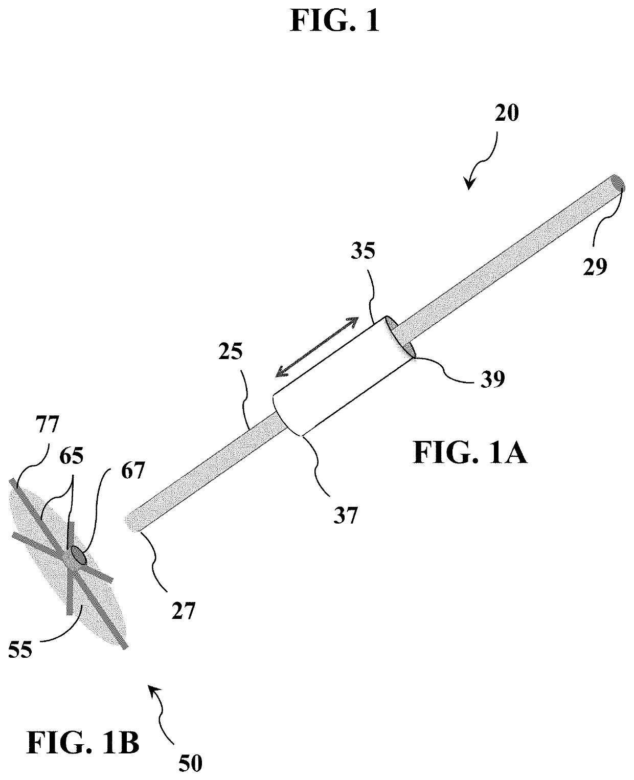
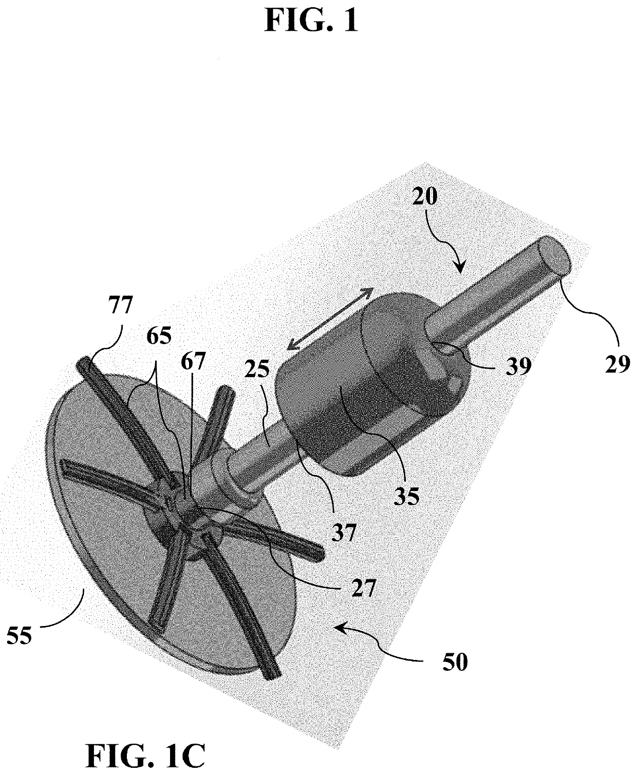
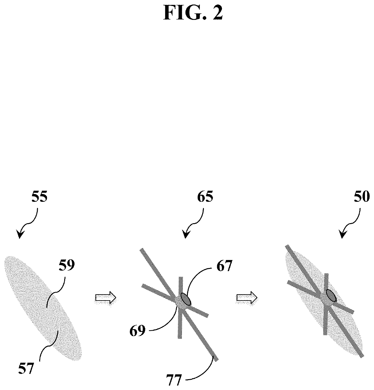
Provided are tissue repair and sealing devices, and methods for the use of tissue repair and sealing devices, for use in both minimally invasive surgical (MIS) procedures and open, non-MIS procedures to rapidly repair tissue fenestrations and create a pressure-resistant, watertight seal in a tissue barrier. Tissue repair and sealing devices disclosed herein comprise an integrated graft and deployable clasp assembly and an applicator assembly having a clasp retain and release member that is slidably connected to a folded, deployable clasp. The applicator assembly places a graft on a tissue inner surface and a deployable clasp on a tissue outer surface to secure the graft to the tissue inner surface to, thereby, repair a tissue fenestration and create a watertight barrier.

Description

CROSS-REFERENCE TO RELATED APPLICATIONS[0001]This U.S. non-provisional patent application was filed on Nov. 18, 2021 as U.S. patent application Ser. No. 17 / 530,365 and claims priority as a continuation from U.S. patent application Ser. No. 17 / 156,162, which was filed on Jan. 22, 2021 and claims the benefit of U.S. Provisional Patent Application No. 62 / 965,722, which was filed on Jan. 24, 2020. The contents of U.S. patent application Ser. No. 17 / 156,162 and U.S. Provisional Patent Application No. 62 / 965,722 are incorporated herein by reference in their entirety.BACKGROUND OF THE DISCLOSURETechnical Field[0002]The present disclosure relates, generally, to the field of medicine, in particular to surgery and surgical procedures, including both minimally invasive surgical (MIS) procedures and open surgical (non-MIS) procedures. Disclosed herein are tissue repair and sealing devices, and methods for their use, which comprise a detachable graft and clasp assembly for repairing tissue fenes...

Claims


Application Information

Patent Timeline
Patent Type & Authority Applications(United States)
IPC IPC(8): A61B17/00A61L31/14A61L27/36A61L27/18A61L27/54A61L27/24A61L27/22A61L27/06A61L27/04A61L31/06A61L31/02
CPCA61B17/0057A61B2017/00623A61L27/3633A61L27/18A61L27/54A61L27/24A61L27/227A61L27/3675A61L27/06A61L27/047A61L27/045A61L31/06A61L31/022A61B2017/00659A61B2017/00615A61L31/14A61B2017/00579A61B2017/00592A61B2017/00867A61B2017/00893A61B2017/00004A61B2017/00716
Inventor MAYBERG, MARC ROBERT
Owner PATCHCLAMP MEDTECH INC
Learn More