Machine learning based processing of magnetic resonance data, including an uncertainty quantification

a technology of magnetic resonance data and uncertainty quantification, applied in the field of machine learning based processing of magnetic resonance data, including uncertainty quantification, can solve the problems of complex mathematical modeling before contrast generation, high cost, and high cost of modeling, so as to avoid limitations and disadvantages of conventional techniques, reduce processing costs, and improve reliability

Pending Publication Date: 2022-06-09
MAX PLANCK GESELLSCHAFT ZUR FOERDERUNG DER WISSENSCHAFTEN EV
View PDF0 Cites 6 Cited by
  • Summary
  • Abstract
  • Description
  • Claims
  • Application Information

AI Technical Summary

Benefits of technology

[0049]It is an objective of the invention to provide an improved method of processing MR data, being capable of avoiding limitations and disadvantages of conventional techniques. In particular, the method is to be capable of processing MR data with a neural network (NN), wherein a reliability of processing MR data with the NN can be evaluated, in particular quantified. Especially, the processing of the MR data, including an error prediction, is to be provided with increased reliability, low processing costs and / or improved ability of recognizing imaging artefacts. It is a further objective of the invention to provide an improved MRI scanner apparatus, being adapted for processing collected MR data and being capable of avoiding limitations and disadvantages of conventional techniques. In particular, the MR scanner apparatus is to be capable of NN based analyzing the MR data, including an uncertainty prediction of the NN output.SUMMARY OF THE INVENTION
[0057]According to a preferred and particularly advantageous example, the at least one image parameter of the sample comprises at least one CEST contrast feature, that is obtained from collected MR data (e. g. unprocessed Z-spectra). Alternatively, other MR contrast features can be created as outlined below. In medical imaging, the invention substantially improves creating parameters of multi-pool Lorentzian fitting of CEST-spectra, e. g. at 3T. Additionally, the at least one uncertainty quantification measure allows radiographers to interpret the generated CEST maps with high confidence.
[0060]According to a preferred embodiment of the invention, the MR data analysis creates a parameter map of the at least one image parameter of the sample, and the at least one uncertainty quantification measure comprises an uncertainty map of an uncertainty quantity of the at least one image parameter. Advantageously, this allows a direct position resolved visualization of the uncertainty quantification measure, e. g. for showing whether the output image parameter map of the sample includes regions of reduced reliability.
[0063]According to a further advantageous embodiment of the invention, the training data of the neural network are provided by training data augmentation. The training data augmentation comprises a numerical enlargement of a given training data distribution of reference samples, e. g. subjects. Applying the training data augmentation has advantages for improving the reliability of uncertainty quantification.
[0065]With more details, training data augmentation has two desirable effects. First, training with noise leads to consistently increased uncertainties when applying a neural network to noise-corrupted data, representing the resulting prediction error more accurately than a network trained without augmentation. Second, including larger simulated B0 shifts in the training data allows better predictions for a larger range of B0 shifts of the input spectra. This resulted in lower prediction errors and consistently lower uncertainties for shifted input spectra when using NN1 compared to NN0. Therefore, including a plausible data augmentation, an advanced uncertainty prediction can be established using the proposed probabilistic predictive model. The uncertainty then indicates for example if input data is either corrupted as in a case where B0 artifacts shifted Z-spectra drastically, or in a case of vessels that can show noise-like pulsation artifacts in the spectral domain that are unique for a certain scan, or any other variation that was not in the training data. This can be seen in the results presented below, e.g. with reference to FIGS. 7 and 8.

Problems solved by technology

Possible artifacts in dynamic CEST MRI due to motion and field alterations.
It is generally known that sophisticated MR contrasts, such as diffusion tensor imaging, quantitative susceptibility mapping or spectroscopic MR imaging, as well as spectrally selective CEST (chemical exchange saturation transfer) imaging, often require complex mathematical modeling before contrast generation.
This modeling can be error-prone and time consuming, because it commonly must be carried out offline and sometimes requires user interaction and expert knowledge for evaluation.
Up to now, such sophisticated evaluations are not possible online in a robust manner, e. g. for medical imaging applications.
For example, for CEST data, both acquisition and subsequent evaluation are prone to artifacts attributed to field inhomogeneity and motion [12].
Last but not least, computations for 3D volume data can take hours depending on the algorithm [4].
However, because deep neural networks (NN) are often considered as “black boxes” with limited insight into the mapping from input to prediction, a question that arises is how accurate and reliable such predictions are.
A neural network that gives seemingly confident predictions for all possible inputs, regardless if reasonably supported by learning or not, may lead to critical failure cases and result in mistrust by potential users.
Due to these limitations, available neural network based MR data analyses cannot be used in medical practice.

Method used


Image

  • Machine learning based processing of magnetic resonance data, including an uncertainty quantification
  • Machine learning based processing of magnetic resonance data, including an uncertainty quantification
  • Machine learning based processing of magnetic resonance data, including an uncertainty quantification

Examples


Embodiment Construction

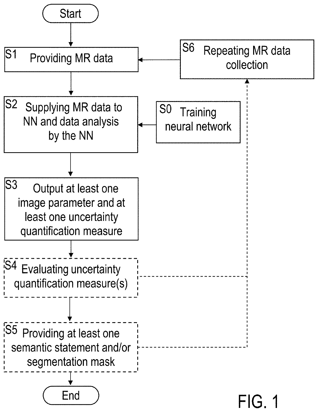
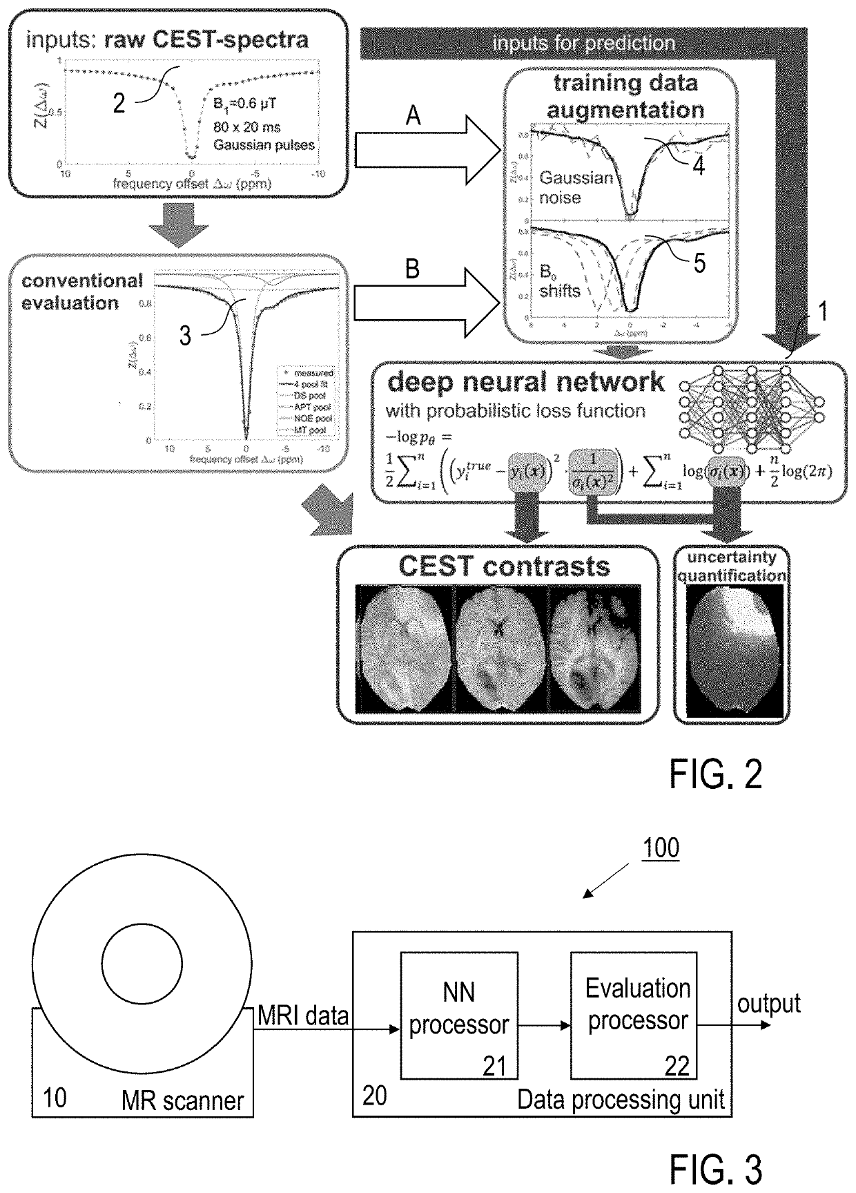
[0077]Embodiments of the invention are described in the following with particular reference to the configuration of the neural network for MR data processing, including creating at least one uncertainty quantification measure. The invention preferably is implemented with an MRI scanner as it is known per se. Accordingly, details of the MRI scanner, the available control schemes thereof, available excitations and read-out sequences, available schemes of MR signal acquisition and types of MR data are not described as they are known per se from prior art. Exemplary reference is made to the processing of CEST data. The implementation of the invention is not restricted to this particular type of data, but rather possible with other MR contrasts, e. g. as mentioned above. Furthermore, exemplary reference is made to providing uncertainty maps as the at least one uncertainty quantification measure. It is to be noted, that a single quantitative error value or a group of error values can be p...


PUM


Abstract

A method of processing magnetic resonance data of a sample under investigation includes the steps of provision of the MR data being collected with an MRI scanner apparatus, and machine learning based data analysis of the MR data by supplying the MR data to an artificial neural network being trained with predetermined training data, wherein at least one image parameter of the sample and additionally at least one uncertainty quantification measure representing a prediction error of the at least one image parameter are provided by output elements of the neural network. Furthermore, a magnetic resonance imaging (MRI) scanner apparatus being adapted for employing the method of processing MR data is described.

Description

FIELD OF THE INVENTION[0001]The invention relates to a method and to an apparatus for processing magnetic resonance (MR) data of a sample under investigation, e. g. a biological organism, like a patient or a test person, or a part thereof, or a material sample to be investigated. Preferably, the processing of MR data is included in a magnetic resonance tomography (MRT) procedure for investigating the sample under investigation. Applications of the invention are available e. g. in the fields of medical imaging or material investigations.TECHNICAL BACKGROUND[0002]In the present specification, reference is made to the following prior art illustrating technical background of the invention and related techniques:[0003][1] Zaiss M, Windschuh J, Paech D, et al. Relaxation-compensated CEST-MRI of the human brain at 7T: unbiased insight into NOE and amide signal changes in human glioblastoma. NeuroImage. 2015; 112:180-188;[0004][2] Windschuh J, Zaiss M, Meissner J E, et al. Correction of B1-...

Claims


Application Information

Patent Timeline
Patent Type & Authority Applications(United States)
IPC IPC(8): G01R33/56G01R33/565G06T7/00
CPCG01R33/5608G01R33/56563G06T2207/20081G06T2207/10088G06T2207/20084G06T7/0012G06T7/11G06T2207/30016G06T2207/30096G01R33/243G01R33/246G01R33/50G01R33/5601G01R33/5605G01R33/485
Inventor ZAISS, MORITZGLANG, FELIXPROKUDIN, SERGEYSCHEFFLER, KLAUS
Owner MAX PLANCK GESELLSCHAFT ZUR FOERDERUNG DER WISSENSCHAFTEN EV
Why Eureka
Try Eureka