Druggable target to treat retinal degeneration

a retinal degeneration and target technology, applied in the field of druggable target to treat retinal degeneration, can solve the problems of degeneration of neural retinal layer and hence vision loss, and no effective treatment for retinal degeneration

Pending Publication Date: 2022-10-27
US DEPT OF HEALTH & HUMAN SERVICES
View PDF0 Cites 1 Cited by
  • Summary
  • Abstract
  • Description
  • Claims
  • Application Information

AI Technical Summary

Problems solved by technology

The dysfunctional RPE has been associated with disease pathology and progression as it is unable to support photoreceptor which leads to the degeneration of neural retinal layer and hence vision loss.
Similarly, to date there are no effective tr

Method used


Image

  • Druggable target to treat retinal degeneration
  • Druggable target to treat retinal degeneration
  • Druggable target to treat retinal degeneration

Examples


example 1

t Competent Human Serum (CC-HS) Induces AMD-Like Cellular Endophenotypes in Mature iPSC-RPE

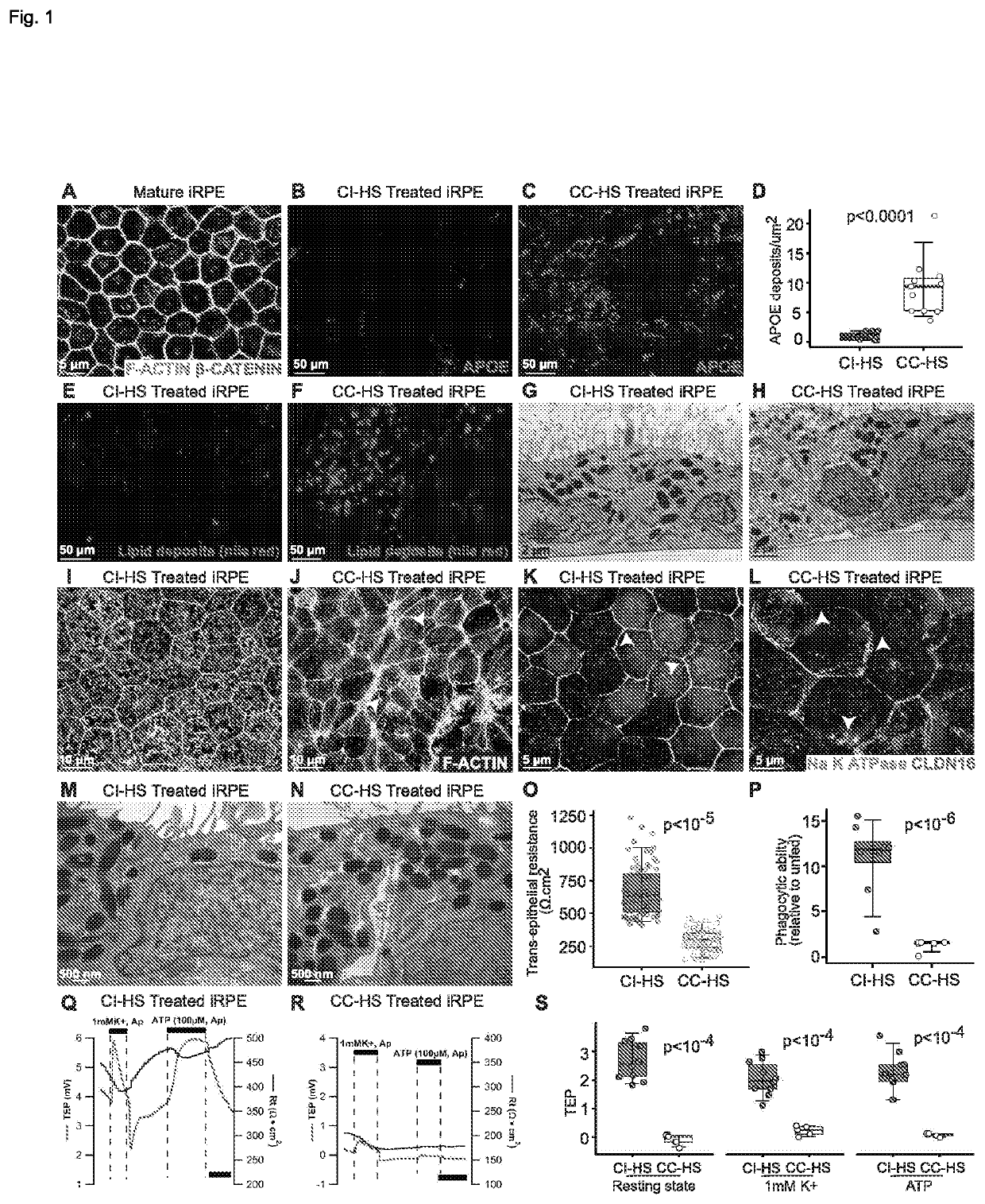
[0152]iPSCs derived from five different healthy individuals were used for this analysis. iPSCs were differentiated into mature RPE cells using previously published protocol (May-Simera et al., 2018, Sharma et al., 2019). Maturity of iRPE cells was confirmed by the presence of β-catenin on the cell membrane, (May-Simera et al., 2018, FIG. 1A) and by progressively increasing trans-epithelial resistance (TER) of the monolayer starting week 3 of culture (p−16, week 3 to weeks 4-6; FIG. 9A). Consistent with published reports for primary RPE cells (Johnson et al., 2011; Pilgrim et al., 2017), a five-fold increase in APOE positive sub-iRPE deposits was observed in CC-HS treated iRPE as compared to CI-HS treated cells (p9E, F). The presence of intracellular lipid deposits was further confirmed by transmission electron microscopy (TEM) of CC-HS treated iRPE cells (yellow arrowheads in FIG. 1H). TEM and...

example 2

ggered AMD Disease Phenotypes are Induced Through C3aR1 and C5aR1 Signaling

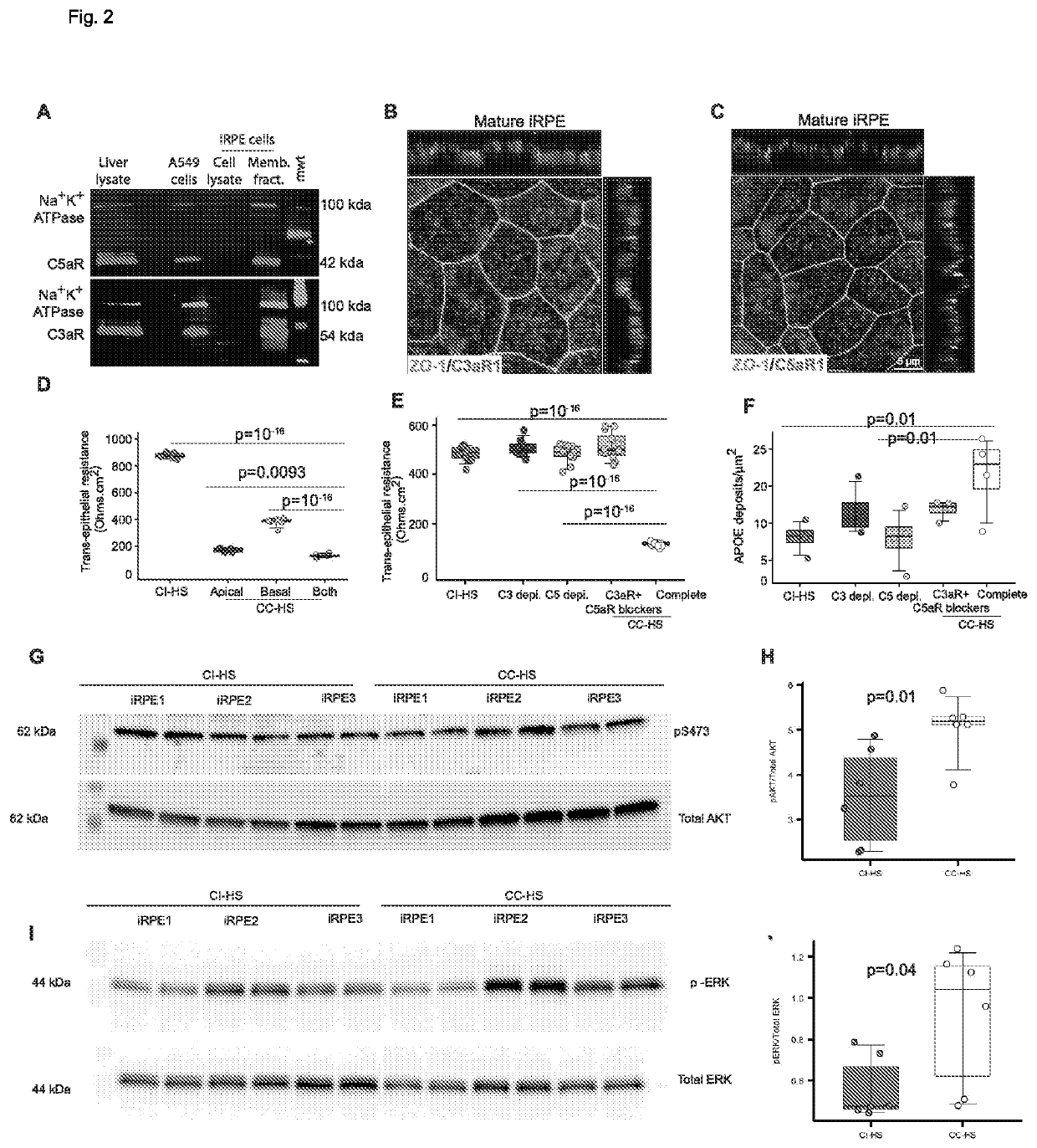
[0155]It was hypothesized that CC-HS triggered AMD cellular phenotypes in iRPE cells are generated by the anaphylatoxin arm of the complement pathway via C3a-C3aR1 and C5a-05aR1 signaling induced intracellular inflammation (Fernandez-Godino and Pierce 2018). RNAseq confirmed the expression of both receptors in iRPE cells with ˜30× higher expression in of C5aR1 as compared to C3aR1 (FIG. 10A). Furthermore, the expression of both receptors increases with CC-HS treatment. Western blot confirmed C3aR1 and C5aR1 receptors localization in the membrane fraction of iRPE cells (FIG. 2A). Their colocalization with the apical membrane marker EZRIN, but not with the basal membrane marker COLLAGEN IV suggests that the receptors for the two complement proteins, C3a and C5a, are predominantly apically located (FIGS. 2B, C, 10B). To verify that CC-HS activates C3aR and C5aR signaling in iRPE cells, samples were checked for p...

example 3

C5aR1 Induced subRPE Deposits are Mediated by Overactivation of NF-KB and Downregulation of Autophagy Pathways

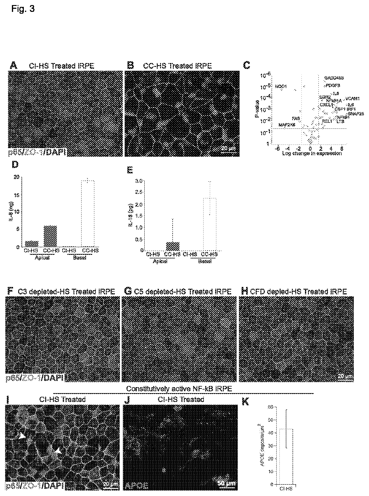
[0156]RNAseq analysis of CI-HS and CC-HS treated iRPE cells revealed dramatically different global gene expression pattern induced by CC-HS treatment (FIG. 11A). Consistent with the effect of anaphylatoxin complement (C3a, C5a) in immune cells, autophagy (p−6) and TNF / NF-KB (p−5) pathways were the most changed by CC-HS treatment of iRPE cells (Freeley et al., 2016; Kumar 2019 Int Rev of Immu; Nguyen et al., 2018; FIG. 11B, C, D). This led us to hypothesize that C3aR1 and C5aR1 signaling in iRPE cells is working through these two pathways. CC-HS treatment indeed caused p65 (RELA) subunit of NF-KB to translocate to the nucleus, suggesting its activation (Rayet and Gelinas 1999, Oncogene; FIG. 3A, B). Nuclear translocation of p65 led to 4-6× increased expression of NF-KB target genes (Tilborghs et al., 2017), as confirmed by RNAseq (FIG. 11B, p−5), qRT-PCR-based validation of s...


PUM


Abstract

This invention relates to novel method of treating or ameliorating a retinal disease or disorder or retinal degradation in a subject and a novel method of restoring retinal pigment epithelium cell compromising the administration of a one or more compounds which modulate Nox4, formation of radical oxygen species, serine protease, a dopamine receptor, NF-kB, mTOR, AMPK, RPE epithelial to mesenchymal transition, RPE dedifferentiation, or one or more Rho GTPases; and kits for administration of the methods.

Description

CROSS-REFERENCE TO RELATED APPLICATIONS[0001]This application claims the benefit of U.S. Provisional Patent Application No. 62 / 899,899 filed on Sep. 13, 2019. The entire contents of this patent application is incorporated herein by reference in its entireSTATEMENT REGARDING FEDERALLY SPONSORED RESEARCH OR DEVELOPMENT[0002]This invention was made with Government support under project numbers Z01 #: EY000532 by the National Institutes of Health, National Eye Institute. The government has certain rights in the invention.BACKGROUND OF THE INVENTION[0003]The retina is a layer of specialized light sensitive neural tissue located at the inner surface of the eye of vertebrates. Light reaching the retina after passing the cornea, the lens and the vitreous humor is transformed into chemical and electrical events that trigger nerve impulses. The cells that are responsible for transduction, the process for converting light into these biological processes are specialized neurons called photorece...

Claims


Application Information

Patent Timeline
IPC IPC(8): A61K31/155A61P27/02A61K9/00
CPCA61K31/155A61P27/02A61K9/0048A61K31/197A61K31/4704A61K31/519A61K31/403A61K31/15A61K31/445A61K31/497A61K31/46A61K31/4535A61K31/55A61K31/438A61K31/201A61K31/416A61K31/12A61K31/5513A61K31/451A61K31/14A61K31/40A61K31/401A61K31/454A61K31/138A61K31/137A61K31/495A61K31/4025A61K31/517A61K31/428A61K31/417A61K31/5685A61K31/496A61K31/42A61K31/265Y02A50/30A61K9/0019
Inventor BHARTI, KAPILBARBOSA SABANERO, KARLA YADIRACHANG, JUSTIN REN YUANJHA, BALENDU SHEKHARSHARMA, RUCHI
Owner US DEPT OF HEALTH & HUMAN SERVICES
Why Eureka
Try Eureka