
Method to independently analyze multiple biological processes in encapsulated 3D cell co-cultures

a cell co-culture and encapsulation technology, applied in the field of multiple encapsulated 3d cell co-culture drug testing or screening method, can solve the problems of poor clinical relevance of cell cultures used, inability to define good candidates with desired biological effects, and high cost of vitro drug testing in terms of time and resources, so as to increase the number of biological processes, increase the repertoire of biological processes, and increase the efficiency and accuracy

Pending Publication Date: 2022-11-03
UNIVERSITY OF GENEVA
View PDF0 Cites 0 Cited by
  • Summary
  • Abstract
  • Description
  • Claims
  • Application Information

AI Technical Summary

Benefits of technology

The present invention provides a method for testing drugs in a way that is more accurate and efficient in predicting how well they will work in humans. This is achieved by using a special co-culturing system that allows multiple biological processes to be measured simultaneously, which increases the clinical relevance of the drug test. This system also utilizes alginate capsules and 3D spheroid cell cultures, which better mimic the behavior of tissue in the body. Additionally, the invention creates a library of reporter genes to measure a wide range of biological processes, which is commonly investigated by pharmaceutical companies.

Problems solved by technology

Because of the large numbers of chemical entities that are screened, in vitro drug testing can be very costly in terms of time and resources.
Despite these large investments, in vitro drug testing is not always successful in defining good candidates with the desired biological effects for a number of reasons, mainly the poor clinical relevance of the cell cultures used.
Therefore, a problem in the current drug screening landscape, which results in the need for a multiplicity of tests to evaluate different properties and effects of potential drug candidates, is the lack of methods that can simultaneously address all of the abovementioned phenomena of cell culture drug testing.
Previous attempts to improve in vitro drug screening and testing have only partially addressed the aforementioned problems, and to the best of Applicant's knowledge have never addressed all of the aforementioned problems simultaneously.
Multiple genes can be detected after the co-culture of different types of cells, however the teaching of that document does not extend to 3D spheroid cell cultures, and the ability to independently analyze the different types of co-cultured cells is not possible.
Furthermore, is not possible to detect in which cells the biological processes have been modulated because the assay readout comprises of soluble protein(s).
While these documents are similar to the current art in that different fluorochomes (quantum dots) can be used to label the capsules, neither of them disclose any means of detecting multiple biological processes per test, nor easily discriminating the various capsules by microscopy because the liposomes are mainly used for genetic diagnosis of tumors.

Method used


Image

  • Method to independently analyze multiple biological processes in encapsulated 3D cell co-cultures
  • Method to independently analyze multiple biological processes in encapsulated 3D cell co-cultures
  • Method to independently analyze multiple biological processes in encapsulated 3D cell co-cultures

Examples


example 1

[0124]Description:

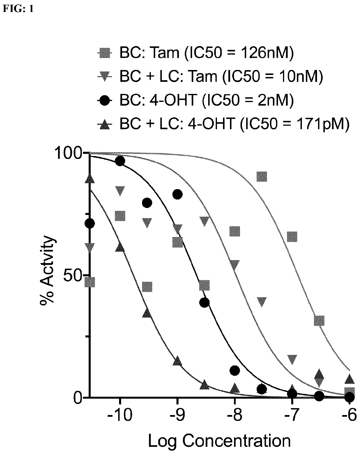
[0125]A monolayer of Breast Cancer (BC) cells stably expressing a luciferase reporter gene under the control of an Estrogen Response Element (MELN cells, Table 1 and 2) is co-cultured or not with encapsulated Liver Cancer (LC) cells and treated with increasing concentrations of Tamoxifen or 4-hydroxytamoxifen for 48 hours. The activity of Estrogen Receptor a in BC cells is determined by measuring the activity of Luciferase (bioluminescence intensity).

[0126]Material and Methods:

[0127]Cell Seeding for BC Cells

[0128]Aspirate the culture medium on the MELN cells (see Table 1)

[0129]Wash once with PBS and then aspirate the PBS

[0130]Add trypsin on the cells and aspirate the excess of trypsin

[0131]Incubate 5 minutes at 37° C.

[0132]Repeatedly pipette white cell culture medium (DMEM without phenol red 10% charcoal-stripped foetal bovine serum) over the cells to detach and then collect them into a tube Seed the cells in a 96-well plate with 10000 cells / well in white cell cult...

example 2

[0163]Description:

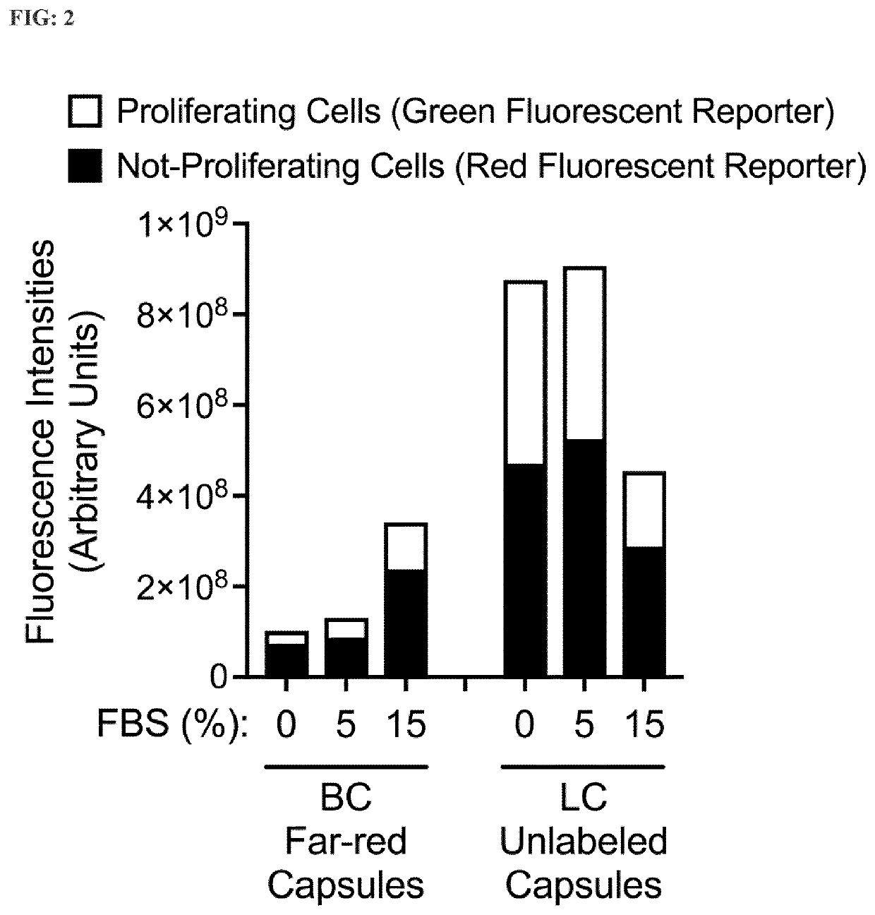
[0164]A co-culture of encapsulated BC cells stably expressing the dual-reporter FUCCI (see Table 2) in far-red-labeled capsules and encapsulated LC cells stably expressing the dual-reporter FUCCI in unlabeled capsules incubated with different serum concentrations. Amounts of active proliferating cells and not-proliferating cells (latent) are determined by quantifying the proportion of red (G1 phase) and green (G2 / S phase) nuclei both in LC cells and in BC cells simultaneously in the same co-culture wells.

[0165]Material and Methods:

[0166]Protocol for Establishing a Stable Reporter Cell Line:

[0167]Seed HEK293 cells in 10 cm-dishes at 4 million cells / dish.

[0168]Incubate for 24 hours at 37° C.

[0169]Cells are transfected with a mix of the plasmids pMD2G, psPAX2 and the lentiviral reporter plasmid of interest (pBOB-EF1-fastFUCCI-puro) by using a PolyEthylenelmine transfection method.

[0170]Add the transfection solution to the cells, and incubate overnight at 37° C.

[0171]P...

example 3

[0208]Description:

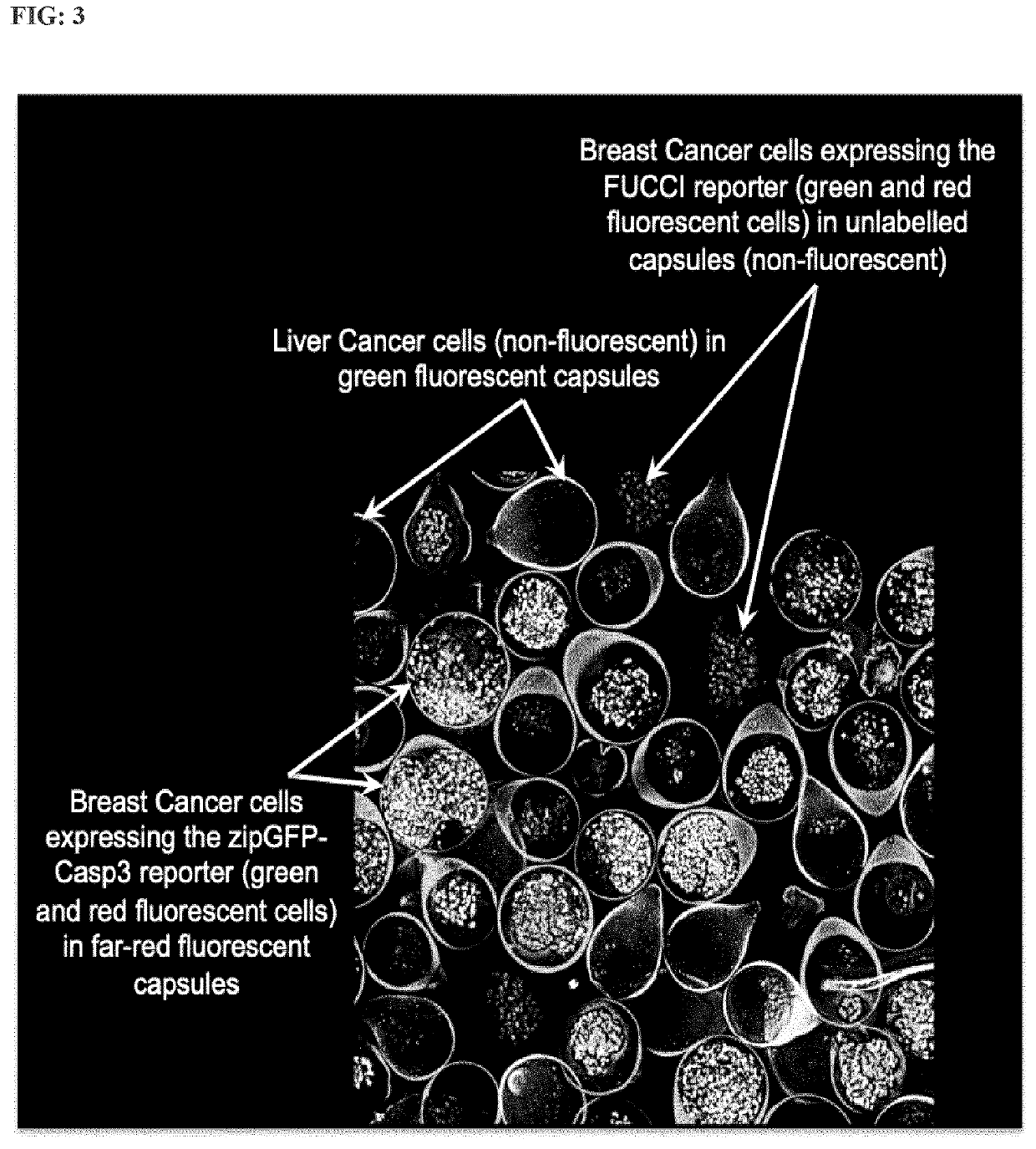
[0209]A co-culture of encapsulated BC cells stably expressing the dual-reporter FUCCI (Table 2) in unlabelled capsules, and encapsulated BC cells stably expressing the reporter zipGFP-Casp3 (Table 2) in far-red-labelled capsules, co-cultured or not with encapsulated LC cells in green-labelled capsules and treated with increasing concentrations of Tamoxifen for 96 hours. Induction of apoptosis is measured by automated confocal microscopy (HCS) in BC cells stably expressing the reporter zipGFP-Casp3 and proliferation is measured at the same time in BC cells stably expressing the dual-reporter FUCCI.

[0210]Materials and Methods

[0211]Protocol for Establishing the Two BC Stable Reporter Cell Lines:

[0212]Same protocol as for Example 2 “Protocol for establishing a stable reporter cell line” except that in addition to make a BC cell line stably expressing the dual-reporter FUCCI, Applicants have also made a BC cell line stably expressing the reporter zipGFP-Casp3 (Table2) w...


PUM

PropertyMeasurementUnit
sizeaaaaaaaaaa
concentrationaaaaaaaaaa
pHaaaaaaaaaa

Abstract

A multiplexed and encapsulated 3D cell co-culture drug testing or screening method which discloses an in vitro drug testing kit suitable for testing the effect of one or more drugs of interest on multiple biological processes in one or more target cell types.

Description

FIELD OF THE INVENTION[0001]The invention relates to a multiplexed and encapsulated 3D cell co-culture drug testing or screening method. The invention also discloses an in vitro drug testing kit suitable for testing the effect of one or more drugs of interest on multiple biological processes in one or more target cell types.BACKGROUND OF THE INVENTION[0002]In vitro screening or testing of molecular entities on biological cells constitutes an essential procedure that is used for the discovery of new pharmaceuticals, and also more recently, during ‘precision medicine’ testing to define the best drug(s) to treat patients. In vitro molecule screening can be used at both the early stages of drug discovery to identify ‘hit’ molecules, or at subsequent stages to define a set of ‘lead’ entities from the hits, which are defined as hits that have multiple favourable characteristics. Because of the large numbers of chemical entities that are screened, in vitro drug testing can be very costly i...

Claims


Application Information

Patent Timeline
Patent Type & Authority Applications(United States)
IPC IPC(8): G01N33/50C12Q1/6897
CPCG01N33/5011C12Q1/6897C12Q1/66G01N33/5008G01N21/6428G01N21/763G01N2500/10G01N2021/6439G01N33/5014G01N33/5041G01N33/5044G01N33/5067C12Q1/18C12Q1/025C12M25/16C12Q2563/159C12Q2563/103C12Q2563/107
Inventor SEGALA, GRÉGORYPEJOSKI, DAVIDROUX, AURÉLIENPICARD, DIDIERMOREAU, DIMITRI VINCENTBOURRAT, BRYAN JOSUÉ
Owner UNIVERSITY OF GENEVA
Learn More