Adeno-Associated Virus Vector Delivery of Muscle Specific Micro-Dystrophin To Treat Muscular Dystrophy

a technology of adenoassociated virus and microdystrophin, which is applied in the direction of drug compositions, peptide/protein ingredients, peptide sources, etc., can solve the problems of muscle function deficits that produce muscular dystrophies (mds), serious impact on quality of life, and membrane fragility

Pending Publication Date: 2022-11-17
RES INST AT NATIONWIDE CHILDRENS HOSPITAL
View PDF0 Cites 0 Cited by
  • Summary
  • Abstract
  • Description
  • Claims
  • Application Information

AI Technical Summary

Benefits of technology

[0012]The present invention is directed to gene therapy vectors, e.g. AAV, expressing the micro-dystrophin gene to skeletal muscles including diaphragm and cardiac muscle to protect muscle fibers from injury, increase muscle strength and reduce and / or prevent fibrosis
[0014]In one embodiment, the invention provides for a rAAV vector comprising a muscle specific control element nucleotide sequence, and a nucleotide sequence encoding the micro-dystrophin protein. For example, the nucleotide sequence encodes a functional micro-dystrophin protein, wherein the nucleotide is, e.g., at least 65%, at least 70%, at least 75%, at least 80%, 81%, 82%, 83%, 84%, 85%, 86%, 87%, 88%, or 89%, more typically at least 90%, 91%, 92%, 93%, or 94% and even more typically at least 95%, 96%, 97%, 98%, 99%, or 100% sequence identity to SEQ ID NO: 1, wherein the protein retains micro-dystrophin activity. The micro-dystrophin protein provides stability to the muscle membrane during muscle contraction, e.g. micro-dystrophin acts as a shock absorber during muscle contraction.
[0038]The invention also contemplates administering a AAV of the invention to a subject suffering from muscular dystrophy who already has developed fibrosis, in order to prevent new fibrosis in these subjects or to reduce fibrosis in these patients. The invention also provides for administering any of the rAAV of the invention to the patient suffering from muscular dystrophy who already has reduced muscle force or has reduced muscle mass in order to protect the muscle from further injury.

Problems solved by technology

Deficits in muscle function produce muscular dystrophies (MDs) that are characterized by muscle weakness and wasting and have serious impacts on quality of life.
These MDs result from membrane fragility associated with the loss of sarcolemmal-cytoskeleton tethering by the DAPC.
Without membrane stabilization from dystrophin or a micro-dystrophin, DMD will manifest uncontrolled cycles of tissue injury and repair ultimately replace lost muscle fibers with fibrotic scar tissue through connective tissue proliferation.
The over-production of fibrotic tissue restricts muscle regeneration and contributes to progressive muscle weakness in the DMD patient.
Finally, AAV-infected cells are not resistant to superinfection.

Method used


Image

  • Adeno-Associated Virus Vector Delivery of Muscle Specific Micro-Dystrophin To Treat Muscular Dystrophy
  • Adeno-Associated Virus Vector Delivery of Muscle Specific Micro-Dystrophin To Treat Muscular Dystrophy
  • Adeno-Associated Virus Vector Delivery of Muscle Specific Micro-Dystrophin To Treat Muscular Dystrophy

Examples


example 1

Generation of the pAAV.MHCK7.Micro-Dystrophin Construct

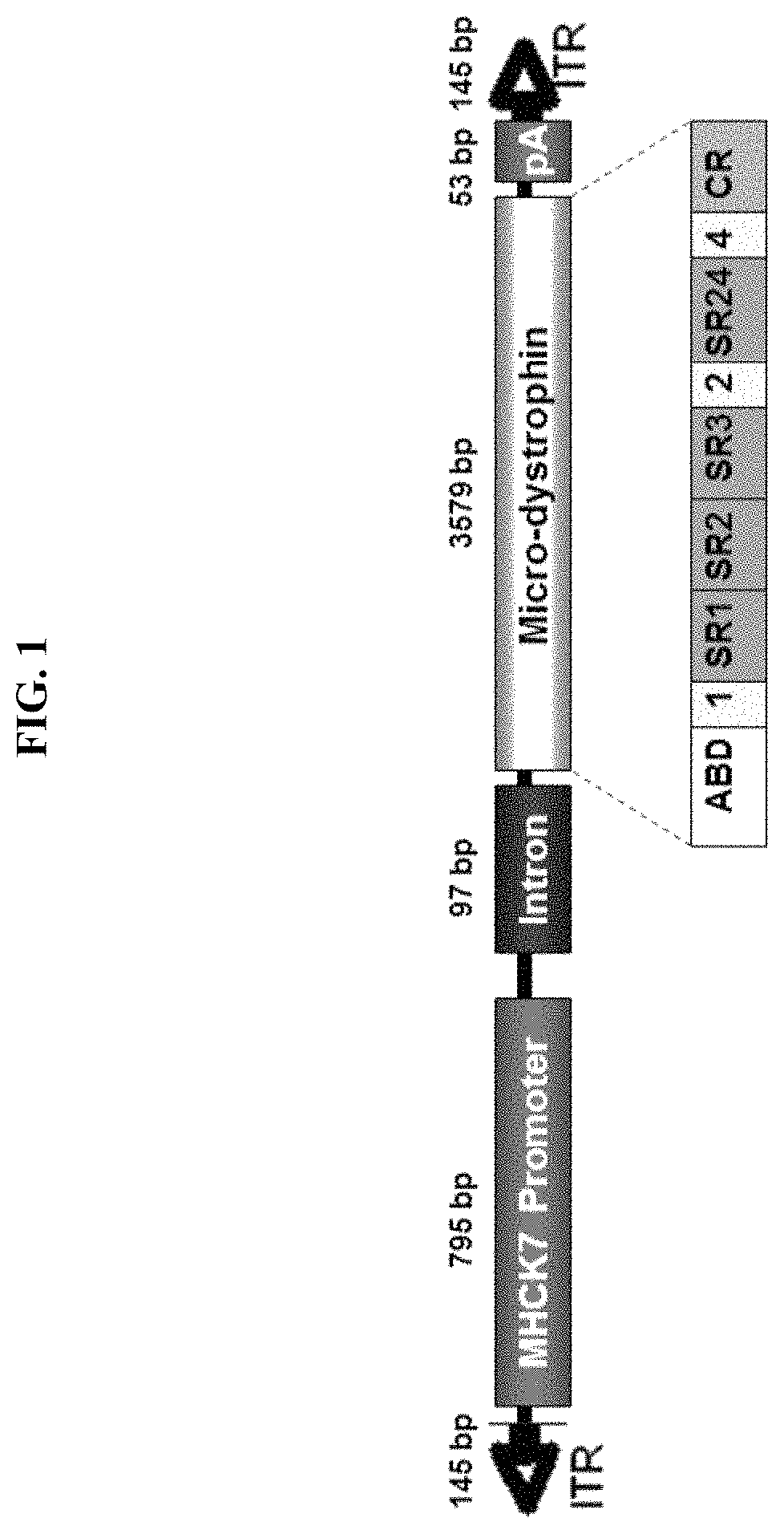
[0119]The pAAV.MHCK7.micro-dystrophin plasmid contains a human micro-dystrophin cDNA expression cassette flanked by AAV2 inverted terminal repeat sequences (ITR) (see FIG. 1). The micro-dystrophin construct was characterized by an in-frame rod deletion (R4-R23), while hinges 1, 2 and 4 and cysteine rich domain remain producing a 138 kDa protein. The expression of the micro-dystrophin protein (3579 bp) was guided by a MHCK7 promoter (795 bp). The plasmid was constructed from the pAAV.MCK.micro-dystrophin plasmid by removing the MCK promoter and inserting the MHCK7 promoter. After the core promoter, the 53 bp endogenous mouse MCK Exon1 (untranslated) is present for efficient transcription initiation, followed by the SV40 late 16S / 19S splice signals (97 bp) and a small 5′UTR (61 bp). The intron and 5′ UTR are derived from plasmid pCMVß (Clontech). The micro-dystrophin cassette had a consensus Kozak immediately in front of the ATG s...

example 2

Intramuscular Expression Studies Using rAAV.MHCK7.Micro-Dystrophin

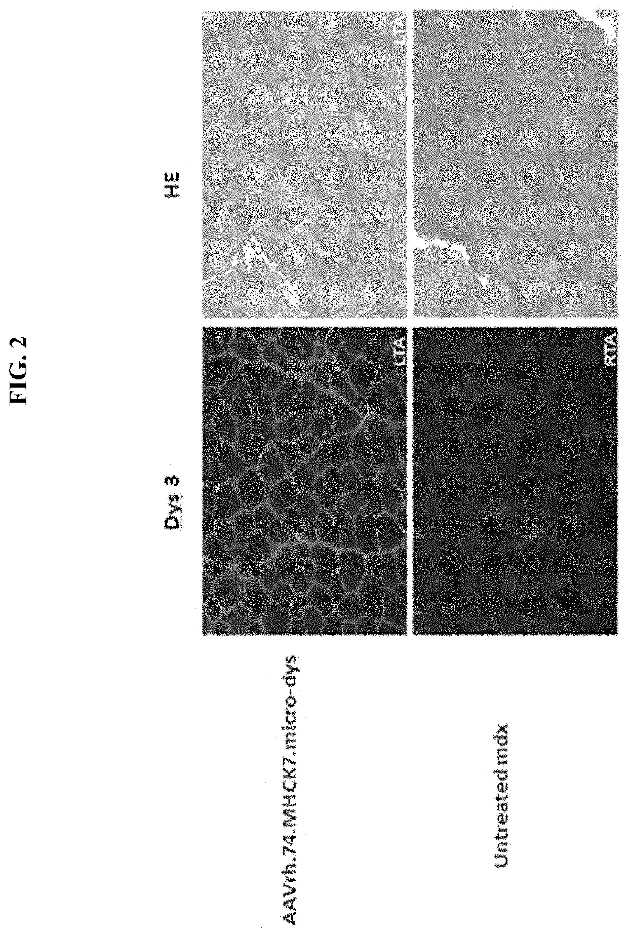
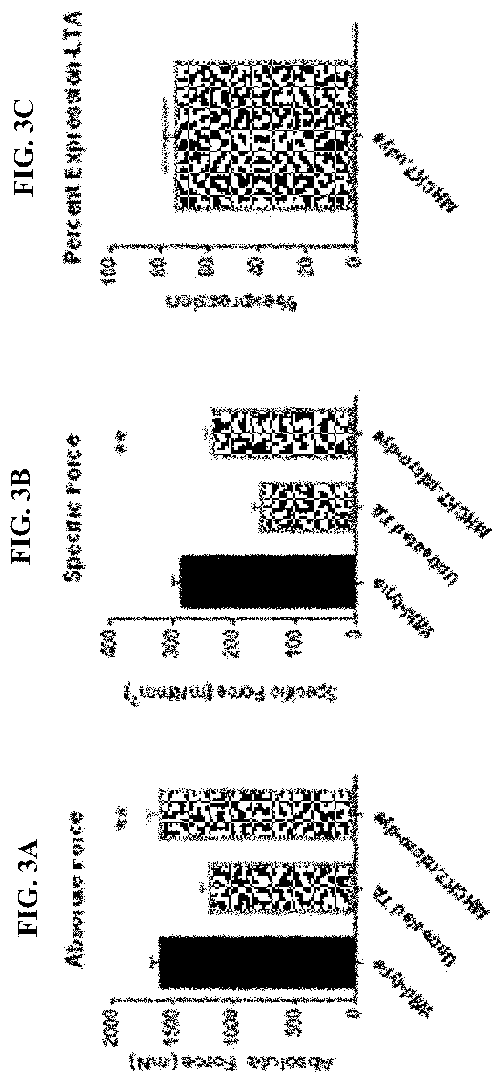
[0121]Expression studies were conducted with the human micro-dystrophin construct (rAAVrh74.MHCK7. micro-dystrophin; described in Example 1) by intramuscular injection. The tibialis anterior muscle of mdx mice (spontaneous Dmdmdx (mutant mice that do not express dystrophin) were injected with 1×10¹¹ vg of the cassette (n=5 per group). Six weeks later the muscles were harvested and stained for dystrophin (Dys3) expression with an N-terminal antibody for dystrophin and hematoxylin and eosin (HE) staining. FIG. 2 shows diffuse gene expression and reduction in centrally located nuclei with 1×10¹¹ vg dose compared to the untreated muscle. Furthermore, a decrease in central nucleation with an increase in average fibers / frame was observed following treatment with micro-dystrophin construct. Expression levels of the rAAVrh74.MHCK7. micro-dystrophin construct were quantified at about 73%.

[0122]In addition to measuring micro-dy...

example 3

Systemic Delivery of rAAVrh.74.MHCK7.Micro-Dystrophin to Mdx Mice

[0123]Cohorts of mdx mice were injected via tail vein with either 2×10¹² vg (8×10¹³ vg / kg) or high dose (planned clinical dose) 6×10¹² vg (2×10¹⁴ vg / kg) of rAAVrh.74.MHCK7.micro-dystrophin at 6 weeks of age. Following 12 weeks of treatment, all muscles were harvested and stained for dystrophin and restoration of DAPC components. Systemically injected (tail vein) mice showed high levels of staining of dystrophin throughout all muscles. FIG. 4A represents the widespread transduction of skeletal, diaphragm and cardiac muscle fibers after a 6×10¹² vg (2×10 14 vg / kg) systemic dose. FIG. 4B shows quantification of the percentage of muscle fibers expressing micro-dystrophin in each tissue. Finally the diaphragm was tested for functional improvement (FIG. 4C). No significant difference was seen at low dose; however there was significant improvement at the high dose. Importantly, FIG. 5 demonstrates other components of the DAPC...


PUM

PropertyMeasurementUnit
timeaaaaaaaaaa
timeaaaaaaaaaa
muscular forceaaaaaaaaaa

Abstract

The invention provides gene therapy vectors, such as adeno-associated virus (AAV) vectors, expressing a miniaturized human micro-dystrophin gene and method of using these vectors to express micro-dystrophin in skeletal muscles including diaphragm and cardiac muscle and to protect muscle fibers from injury, increase muscle strength and reduce and / or prevent fibrosis in subjects suffering from muscular dystrophy.

Description

[0001]This application is a U.S. National stage of International Application No. PCT / US2018 / 022881 filed Mar. 16, 2018 which claims priority to U.S. Provisional Patent Application No. 62 / 473,148, filed Mar. 17, 2017 which is incorporated by reference herein in its entirety.[0002]This invention was made with government support under grant number NS055958 awarded by the National Institutes of Health / national Institute of Neurological Disorders and Stroke. The government has certain rights in the invention.INCORPORATION BY REFERENCE OF MATERIAL SUBMITTED ELECTRONICALLY[0003]This application contains, as a separate part of the disclosure, a Sequence Listing in computer-readable form which is incorporated by reference in its entirety and identified as follows: Filename: 51475_Seqlisting.txt; Size: 29,608 bytes, created; Jun. 3, 2022.FIELD OF INVENTION[0004]The invention provides gene therapy vectors, such as adeno-associated virus (AAV) vectors, expressing a miniaturized human micro-dyst...

Claims


Application Information

Patent Timeline
Patent Type & Authority Applications(United States)
IPC IPC(8): C12N15/86A61P21/00A61K9/00A61K38/17C12N7/00
CPCC12N15/86A61P21/00A61K9/0029A61K38/1709C12N7/00A61K48/00A01K2227/105A01K2267/0306C07K14/47C12N2750/14143C12N2830/008A01K67/0275A61K48/0058
Inventor RODINO-KLAPAC, LOUISEMENDELL, JERRY R.
Owner RES INST AT NATIONWIDE CHILDRENS HOSPITAL
Why Eureka
Try Eureka